Original Article
Postoperative hyperlactatemia is linked to acute kidney
injury after type A aortic dissection surgery: a
retrospective study
Caixia Ruan, Wei Shang, Yazhou Liu, Chenglong Li, Hong Wang, Nan Liu, Xiaotong Hou
Center for Cardiac Intensive Care, Beijing Anzhen Hospital, Beijing Institute of Heart, Lung and Blood Vessel Dis-eases, Capital Medical University, Beijing 100029, China
Received March 7, 2016; Accepted June 5, 2016; Epub July 15, 2016; Published July 30, 2016
Abstract: Purpose: Acute kidney injury (AKI) could increase the risk of mortality after cardiac surgery. We aimed to explore the predictive value of hyperlactatemia in AKI in patients undergoing type A aortic dissection (TA-AAD) opera-tion. Methods: A total of 337 consecutive patients who underwent TA-AAD surgery in Beijing Anzhen Hospital from February 2009 to December 2013 were included into this study. Based on blood lactate level at 6 hours after
sur-gery (median, 4.1 mmol/L), patients were divided into two groups: high (≥4.1 mmol/L) and low lactic (<4.1 mmol/L)
groups. We examined the association between risk factors and AKI, especially the impact of postoperative lactate level on the incidence of AKI. Results: Of 337 patients, 152 (45.1%) patients had early postoperative AKI. 29 (8.6%) patients needed Continuous Renal Replacement Therapy (CRRT). There were 164 patients in the high lactate group and 173 patients in the low lactate group. Additionally, the rates of the AKI Stage 2-3 were also markedly different
between the two groups (P<0.05). Furthermore, postoperative lactate level at 6 h was an effective predicator for AKI (OR, 1.284; 95% CI: 1.129-1.460; P<0.001; AUC, 0.767) and RRT (OR, 1.295; 95% CI: 0.737-0.891; P<0.001; AUC, 0.814). Lactate level of 5.05 mmol/L was the boundary value for AKI Stage 2-3 (sensitivity 71.0%, specific
-ity 69.3%). Meanwhile, lactate level of 7.0 mmol/L was the boundary value for CRRT (sensitiv-ity 73.9%, specific-ity
79.5%). Conclusions: Our study found that early postoperative lactate level was an effective predictor of AKI and CRRT for patients with acute type A aortic dissection.
Keywords: Hyperlactatemia, type A aortic dissection, acute kidney injury, renal replacement therapy
Introduction
Acute kidney injury (AKI) is a common serious complication for patients following the aorta operation. It was reported that the incidence of AKI was approximately 28.6%-54.0%, of which 1.7%-11% patients had to receive renal replace-ment therapy (RRT) [1]. Moreover, AKI was an independent predictor of increased risk of mor-tality irrespective of the perioperative risk fac-tors [2]. In a Sweden nationwide population-based study of 27,929 patients undergoing cardiac surgery, Rydén et al showed that AKI was associated with long-term risk of mortality and myocardial infarction [3]. Additionally, Pickering et al found that the pooled rate of car-diopulmonary bypass (CPB)-related AKI was 18.2%, and of RRT was 2.1%. Moreover, AKI was associated with a more than 2-fold in- crease in early mortality [4]. Hyperlactatemiain patients who have undergone type A aortic
dis-section (TA-AAD) surgeryis fairly common andis associated with tissue hypoxia [5], which is associated with increased risk of AKI [6, 7]. Several studies have confirmed that postopera-tive hyperlactatemia was related to incidence of AKI in patients with cardiac surgery. However, no study evaluates the predictive value of hyperlactatemia for AKI and RRT in patients with aortic dissection surgery. Therefore, our study is to evaluate the relationship of the post-operative lactate level with the rate of AKI and RRT.
Materials and methods
Study population
owing to incomplete data. Our study was approved by the Beijing Anzhen Hospital Review Board. On the basis of the Stanford classifica-tion, acute type A dissection was separated into 3 subtypes [8, 9]. Subtype A1: normal aor-tic root type; Subtype A2: mild-to-moderately involved aortic root type; and Subtype A3: severely involved aortic root type. Additionally, according to the pathological characteristics of aortic arch, it divided into subtype C (complex type) and subtype S (simple type). Meanwhile, there are several different operation types, including aortic root plastic or replacement; ascending aorta replacement; partial or full aortic arch replacement and aortic arch replacement plus stented elephant trunk implantation surgery.
CPB is established based on different surgery circumstances, including moderate hypother-mal CPB, hypotherhypother-mal CPB, deep hypothermia circulatory arrest (DHCA) and selective cerebral perfusion (SCP). Arteriovenous cannulation methods varied according to different types of surgeries. When lesions affect aortic arch, cerebral perfusion or separated perfusion for upper and lower body was performed to protect brain function.
After going back to ICU, the mechanical ventila-tion mode was set as control mode ventilaventila-tion: rate was 12/min; tidal volume 6-8 mL/kg; PEEP 4-8 mmHg, FiO2 40%-60% to keep SAT ≥97%. Additionally, we adjusted the amount vasoac-tive drugs, such as dopamine, dobutamine, epi-nephrine, norepinephrine to keep hemodynam-ic stable.
KDIGO criteria
AKI was defined with the Kidney Disease Im- proving Global Outcomes Work Group (KDIGO) criteria [10], including the increase of serum creatinine (sCr) 0.3 mg/dl within 48 h, or increase of sCr by 1.5-fold above baseline, known orassumed to have occurred within 7 days. Meanwhile, participants with AKI was divided into 3 stages by postoperative sCr level. Stage 1: sCr≥1.5 times baseline or sCr≥0.3 mg/dL increase; Stage 2: sCr≥2.0 times base-line; Stage 3: ≥3.0 times baseline or increase of sCr to ≥4.0 mg/dl or initiation of RRT.
Continuous renal replacement therapy (CRRT)
The indications of CRRT include volume over-load, serious kidney dysfunction and
electro-lyte disturbances. Based on the participants’ renal function, cardiopulmonary function, in- flammatory response, water-electrolyte status and acid-base balance, surgeons, ICU doctors and renal physicians will decide whether the RRT was performed. For patients with CRRT, we used the continuous veno venous hemo dia fil-tration (CVVHDF) mode with the Prismaflex sys-tem (Barkey Gambro, Sweden). The treatment mode, dose, anticoagulation methods and dehydrated volume were performed according to the patients’ conditions.
Data collection
We used the BECKMAN COULTER-AU5400 au- tomatic biochemical analyzer to assess se- rum creatinine, urea nitrogen and alanine ami-notransferase (ALT). Additionally, RADIOMETER-ABL800 blood gas analyzer was used to detect lactate level and other items. We also recorded patients’ central venous pressure (CVP), mean arterial pressure (MAP), arterial blood gas anal-ysis (including PaO2, glucose, lactate) at 6 h post-operation, in-hospital rate of AKI and CRRT.
Statistical analysis
All statistical analysis was performed with SPSS 19.0 (SPSS, Inc., Chicago, Ill). Continuous variables were expressed as mean ± SD or the median with interquartile range. Independent continuous variables were compared with 2-ta- iled Student t tests. Categorical variables were expressed as frequency or ratio and compared with the Pearson c2 statistic. Stepwise
multi-variate logistic regressions were used to ana-lyze the predictors of AKI and RRT. Receiver operating characteristic (ROC) curves were con-structed, and the area under the ROC curve (AUC) was used to assess discriminant ability of the lactate levels measured 6 hours after sur-gery to predict the rate of AKI and RRT. The cut-off was the value that maximized the sensitivity and specificity. P<0.05 was statistically signi- ficant.
Result
Study population
(21.2%). According to serum lactate level within 6 h after operation (median, 4.1 mmol/L), all participants were separated into high lactic
[image:3.612.91.364.96.408.2]1.000-1.001, P<0.001) were associated with the occurrence of AKI, and the lactate are the risk factors for AKI.
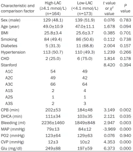
Table 1. The Characteristics of the high-LAC versus low-LAC groups
Characteristic and comparison factor
High-LAC
(≥4.1 mmol/L)
(n=164)
Low-LAC
(<4.1 mmol/L)
(n=173)
t value or χ2 value
P value
Sex (male) 129 (48.1) 139 (51.9) 0.076 0.783
Age (year) 49.0±10.9 47.0±11.1 1.678 0.094
BMI 25.8±3.4 25.6±3.7 0.385 0.701
Smoking 84 (49.4) 86 (50.6) 0.112 0.738
Diabetes 5 (31.3) 11 (68.8) 2.004 0.157
Hypertension 113 (50.7) 110 (49.3) 1.239 0.266
CHD 2 (25.0) 6 (75.0) 1.814 0.178
Stanford 8.420 0.394
A1C 54 49
A2C 49 42
A3C 66 64
A1S 2 4
A2S 1 1
A3S 2 3
CPB (min) 202±53 184±48 3.149 0.002
DHCA (min) 111±34 103±35 2.121 0.035
Bleeding (ml) 2236±1460 1849±848 2.947 0.003
MAP (mmHg) 79±13 84±12 -3.969 0.000
PO2 (mmHg) 123±64 129±63 0.076 0.940
CVP (mmHg) 12±3 10±2 4.353 0.000
Glu (mg/dl) 249±88 197±59 6.373 0.000
LAC, lactate; BMI, body mass index; CHD, coronary heart disease; CPB, cardio-pulmonary bypass; DHCA, deep hypothermal circulatory arrest; LVEF, left ven-tricle eject fraction; MAP, mean arterial pressure; CVP, central venous pressure; Glu, glucose; Data are mean ± standard deviation or n (%).
Table 2. Outcomes and comparisons of high- versus low-LAC groups
Characteristic (≥4.1 mmol/L)High-LAC
(n=164)
Low-LAC
(<4.1 mmol/L)
(n=173)
t value or χ2 value
P value
Ventilation time (h) 68±103 38±50 3.472 0.001
ICU LOS (day) 4.6±6.3 2.5±2.4 4.143 0.000
Hospital LOS (day) 23.4±15.3 17.9±8.0 3.640 0.000
Hospital mortality 18 (94.7) 1 (5.3) 17.228 0.000
CRRT (n/%) 26 (89.7) 3 (10.3) 21.51 0.000
AKI 92 (60.5) 60 (39.5) 16.04 0.000
Stage 1 40 (48.2) 43 (51.8) 0.004 0.947
Stage 2 28 (66.7) 14 (33.3) 6.334 0.012
Stage 3 24 (88.9) 3 (11.1) 19.163 0.000
ICU, intensive-care unit; LOS, length of stay; CRRT, continuous renal replace -ment therapy; AKI, acute renal injury; Data are median (interquartile range), mean ± standard deviation or n (%).
(≥4.1 mmol/L) and low lactic (<4.1 mmol/L) groups. The char-acteristics of two groups are illustrated in Table 1. There were no significant differences bet- ween two groups in age, BMI, rates of smoking, diabetes, coro-nary heart disease, hypertension and the Stanford types of aorta dissection. However, the two gro- ups were statistically different in duration of CPB (202±53 min vs. 184±48 min), DHCA (111±34 min vs. 103±35 min), blood loss (2236±1460 mL vs. 1849±848 mL), MAP (79±13 mmHg vs. 84± 12 mmHg), CVP (12±3 mmHg vs. 10±2 mmHg) and blood gluco- se (249±88 mmol/L vs. 197±59 mmol/L).
Outcomes
AKI occurred in 152 (45.1%) patients, of which 29 patients (8.6%) needed CRRT. The aver-age length of CRRT was 5.9±3.1 d, and average length in ICUwas 3.5±4.8 d. There were significant differences between the two groups in the length of intubation (68±103 h vs. 38±50 h), ICU length of stay (4.6±6.3 d vs. 2.5±2.4 d), hospital length of stay (23.4±15.3 d vs. 17.9±8.0 d) and rate AKI (60.5% vs. 39.5%), CRRT (89.7% vs. 10.3%), stage 2 AKI (66.7% vs. 33.3%) and stage 3 AKI (88.8% vs. 11.1%). However, the risk of Stage 1 AKI was comparable between the two groups. The outcomes are illustrated in Table 2.
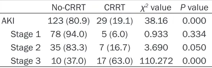
[image:3.612.91.362.495.649.2]The comparisons of the stage of AKI versus RRT in Table 4. there were significant differenc-es observed between the No-CRRT versus CRRT groups in AKI (80.9% vs. 19.1%, respec-tively, P=0.001), stage 2 of AKI (83.3% vs. 16.7%, P=0.050) and stage 3 of AKI (37.0% vs. 63.0%, P<0.001).
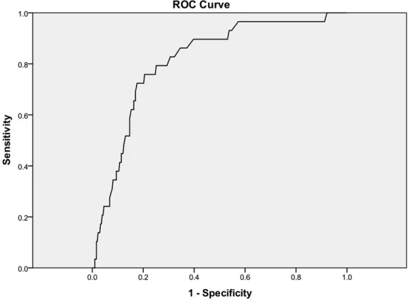
The receiver operating characteristics (ROC) curve of lactate predicting stage 2 to stage 3 of AKI after surgery of aortic dissection type A was illustrated in Figure 1. The AUC was 0.767 (OR 1.284; 95% CI 1.129-1.460, P<0.001). Lactate levels of 5.05 mmol/L was the boundary value of AKI (sensitivity 71.0%, specificity 69.3%). Additionally, the AUC of lactate predicting CRRT 0.814 (95% CI 0.737-0.891, P<0.001). Lactate levels of 7.0 mmol/L was the boundary value of CRRT (sensitivity, 75.9%; specificity, 79.5%) (Figure 2).
Discussion
In consistent with previous studies, our study demonstrated that lactate levels at 6 hours
after aortic dissection surgery was significantly related to postoperative adverse events, includ-ing AKI and CRRT. A threshold of 5.05 mmol/L at 6 hours after surgery is independently asso-ciated with the risk of AKI. Additionally, a lac-tate level of 7.0 mmol/L identifies patients needing CRRT. More studies are needed to con-firm our findings.
Hyperglycemia was associated with increased risk of AKI. Cardiac surgery is the second lead-ing causes of AKI in the ICU, just followlead-ing sep-sis. Although the pathophysiology of AKI follow-ing cardiac surgery is multifactorial, the do- minant mechanism of injury is thought to be intraoperative ischemia-reperfusion injury. Si- nce lactate is a sensitive biomarker of tissue hypoxia, it can be used as a marker of AKI. Dividing 117 patients in the AKI group (n=17) and non-AKI group (n=100), Zhang et al report-ed that normalizreport-ed lactate load was indepen-dently associated with postoperative AKI in patients undergoing CPB [11]. The AUC was 0.63 (95% CI 0.47-0.79). The best sensitivity and specificity were 41.2% and 87% at the cut-off point of 4.4 mmol/L. Moreover, they found that at different time points, serum lactate showed markedly different diagnostic perfor-mance. While lactate measured early may be timely enough but also lacks accuracy, and lac-tate measured late may be accurate but also may be too late. They found that the time point of 8.4 hours after ICU entry showed better diag-nostic performance (AUC 0.73; 95% CI, 0.64-0.81) [11]. Using a modified RIFLE (risk, injury, failure, loss of kidney function, and end-stage renal failure), Lopez-Delgado and coworkers demonstrated that serum lactate 24 hours after admission is an independent risk factor for AKI [12]. However, patients enrolled in those studies are mainly with coronary artery bypass surgery (CABG) or valve surgery. Meanwhile, variable adherence to AKI definitions (RIFLE, AKIN and KDIGO criteria) can lead to significant differences in estimates of test discrimination. In consistent with these studies, we confirmed that postoperative hyperlactatemia was related to AKI according to KDIGO criteria. However, we recommended that lactate level (5.05 mmol/L) at 6 h after aortic dissection surgery was an effective predictor of AKI.
[image:4.612.90.296.94.266.2]Hyperlactatemia was also related to severe AKI. For patients with severe AKI, several inde-pendently validated models have been devel-oped to predict RRT after cardiac surgery. Of Table 3. Correlation analysis of factors may
ef-fect LAC behaviors
Variable P value R value
Bleeding <0.001 0.239
Age 0.509 0.036
BMI 0.04 0.116
LVEF 0.231 -0.72
CPB <0.001 0.206
Clamping 0.015 0.133
DHCA 0.74 0.018
MAP <0.001 -0.209
CVP <0.001 0.336
PO2 0.162 -0.076
ALT 0.001 0.180
GLU <0.001 0.503
LVEF, left ventricle eject fraction; ALT, alanine aminotrans-ferase; GLU, glucose.
Table 4. The comparisons of the stage of AKI in CRRT
No-CRRT CRRT χ2 value P value
AKI 123 (80.9) 29 (19.1) 38.16 0.000
Stage 1 78 (94.0) 5 (6.0) 0.933 0.334 Stage 2 35 (83.3) 7 (16.7) 3.690 0.050 Stage 3 10 (37.0) 17 (63.0) 110.272 0.000
[image:4.612.91.298.334.401.2]these, the Thakar score have demonstrated good to excellent discrimination in both the original derivation and validation cohorts (AUC 0.81 vs. 0.82, respectively) and also in later independent validations studies (AUC 0.86 and 0.82, respectively) for AKI requiring dialysis [13-15]. However, whether new biomarkers,
biomarkers NGAL and KIM-1 demonstrated AUC<0.7, whereas cystatin c had AUC<0.75. In the 24-hour postoperative period, the compos-ite AUC for postoperative urine cystatin C, NGAL, and IL-18 were ≤0.7. Similarly, the com-posite AUC for postoperative blood NGAL and cystatin-c were <0.7 [18]. Recently, growth
[image:5.612.91.383.76.294.2]dif-Figure 1. ROC curve of LAC alerting stage 2 and stage 3 of AKI after the opera-tion of A type aortic dissecopera-tion. ROC, receiver operating characteristic; LAC, lactate; AKI, acute kidney injury.
Figure 2. ROC curve of LAC alerting CRRT after the operation of A type aortic dissection. ROC, receiver operating characteristic; CRRT, continuous renal re-placement therapy; LAC, lactate.
such as Cyctatin C, neutr- ophil gelatinase-associated lipocalin (NGAL), kidney inju-ry marker 1 (KIM-1), urine IL-18 and B-type peptide (BNP) might better predict severe AKI has not been addressed in large studies. Using the lactate alone, our study showed that a lactate level of 7.0 mmol/L at 6 h after surgery could identify patients with severe AKI. The AUC of predicting stage 2 to stage 3 of AKI after sur-gery was 0.767 and the AUC of predicting CRRT was 0.814. Furthermore, we also demonstrated that higher lactate levels (7.0 mmol/L) was associated with incre- ased risk of CRRT, which was confirmed by another studies conducted in pediat-ric patients undergoing CPB [16].
[image:5.612.90.383.361.575.2]ferentiation factor 15 has been found to be associated with the risk of AKI. Previous stud-ies have shown that pre-operative blood GDG-15 levels significantly improve the prognostic value the the EuroSCORE for mortality after car-diac surgery [19]. Guenancia et al found that GDF-15 was the best pre-operative biomarker to predict AKI (AUC, 0.83) compared with eGFR (AUC 0.67) and NT-proBNP (AUC 0.62). More- over, pre-operative GDF-15 was also markedly better than the EuroSCORE in predicting AKI (AUC 0.62) [20].
There are several effective therapeutic agents for the prevention and treatment of AKI after cardiac surgery. Maintaining adequate hydra-tion and optimal MBP, avoiding tissue hypoper-fusion, and minimizing exposure to nephrotoxic agents are effective strategies that help decrease renal injury. Preoperative hydration with half isotonic saline (1 ml/kg/h) adminis-tered for 12 hours seems to be beneficial in patients with moderate to severe kidney dis-ease (GFR<45 ml/min/1.73 m2) affected a 53%
reduction in AKI [21]. Additionally, conservative hydration strategy in the immediate postopera-tive period was associated with more ventila-tor-free and ICU-free days without an increase in AKI [22]. Moreover, hyperglycemia induces oxidative stress and stimulates reactive oxygen species. The Society for Thoracic Surgeon guideline recommend the blood glucose level less than 180 mg/dL during cardiac surgery. The use of angiotensin-converting enzyme in- hibitors, angiotensin receptor blockers, statins, diuretics, natriuretic peptides, fenoldopam, dopamine and N-acetylcysteine in the preven-tion of AKI remains uncertain [23]. Therefore, more effective strategies and drugs are need-ed to prevent and treat AKI.
Limitations
There are several limitations in this study. First, this is a retrospective study, and more high quality studies are needed to confirm our find-ings. Second, the sample size of participants was relatively small, which will likely have reduced the statistical power for data analysis. Third, all patients enrolled in our study was operated by several different surgeons, which may influence the result. Finally, patients were followed by short-term, and more long-term fol-low-up data and hard endpoints, such as mor-tality are needed to explore the relationship
between hyperlactatemia and clinical outcome. Therefore, large studies with longer follow-up and careful matching of key clinical variables are needed.
Conclusions
We found that hyperlactatemiaat 6 hours after type-A aortic dissection surgery was related to postoperative adverse events, including AKI and CRRT. A threshold of 5.05 mmol/L at 6 hours after surgery is independently associat-ed with the risk of AKI. Additionally, a lactate level of 7.0 mmol/L identifies patients needed CRRT. Longer follow-up studies are needed to confirm our findings.
Disclosure of conflict of interest
None.
Address correspondence to: Dr. Xiaotong Hou, Center for Cardiac Intensive Care, Beijing Anzhen Hospital, Beijing Institute of Heart, Lung and Blood
Vessel Diseases, Capital Medical University, No. 2,
Anzhen Road, Chaoyang District, Beijing 100029, China. Tel: +8618911662932; E-mail: xt.hou@ ccmu.edu.cn
References
[1] Macedo E, Mehta RL. Preventing Acute Kidney Injury. Crit Care Clin 2015; 31: 773-784. [2] Kandler K, Jensen ME, Nilsson JC, Møller CH,
Steinbrüchel DA. Acute kidney injury is inde-pendently associated with higher mortality af-ter cardiac surgery. J Cardiothorac Vasc Anesth 2014; 28: 1448-1152.
[3] Rydén L, Ahnve S, Bell M, Hammar N, Ivert T,
Sartipy U, Holzmann MJ. Acute kidney injury after coronary artery bypass grafting and long-term risk of myocardial infarction and death. Int J Cardiol 2014; 172: 190-195.
[4] Pickering JW, James MT, Palmer SC. Acute kid-ney injury and prognosis after cardiopulmo-nary bypass: a meta-analysis of cohort studies. Am J Kidney Dis 2014; 65: 283-293.
[5] Okorie ON, Dellinger P. Lactate: biomarker and potential therapeutic target. Crit Care Clin 2011; 27: 299-326.
[6] Zhang Z, Ni H. Normalized lactate load is as-sociated with development of acute kidney in-jury in patients who underwent cardiopulmo-nary bypass surgery. PLoS One 2015; 10: e0120466.
dissection. Ann Thorac Surg 2012; 94: 766-771.
[8] Lansman SL, McCullough JN, Nguyen KH, Spielvogel D, Klein JJ, Galla JD, Ergin MA, Gri-epp RB. Subtypes of acute aortic dissection. Ann Thorac Surg 1999; 67: 1975-1978. [9] Sun L, Qi R, Zhu J, Liu Y, Chang Q, Zheng J.
Repair of Acute Type A Dissection: Our Experi-ences and Results. Ann Thora Surg 2011; 91: 1147-1152.
[10] Kidney Disease: Improving Global Outcomes (KDIGO) Acute Kidney Injury Work Group. KDI-GO clinical practice guideline for acute kidney injury. Kidney Int Suppl 2012; 2: 1-138. [11] Zhang Z, Ni H. Normalized lactate load is
as-sociated with development of acute kidney in-jury in patients who underwent cardiopulmo-nary bypass surgery. PLoS One 2015; 10: e0120466.
[12] Lopez-Delgado JC, Esteve F, Javierre C, Torrado H, Rodriguez-Castro D, Carrio ML, Farrero E, Skaltsa K, Mañez R, Ventura JL. Evaluation of Serial Arterial Lactate Levels as a Predictor of Hospital and Long-Term Mortality in Patients After Cardiac Surgery. J Cardiothorac Vasc Anesth 2015; 29: 1441-1453.
[13] Thakar CV, Arrigain S, Worley S, Yared JP, Paga-nini EP. A clinical score to predict acute renal failure after cardiac surgery. J Am Soc Nephrol 2015; 16: 162-168.
[14] Mehta RH, Grab JD, O’Brien SM, Bridges CR, Gammie JS, Haan CK, Ferguson TB, Peterson ED; Society of Thoracic Surgeons National Car-diac Surgery Database Investigators. Bedside tool for predicting the risk of postoperative di-alysis in patients undergoing cardiac surgery. Circulation 2006; 114: 2208-2216.
[15] Wijeysundera DN, Karkouti K, Dupuis JY, Rao V, Chan CT, Granton JT, Beattie WS. Derivation
and validation of a simplified predictive index
for renal replacement therapy after cardiac surgery. JAMA 2007; 297: 1801-1809.
[16] Maarslet L, Møller MB, Dall R, Hjortholm K, Ravn H. Lactate levels predict mortality and need for peritoneal dialysis in children under-going congenital heart surgery. Acta Anaesthe-siol Scand 2012; 56: 459-464.
[17] Obermüller N, Geiger H, Weipert C, Urbschat A.
Current developments in early diagnosis of
acute kidney injury. Int Urol Nephrol 2014; 46:
1-7.
[18] Ho J, Tangri N, Komenda P,Kaushal A, Sood M, Brar R, Gill K, Walker S, MacDonald K, Hiebert BM, Arora RC, Rigatto C. Urinary, Plasma, and Serum Biomarkers’ Utility for Predicting Acute
Kidney Injury Associated With Cardiac Surgery in Adults: A Meta-analysis. Am J Kidney Dis 2015; 66: 993-1005.
[19] Heringlake M, Charitos EI, Gatz N, Käbler JH, Beilharz A, Holz D, Schön J, Paarmann H, Pe-tersen M, Hanke T. Growth differentiation fac-tor 15: a novel risk marker adjunct to the
Eu-roSCORE for risk stratification in cardiac
surgery patients. J Am Coll Cardiol 2013; 61: 672-681.
[20] Guenancia C, Kahli A, Laurent G, Hachet O, Malapert G, Grosjean S, Girard C, Vergely C, Bouchot O. Pre-operative growth differentia-tion factor 15 as a novel biomarker of acute kidney injury after cardiac bypass surgery. Int J Cardiol 2015; 197: 66-71.
[21] Zacharias M, Mugawar M, Herbison GP, Walker RJ, Hovhannisyan K, Sivalingam P, Conlon NP. Interventions for protecting renal function in the perioperative period. Cochrane Database Syst Rev 2013; 9: CD003590.
[22] Society of Thoracic Surgeons Blood Conserva-tion Guideline Task Force, Ferraris VA, Brown JR, Despotis GJ, Hammon JW, Reece TB, Saha SP, Song HK, Clough ER; Society of Cardiovas-cular Anesthesiologists Special Task Force on Blood Transfusion, Shore-Lesserson LJ, Good-nough LT, Mazer CD, Shander A, Stafford-Smith M, Waters J; International Consortium for Evi-dence Based Perfusion, Baker RA, Dickinson TA, FitzGerald DJ, Likosky DS, Shann KG. 2011 update to the Society of Thoracic Surgeons and the Society of Cardiovascular Anesthesiol-ogists blood conservation clinical practice guidelines. Ann Thorac Surg 2011; 91: 944-982.
[23] Alsabbagh MM, Asmar A, Ejaz NI, Aiyer RK, Kambhampati G, Ejaz AA. Update on clinical