Original Article
Ginsenoside Rg1 activates bone marrow-derived
dendritic cells in mice and acts as an effective
anti-infection vaccine adjuvant
Hong Zhou, Ze Zhang, Yu Sun, Yan Li, Meiling Zhang, Yanjun Li
Department of Respiration, First Affiliated Hospital of Harbin Medical University, Harbin 150000, China
Received June 16, 2016; Accepted August 22, 2016; Epub November 15, 2016; Published November 30, 2016
Abstract: Background: Ginsenoside, the active ingredient extracted from Panax ginseng, is reported to enhance immune response. In the present study, we explored whether gisenoside could activate the dendritic cells and work as an immune adjuvant in anti-infectious vaccines. Methods: The mouse dendritic cells were obtained from bone
marrow cells being treated with GMSF and IL-4. The inflammatory cytokines were detected by ELISA assay. The
transcript levels of chemokines were detected by real time PCR. Lymphoproliferative activity was detected by MTT assay. Pneumolysin antigen (PN) specific antibody titers were detected by ELISA assay. Results: The Rg1 promoted the secretion of inflammatory cytokines in a dose dependent manner, including tumor necrosis factor-α, interleukin-12p70 (IL-12 p70), IL-6 and IL-1β. Moreover, it also increased the transcript levels of chemokines, such as IP-10, RANTES, MCP-1 and IL-8. In the animal model, BalB/c mice were immunized subcutaneously with 50 μg of Rg1 plus 50 μg of PN, 50 μg of Rg1, or 50 μg of PN alone. The mice were booster immunized after 1 week. After the final immunization, lymphoproliferative activity, PN-specific antibody titers and cytokines secretion were determined. The
results showed that the splenocytes from mice immunized with Rg1 in combination with PN had a higher
lympho-proliferative activity, developed a higher level of PN-specific antibody titer and produced higher levels of INF-γ and
IL-4, compared with the other groups (**P<0.01). Conclusion: Thus, our results demonstrated that ginsenoside Rg1 could effectively activate innate immune cells, especially dendritic cells and exert a potent adjuvant effects to elicit anti-infection immunity. It could have a potential to elicit immune effects to anti-tumor or infectious diseases.
Keywords: Ginsenoside, adjuvant, immune response, anti-infection
Introduction
Nowadays, vaccines are widely used to prevent
various infectious diseases, including influen -za, hepatitis B, hepatitis A, meningitis or even cervical cancer, etc [1-3]. However, there are still no effective vaccines to cure malaria, tuberculosis, AIDS and cancer [4-6]. Thus, new and effective vaccines are urgently to be found and explored, as well as the effective adjuvant with low toxicity. Traditional adjuvants are
usu-ally used to highlight the antigen-specific anti -body titers and mostly polarize the immune sys-tem towards Th2 responses [7, 8]. Recently, new generation of adjuvants was studied, such as saponins [9, 10], cytokines [11, 12], heat shock family proteins [13] and Toll-like receptor agnists [14-16], and so on. The new generation
of adjuvants can activate innate immune response, which leads to the activation of pro-fessional antigen-presenting cells (APCs), or changes the type of immune response [17]. Ginseng is a kind of precious Chinese medicine, with high medicinal value, and has been used for a long history in China [18, 19]. It is used to treat diabetes, hypertension, hyperlipidemia, and heart failure when administered in the doses recommended in China [20-22]. One of the possible mechanisms was due to
ulated the immune response in vivo with low haemolytic effect [27].
In the present study, we explored whether Rg1 could stimulate protective adaptive immune responses and identify its potential as a vac-cine adjuvant. We used recombinant pneumoly-sin antigen (PN) as a candidate vaccine anti-gen, which had antigenicity without hemolytic activity, to evaluate the adjuvant effects of Rg1 in vitro and in vivo.
Materials and methods BM-derived DCs culture
Bone marrow-derived DCs were generated as
described in the paper [28]. Briefly, bone mar -row cells were obtained from C57BL/6 mice, and cultured in 6-well plates at 3.5 × 10⁶ cells/ well in 4 mL of complete RPMI 1640 supple-mented with 10% FCS. Additionally, the com-plete RPMI 1640 was also including 2 mM
L-glutamine, 100 μg/mL of streptomycin, 100 U/mL penicillin, 50 μM 2-ME (Life Technologies,
Invitrogen, USA) in the presence of recombi-nant mouse GM-CSF (10 ng/mL; R&D Systems, Heidelberg, Germany) and recombinant mouse IL-4 (3 ng/mL; R&D Systems, Heidelberg, Germany). Roswell Park Memorial Institute (RPMI) 1640 medium (Cat. No. 11875093) was
obtained from ThermoFisher Scientific
Cor-poration. The cells were cultured at 37°C in a
humidified incubator with 5% CO2. Every twice day, half of the medium was removed and replaced with fresh medium. The BM-derived DCs were cultured for 7 days for the next experiments.
Identification of BM-derived DCs by FACS
analysis
The DCs were treated with 10 ng/mL of recom-binant mouse GM-CSF and 3 ng/mL recombi-nant mouse IL-4 for 7 days. The cells were
col-After 5 days of culture, BM-derived mouse DCs were adjusted to 5 × 10⁵ cells/mL and treated with with increasing concentrations of Rg1 (0.1
µg/mL, 0.5 μg/mL, 2.5 μg/mL and 12.5 µg/
mL) for 48 h. Culture supernatants were har-vested at 37°C and centrifuged for cytokines
analysis by ELISA. The concentrations of TNF-α, IL-6, IL-1β and IL-12 p70 were determined
according to the manufacturer’s protocol (Neobioscience, Beijing, China). In another experiment, the splenocytes were restimulated
with 1 μg/mL of PN for 24 h. The levels of IFN-γ
and IL-4 in supernatants were measured by ELISA assay according to the manufacturer’s protocol (Neobioscience, Beijing, China). All of the samples were analyzed in duplicate for cytokine levels.
RNA preparation and real-time RT-PCR
Immature BM-DCs were treated with 2.5 μg/mL
of Rg1 for 24 hours for detection of
chemo-kines, including interferon-γ-inducible protein
10 (IP-10), regulated upon activation normal T-cell expressed and secreted (RANTES), mono-cyte chemoattractant protein-1 (MCP-1) and
IL-8. Briefly, total RNA was isolated by an
RNApure kit (Bioteke, China) according to the kit protocols. The RNA was retrotranscribed with MLV-reverse transcriptase (Invitrogen, USA). The reaction condition of real-time PCR (ABI Prism 7500, USA) was 40 cycles of 95°C for 12 s and 60°C for 1 min with SYBR Green. The comparative Ct method was used to
quan-tify transcripts, normalizing for β-actin. All the
ATGAATTCTCAGCCCTCTTCAAAAACTTCTC-3’,
β-actin, 5’-AGAGGGAAATCGTGCGTGAC-3’ and
5’-CAATAGTGATGACCTGGCCGT-3’.
Antigen, adjuvant and mice immunization Ginsenosides Rg1 was purchased from Hongjiu Biotechnology (Jilin, China). The mutant pneu-molysin antigen (PN) used was purchased from HaiGui Biosciences Corporation (Shanghai,
Splenocyte proliferation
One week after the final immunization, spleno -cytes were prepared as described in the paper [29]. The cells were adjusted to 2 × 10⁵ cells/ mL in RPMI 1640 supplemented with 10% FCS
and restimulated with 1 μg/mL PN for 48 h at
[image:3.612.92.520.73.253.2]37°C in a 5% CO2 humid incubator. Cell prolif-eration was then determined with the MTT assay [30].
Figure 1. Identification of bone marrow-derived DCs in C57BL/6 mice. A. The bone marrow-derived dendritic cells
were cultured as described in Material and Methods. The bone marrow cells were treated with GM-CSF plus IL-4
for 7 days to obtain the BM-derived DCs. The BM-derived DCs were observed by microscopy (original magnification,
× 100). B. The BM-DCs were stained with MHC II+ and CD11c+ antibodies and the identification of BM-DCs were
analyzed by FACS on day 7.
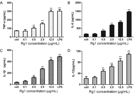
Figure 2. Ginsenoside Rg1 promotes the secretion of cytokines in bone mar-row-derived DCs. Cultured BM-DCs were treated with increasing concentra-tions of Rg1 (0.1 µg/mL, 0.5 μg/mL, 2.5 μg/mL and 12.5 µg/mL) for 24 h.
The levels of TNF-α (A), IL-6 (B), IL-1β (C) and IL-12 p70 (D) in supernatants
were measured by ELISA assay. Data are shown as mean concentrations (pg/mL) ± SD. **P<0.01, compared with untreated cells.
China). Twenty-four C57BL/6 male mice (6 to 8 weeks old)
were kept in specific patho -gen-free conditions. They were randomly divided into four groups of 6 mice each. The groups were as following: Rg1 in combination with PN anti-gen immunized group, PN immunized group, Rg1 nized group and PBS immu-nized group. Animals were injected subcutaneously twice
on days 1 and 14, with 20 μg PN antigen alone, 20 μg of Rg1 alone, 20 μg of PN anti
-gen plus 20 μg of Rg1, or with
PBS. The experiment was approved by the Instituti- onal Animal Care and Use
Committee of first affiliated
[image:3.612.92.372.336.533.2]Results
Identification of bone marrow-derived DCs in C57BL/6 mice
The dendritic cells were obtained and cultured from bone marrow cells of C57BL/6 mice as
in BM-derived DCs, real time PCR assay was performed to test the mRNA levels of chemo-kines. The dendritic cells were treated with 2.5
μg/mL of Rg1 for 24 hours and the m RNA lev
-els of interferon-γ-inducible protein 10 (IP-10),
regulated upon activation normal T-cell expre- ssed and secreted (RANTES), monocyte che-Figure 3. Ginsenoside Rg1 increases the transcription levels of chemokines
in bone marrow-derived DCs. Cultured BM-DCs were treated with 2.5 μg/mL
of Rg1 for 24 hours. The transcription levels of IP-10 (A), RANTES (B), MCP-1 (C) and IL-8 (D) were detected by real time PCR assay. Untreated cells were used as negative controls and LPS-treated cells were used as positive con-trols. **P<0.01, compared with untreated cells.
[image:4.612.92.375.71.287.2]rate of bone marrow-derived dendritic cells. As shown in Figure 1B, 79.84% of the den-dritic cells exhibited the
char-acteristic DC-specific marker
CD11c+.
Ginsenoside Rg1 promotes the secretion of cytokines in bone marrow-derived DCs In order to test whether gin-senoside Rg1 could promote the secretion of cytokines,
such as TNF-α, IL-6, IL-1β and
IL-12, DCs were treated with increasing concentrations of Rg1 for 24h and ELISA assay was performed according to the kit protocols. As shown in Figure 2, Rg1 induced a dose-dependent increase in cyto-kines production in BM-deriv- ed DCs. All the results demon-strated that ginsenoside Rg1 could promote the produc- tion of cytokines in dendritic
cells and induce inflammatory
response to activate innate immune responses.
Ginsenoside Rg1 increases the transcription levels of chemokines in bone marrow-derived DCs
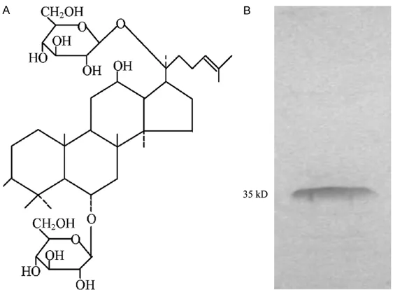
To test whether Rg1 promotes the secretion of chemokines Figure 4. The adjuvant and antigen. A. Chemical structure of ginsenoside
Rg1. The molecular formula is C42H72O14 and the molecular weight
is 801.02. B. The PN antigen was identified by SDS-PAGE. The molecular
[image:4.612.92.373.376.583.2]moattractant protein-1 (MCP-1) and IL-8 were detected. As shown in Figure 3, treatment with
Rg1 significantly increased the transcription
levels of IP-10, RANTES, MCP-1 and IL-8 in BM- derived DCs. The results demonstrated that DCs could induce and regulate the activation of T-lymphocytes by secreting chemokines to recruit them.
PN-specific splenocyte significantly proliferates in PN-Rg1 immunized mice
In order to test whether Rg1 could be used as the vaccine adjuvant, we selected a special antigen in combination with Rg1 to immunize
the C57BL/6 mice. Specifically, the C57BL/6
mice were randomly divided into four groups including PN in combination with Rg1 (PN-Rg1 group), PN alone (PN group), Rg1 alone (Rg1 group) or PBS control (PBS group). The mice were immunized twice on days 0, and 14. The chemical structure of Rg1 was shown in Figure 4A. The purity of antigen was more than 99% (Figure 4B). One week after final immunization,
splenocytes from each group were in vitro-stim-ulated with special antigen PN, irrelevant anti-gen OVA or positive control antianti-gen ConA for 24 h. As shown in Figure 5A, in vitro stimulated
[image:5.612.104.520.63.207.2]with PN antigen, the splenocytes significantly
Figure 5. Anti-PN serum IgG titer increases in PN-Rg1 immunized mice. Mice were cultured and immunized as
de-scribed in Materials and Methods. Seven days after the final inoculation, peripheral blood samples were collected
from the tails in each mouse. The serum antibody titers were measured by ELISA assay. Each group contained 6 mice.
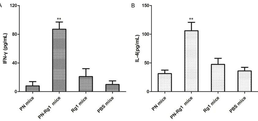
Figure 6. Serum IFN-γ and IL-4 levels are significantly up-regulated in PN-Rg1-immunized mice. The mice were cul
[image:5.612.98.519.282.479.2]group by ELISA assay. As shown in Figure 5B, the serum levels of anti-PN IgG titer were
sig-nificantly higher in PN-Rg1 group than that in
PN group or PBS group (**P<0.01). There was
no significant difference between PN-immunized
group and Rg1-immunized group or PBS-immunized group (P>0.05). All the results
obvi-ously demonstrated that Rg1 worked as an effi
-cient vaccine adjuvant to prime antigen-specific
immune response.
Serum IFN-γ and IL-4 levels are significantly up-regulated in PN-Rg1-immunized mice
Furthermore, we detected the cytokines secre-tion levels in serum of PN-Rg1 group, PN group,
Rg1 group and PBS group. Briefly, splenocytes
from the immunized mice were in
vitro-stimulat-ed with 1 μg/mL of PN for 24 h, the serum IFN-γ
and IL-4 levels were determined by ELISA assay. As shown in Figure 6, the mean value of IFN-γ and IL-4 in the PN-Rg1 group was significantly
higher than in the PN group, Rg1 grouu or PBS group (P<0.01). All the results obviously revealed that Rg1, as a vaccine adjuvant, could
induce the PN-specific cellular and humoral
immune responses. Discussion
Streptococcus pneumoniae (Spn) is a key pathogen for pneumonia, meningitis and otitis media [31-33]. It is also the main pathogen to induce diseases in immunocompromised pati- ents, or infants, young children and the elderly diseases in the world [34-36]. With the discov-ery of resistant strains of the streptococcus pneumoniae, the effects of the vaccine in pre-vention of infection are increasingly important [37, 38]. In the present study, we used pneumo-coccal hemolysin antigen (PN) as the candidate antigen, which was encoded by pneumococcal
concentrations of Rg1 to treat BM-derived DCs in vitro. The results obviously showed that Rg1 could enhance the cytokines production,
including TNF-α, IL-6, IL-1β and IL-12. The Rg1-induced inflammatory cytokines secretion
could effectively activate innate immune responses. Moreover, Rg1 also increased the transcription of chemokines, including IP-10, RANTES, MCP-1 and IL-8. IP-10 is a C-X-C che-mokine that activated T cells and macrophages
and attracted T cells into the inflammatory site.
RANTES activates human basophil
granulo-cytes and plays a regulatory role in inflamma -tory processes. MCP-1 (CCL2) is one of the important chemokines that promote migration
and infiltration of monocytes or macrophages.
Thus, Rg1 acted as a new adjuvant, which played an important role to activate the innate immune responses by promoting the cytokines and chemokines.
Furthermore, we tested the adjuvant activity in a mouse model. As the expected, the mice immunized with PN in combination with Rg1 showed a higher level of anti-PN IgG titers, as
well as the more antigen-specific splenocyte
proliferation activity. Additionally, the spleno-cytes from mice immunized with Rg1 in
combi-nation with PN produced higher levels of IFN-γ
and IL-4 when in vitro stimulated with PN pro-tein. All the results demonstrated that Rg1, as an adjuvant of PN antigen, promoted the mixed Th1/Th2 immune response in vivo. This was consistent with that ginsenoside Rg1 and alu-minum hydroxide synergistically promoted immune responses to ovalbumin in BALB/c mice [39]. In conclusion, Rg1 may be used as an effective new vaccine adjuvant to enhance the innate and adaptive immune responses. Disclosure of conflict of interest
Address correspondence to: Dr. Hong Zhou, Depart- ment of Respiration, The First Affiliated Hospital of
Harbin Medical University, The Seventh Road, Qunli District 2075, Harbin 150000, China. E-mail: zhouhongred@163.com
References
[1] Pagliusi S, Leite LC, Datla M, Makhoana M, Gao Y, Suhardono M, Jadhav S, Harshavardhan GV and Homma A. Developing countries vac-cine manufacturers network: doing good by making high-quality vaccines affordable for all. Vaccine 2013; 31 Suppl 2: B176-183.
[2] Gautret P, Botelho-Nevers E, Brouqui P and Parola P. The spread of vaccine-preventable diseases by international travellers: a public-health concern. Clin Microbiol Infect 2012; 18 Suppl 5: 77-84.
[3] Hyde TB, Dentz H, Wang SA, Burchett HE, Mounier-Jack S, Mantel CF; New Vaccine Introduction Impact Published Literature Working Group. The impact of new vaccine in-troduction on immunization and health sys-tems: a review of the published literature. Vaccine 2012; 30: 6347-6358.
[4] Skinner MA, Laidlaw SM, Eldaghayes I, Kaiser P and Cottingham MG. Fowlpox virus as a re-combinant vaccine vector for use in mammals and poultry. Expert Rev Vaccines 2005; 4: 63-76.
[5] Andre FE. How the research-based industry ap-proaches vaccine development and establish-es prioritiestablish-es. Dev Biol (Basel) 2002; 110: 25-29.
[6] Stewart TJ, Drane D, Malliaros J, Elmer H, Malcolm KM, Cox JC, Edwards SJ, Frazer IH and Fernando GJ. ISCOMATRIX adjuvant: an adjuvant suitable for use in anticancer vac-cines. Vaccine 2004; 22: 3738-3743.
[7] Camussone CM, Veaute CM, Pujato N, Morein B, Marcipar IS and Calvinho LF. Immune re-sponse of heifers against a Staphylococcus aureus CP5 whole cell and lysate vaccine for-mulated with ISCOM Matrix adjuvant. Res Vet Sci 2014; 96: 86-94.
[8] Quintilio W, Kubrusly FS, Iourtov D, Miyaki C, Sakauchi MA, Lucio F, Dias Sde C, Takata CS, Miyaji EN, Higashi HG, Leite LC and Raw I. Bordetella pertussis monophosphoryl lipid A
as adjuvant for inactivated split virion influen -za vaccine in mice. Vaccine 2009; 27: 4219-4224.
[9] Waite DC, Jacobson EW, Ennis FA, Edelman R, White B, Kammer R, Anderson C and Kensil CR. Three double-blind, randomized trials eval-uating the safety and tolerance of different for-mulations of the saponin adjuvant QS-21. Vaccine 2001; 19: 3957-3967.
[10] Fernandez-Tejada A, Chea EK, George C, Pillarsetty N, Gardner JR, Livingston PO, Ragupathi G, Lewis JS, Tan DS and Gin DY. Development of a minimal saponin vaccine ad-juvant based on QS-21. Nat Chem 2014; 6: 635-643.
[11] Wright AK, Christopoulou I, El Batrawy S, Limer J and Gordon SB. rhIL-12 as adjuvant aug-ments lung cell cytokine responses to pneu-mococcal whole cell antigen. Immunobiology 2011; 216: 1143-1147.
[12] Rose WA 2nd, Okragly AJ, Patel CN and Benschop RJ. IL-33 released by alum is re-sponsible for early cytokine production and has adjuvant properties. Sci Rep 2015; 5: 13146.
[13] Wendling U, Paul L, van der Zee R, Prakken B, Singh M and van Eden W. A conserved myco-bacterial heat shock protein (hsp) 70 se-quence prevents adjuvant arthritis upon nasal administration and induces IL-10-producing T cells that cross-react with the mammalian self-hsp70 homologue. J Immunol 2000; 164: 2711-2717.
[14] van Haren SD, Ganapathi L, Bergelson I, Dowling DJ, Banks M, Samuels RC, Reed SG, Marshall JD and Levy O. In vitro cytokine induc-tion by TLR-activating vaccine adjuvants in hu-man blood varies by age and adjuvant. Cytokine 2016; 83: 99-109.
[15] Finocchiaro LM, Fondello C, Gil-Cardeza ML, Rossi UA, Villaverde MS, Riveros MD and Glikin GC. Cytokine-Enhanced Vaccine and Interferon-beta plus Suicide Gene Therapy as Surgery Adjuvant Treatments for Spontaneous Canine Melanoma. Hum Gene Ther 2015; 26: 367-376.
[16] Matsumoto M, Tatematsu M, Nishikawa F, Azuma M, Ishii N, Morii-Sakai A, Shime H and
Seya T. Defined TLR3-specific adjuvant that in
-duces NK and CTL activation without signifi -cant cytokine production in vivo. Nat Commun 2015; 6: 6280.
[17] Bielinska AU, Makidon PE, Janczak KW, Blanco LP, Swanson B, Smith DM, Pham T, Szabo Z, Kukowska-Latallo JF and Baker JR Jr. Distinct pathways of humoral and cellular immunity in-duced with the mucosal administration of a nanoemulsion adjuvant. J Immunol 2014; 192: 2722-2733.
[18] Hon CC, Chow YC, Zeng FY and Leung FC. Genetic authentication of ginseng and other traditional Chinese medicine. Acta Pharmacol Sin 2003; 24: 841-846.
highfructose diet induced metabolic syn-drome. BMC Complement Altern Med 2016; 16: 98.
[23] Cho IH. Effects of Panax ginseng in neurode-generative diseases. J Ginseng Res 2012; 36: 342-353.
[24] Han H, Chen Y, Bi H, Yu L, Sun C, Li S, Oumar SA and Zhou Y. In vivo antimalarial activity of ginseng extracts. Pharm Biol 2011; 49: 283-289.
[25] Qu DF, Yu HJ, Liu Z, Zhang DF, Zhou QJ, Zhang HL and Du AF. Ginsenoside Rg1 enhances im-mune response induced by recombinant Toxoplasma gondii SAG1 antigen. Vet Parasitol 2011; 179: 28-34.
[26] Su F, Yuan L, Zhang L and Hu S. Ginsenosides Rg1 and Re act as adjuvant via TLR4 signaling pathway. Vaccine 2012; 30: 4106-4112. [27] Sun HX, Chen Y and Ye Y. Ginsenoside Re and
notoginsenoside R1: Immunologic adjuvants with low haemolytic effect. Chem Biodivers 2006; 3: 718-726.
[28] Li J, Guo J, Su Z, Hu M, Liu W and Wei Q. Calcineurin subunit B activates dendritic cells and acts as a cancer vaccine adjuvant. Int Immunol 2011; 23: 327-334.
[29] Hu M, Su Z, Yin Y, Li J and Wei Q. Calcineurin B subunit triggers innate immunity and acts as a novel Engerix-B HBV vaccine adjuvant. Vaccine 2012; 30: 4719-4727.
[30] Mosmann T. Rapid colorimetric assay for cel-lular growth and survival: application to prolif-eration and cytotoxicity assays. J Immunol Methods 1983; 65: 55-63.
[34] Escolano-Martinez MS, Domenech A, Yuste J, Cercenado MI, Ardanuy C, Linares J, de la Campa AG and Martin-Galiano AJ. DiiA is a novel dimorphic cell wall protein of Strep- tococcus pneumoniae involved in invasive dis-ease. J Infect 2016; 73: 71-81.
[35] Straume D, Stamsas GA and Havarstein LS. Natural transformation and genome evolution in Streptococcus pneumoniae. Infect Genet Evol 2015; 33: 371-380.
[36] Ahmadi K, Akya A, Numanpour B, Salimi A and Veisi-Raigani A. Frequency of Streptococcus pneumoniae infection in patients with suspect-ed meningitis in Imam Reza Hospital of Kermanshah in the west of Iran. Iran J Microbiol 2015; 7: 12-17.
[37] Staceviciene I, Petraitiene S, Vaiciuniene D, Alasevicius T, Kirsliene J and Usonis V. Anti- biotic resistance of Streptococcus pneumoni-ae, isolated from nasopharynx of preschool children with acute respiratory tract infection in Lithuania. BMC Infect Dis 2016; 16: 216. [38] Swedan SF, Hayajneh WA and Bshara GN.
Genotyping and serotyping of macrolide and multidrug resistant Streptococcus pneumoni-ae isolated from carrier children. Indian J Med Microbiol 2016; 34: 159-165.