Original Article
The role of human prostate cancer specific binding
peptide on the malignant phenotype of PC-3M cell
Xiaona Chang1*, Yanpeng Li2*, Chenchen Tang1, Bo Ren1, Bo Fang1, Xiangxiang Li1, Lijuan Zhou1, Baihui Li1, Wei Li1, Yilei Li1
1The Key Laboratory of Pathobiology, Ministry of Education, The College of Basic Medical Sciences, Jilin University,
Changchun 130021, Jilin, P. R. China; 2The high School Attached to Jilin University, Changchun 130021, Jilin, P.
R. China. *Equal contributors.
Received November 29, 2016; Accepted May 7, 2017; Epub June 15, 2017; Published June 30, 2017
Abstract: The objective of this study was to investigate the effect of a novel short peptide (B08: human prostate cancer specific binding peptide) on the malignant phenotype of the human prostate cancer cell line PC-3M. We developed a phage-displayed 7-mer peptide library to screen the target peptides that were specifically bound to PC-3M cells with subtractive panning from normal prostate cells and PC-3 prostate cancer cells. B08 was found to have high affinity to highly metastatic PC-3M cells. In order to explore the function of B08 on PC-3M cells, cell growth assay MTS and colony formation assay were used to determine the effect of B08 on cell proliferation. Flow cytometry (FCM) was employed to explore the apoptotic effect of B08 on PC-3M cells. The wound healing assay and transwell invasion assay were done to evaluate changes in the abilities of cell migration and invasion respectively. VEGFA at mRNA and protein levels were detected by PCR and western blotting. We found that B08 inhibited the prolifera-tion and reduced the rate of colony formaprolifera-tion of PC-3M cells, but did not cause a higher apoptotic rate. Addiprolifera-tional analysis showed that B08 significantly inhibited the migration and invasion of PC-3M cells. Then we also found that B08 decreased the VEGFA mRNA and protein of PC3M cells by PCR and western blotting. In conclusion, B08 can inhibit the malignant phenotype of PC-3M, which may via down-regulating the VEGF signaling pathway in vitro. Thus, peptide B08 may be an effective targeted therapeutic agent for the treatment of prostate cancer.
Keywords: Prostate cancer, specific binding peptide, malignant phenotype, PC-3M
Introduction
Prostate cancer, the most common cancer in the male urinary and reproductive system [1], is
difficult to be treated after the occurrence of
local invasion and distal metastasis. Moreover, few targets associated with metastasis and
malignancy have been identified. The five-year
relative survival rate of early stage prostate cancer is >99% while that of advanced meta-static disease is only 28% [2]. However, the effects of current clinical chemotherapy are not
satisfied. Therefore, novel reagents are urgent -ly needed for the effective targeted therapeutic agent of prostate cancer.
In the early reports, the short peptide can not
only specifically bond with malignant tumor
cells directly but also provide drug targeted therapy of effective carrier [3]. In addition, it
can improve the local concentration of drug and reduce the adverse reaction of chemotherapy medicine at the same time [3]. Several
strate-gies have been used for the identification of
tumor-associated proteins, such as serological analysis of recombinant cDNA expression librar-ies, ribosome display, tumor-specific antibody
cloning, and phage antibody libraries [4]. In our previous study, we developed a phage-dis-played 7-mer peptide library to screen the
tar-get peptides that were specifically bound to
PC-3M cells with subtractive panning from nor-mal prostate cells and PC-3 prostate cancer cells. A novel short peptide (B08: human
pros-tate cancer specific binding peptide) was found to have high affinity to highly metastatic PC-3M
peptide B08 on the malignant phenotype of PC-3M may have important theoretical and
practical significance for the further research of specific short peptide research.
However, the effects of B08 on the malignant potential of PC-3M cells remain uncovered and the potential molecular mechanisms which B08 involves in are still unclear. In order to explore the possibility of developing B08 as a therapeutic agent and to clarify its potential pathway, we investigated the effects and the molecular mechanisms of B08 on PC-3M cells. The present study was conducted by evaluating the assessments of B08 on the proliferation, migration and invasion abilities of PC-3M cells and the involvement of the VEGF signaling in vitro.
Materials and methods
Cell lines and cell culture
The human prostatic cancer cells PC-3M were obtained from the key laboratory of pathobiol-ogy, Ministry of Education, Jilin University and routinely cultured in RPMI medium 1640 (GIBCO, USA) supplemented with 10% fetal
bovine serum (FBS) in a humidified 37°C incu -bator containing 5% CO2.
Cell growth assay
The PC-3M cells were cultured in 96-well cul-ture plates at a cell density of 5000 cells/well, in 1640 containing 10% FBS. Following adher-ence overnight, the medium was replaced and the cells were incubated with different concen-trations (4*1010, 8*1010, 1.2*1011, 1.6*1011
pfu) of peptide B08 and control peptide 08K for 24 h. Viable proliferating cells were detected by the CellTiter 96®AQ
ueous One Solution Cell Proli- feration Assay(a). Cell viability was expressed as optical density (OD), which was detected by an Automatic microplate reader (TEAN, Swiss) at a 490 nm wavelength. The inhibitory rate of cell proliferation was calculated. Seven inde-pendent experiments were performed over multiple days.
Flow cytometry (FCM) detect the apoptosis
Cell cycle analysis was performed by using propidium iodide (PI) assay. PC-3M cells were seeded at 3×10⁴ cells/well into six well plates for 24 h, then treated with 4*1010 pfu peptide B08, and 4*1010 pfu peptide 08K for 24 h. Following incubation, cells were treated with PI
(50 μg/mL), annexin V-FITC conjugate (5 μg/
mL) and dispersed in PBS (1%, pH 7). Each analysis was performed at least thrice (n = 3) and a count of a minimum of 10,000 events was taken for each analysis. Data are repre-sented as mean ± S.D.
Colony formation assay
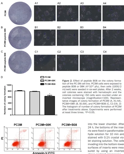
Cells (1000/2 ml/well) were seeded in six well plates, and treated with 4*1010 pfu peptide B08 and 08K for 2 weeks to form colonies. The formed colonies were stained with hematoxylic, and the colonies containing >50 cells were counted under an inverted microscope.
Migration assay
The ability of cells migration ability in a mono-layer culture was determined using the wound
healing assay. Cells were grown to full conflu -ence in 24-well plates and scratches were
per-formed using a 100 μL tip. The medium was
removed, and cells were washed with PBS and medium replaced by 1% FBS RPMI medium 1640 containing peptide B08, 08K (1.2*1011). Scratch closure was analyzed under the micro-scope and images were captured at different time points.
Invasion assay
[image:2.612.92.289.73.171.2]PC-3M cells were treated with peptide B08, 08K (1.2*1011 pfu) for 24 h. Then cells were seeded 8×10³ cells per well in 200 μl 1% FBS RPMI medium 1640 supplemented with B08, 08K (1.2*1011 pfu) into the upper chamber of
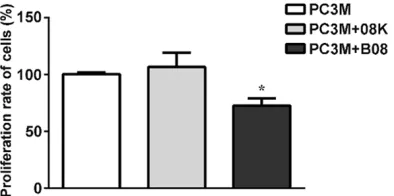
Figure 1. Effect of peptide B08 on cell proliferation of PC-3M cells. After PC3M cells were treated with pep-tide B08 (4*1010 pfu) or mock-vehicle peptide 08K
(4*1010 pfu) or without any peptide, the OD value
the transwell in 24-well plates (membrane pore
size, 8 μm; Corning Incorporated; Corning, NY,
USA) with Matrigel (BD Pharmingen), and 500
μl 15% FBS RPMI medium 1640 was added
twice in ice-cold PBS and subsequently lysing them by using RIPA buffer containing 1% PMSF.
Protein concentration was quantified with the
[image:3.612.89.521.67.603.2]BCA Protein Kit (Beyotime) and equal amounts
Figure 2. Effect of peptide B08 on the colony forma-tion of the PC-3M cell line. PC3M cells were exposed to peptide B08 or 08K (4*1010 pfu), then cells (1000/2
ml/well) were seeded in six-well plates. After 2 weeks, cell colonies were stained with hematoxylin and the colonies containing >50 cells were counted under an inverted microscope (magnification×100). Represen -tative images of colony formation of PC3M (A, A1-A4), PC3M+08K (B, B1-B4), and PC3M+B08 (C, C1-C4). D: The histogram of number of colony formation of PC3M after treatments above. Experiments were performed at least three times. *P<0.05.
Figure 3. Effect of peptide B08 on PC-3M cells apoptosis. Annexin V-FITC/PI assay was used to analyze the percentages of apoptotic PC-3M cells, PC-3M cells treated with peptide 08K, PC-3M cells treated with peptide B08. The apoptotic rates were 6.11%, 5.31%, 6.49%, respectively.
into the lower chamber. After 24 h, the bottoms of the inse-
rts were fixed in paraformalde -hyde solution for 10 min and stained with 0.1% crystal vio-let staining solution. The cells invading into the bottom-lower surfaces of inserts were mea-sured by using an inverted phase contrast microscope.
Protein extraction and west-ern blotting
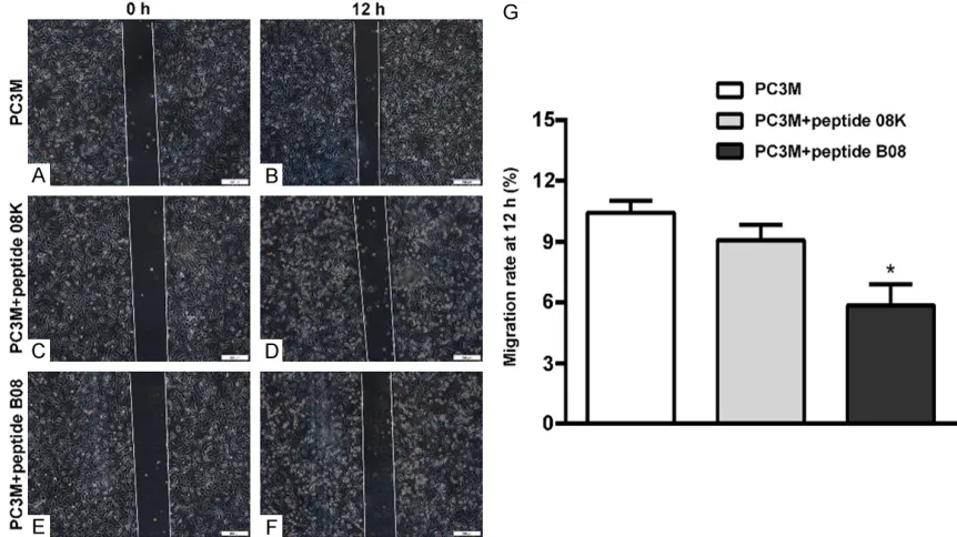
Figure 4. Effect of peptide B08 on the migration of PC-3M cells. Migrated cells were photographed at 0 h and 12 h. A and B: Representative images of migrated cells treated with no any peptide. C and D: Representative images of migrated cells treated with peptide 08K. E and F: Representative images of migrated cells treated with peptide B08. G: The histogram of migrated rate at 12 h of PC3M after treatments above. Experiments were performed at least three times. (magnification×100). *P<0.05.
of proteins were resolved onto a 12% SDS-PAGE, transferred a to a PVDF membrane (Millpore) and probed with primary antibody
anti-VEGFA, β-actin. Membranes were then
incubated with the horseradish peroxidase (HRP)-conjugated secondary antibody (1:2,000) and detection was performed with enhanced chemiluminescence (ECL Kit). Protein levels
were normalized to β-actin.
RNA extraction, standardization of the one-step duplex RT-PCR
Total RNA was extracted from PC-3M cells using Trizol reagent according to the manufac-turer’s instructions (Invitrogen). The StarScript II One-step RT-PCR Kit (GenStar) carried out in
20 μL reaction volume comprising of 1 μg of RNA, 0.4 μM of forward and reverse primers of GAPDH and VEGFA, 4 μl 5X reaction buffer, 1.5 μl One-step RT/Taq Mix, add DEPC-ddH2O to 20
μl. The reaction condition for the thermal cycles were 42°C for 30 min, 94°C for 2 min followed by 35 cycles of 94°C for 30 s, 55°C for 30 s and 72°C for 30 s respectively and final extension at 72°C for 7 min. After the PCR, the products
were electrophoresed on 2% agarose (Sigma-Aldrich, St. Louis, MO, USA) gel stained with
ethidium bromide and visualized under gel
doc-umentation system (Tanon). The specific prim -ers used were listed as follows: GAPDH forward: TGTTGCCATCAATGACCCCTT, reverse: CTCCAC- GACGTACTCAGCG; VEGFA forward: GGTGGGGT- CATGTGTGTGG, reverse: AGGTCTTGTTCGCTGC- CTGA.
Statistical analysis
Data are expressed as the means ± S.D. An unpaired two-tailed t test analysis was used to analyze the data from different groups. A value of P<0.05 was considered to indicate a
statisti-cally significant difference. All analyses were
performed with SPSS version 19.0 (SPSS Inc., Chicago, IL, USA).
Results
Effects of peptide B08 on PC-3M cells prolif-eration in vitro
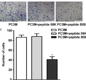
Figure 5. Effect of peptide B08 on the invasion of PC-3M cells. Invasion as-say was performed after 24 h incubation. A: Representative images of inva-sive cells treated with no any peptide. B: Representative images of invainva-sive cells treated with peptide 08K. C: Representative images of invasive cells treated with peptide B08. D: The histogram of number of invasive cells after treatments above. Experiments were performed at least three times. (mag-nification×100). *P<0.05.
Figure 6. Effect of peptide B08 on the level of VEGFA protein. A: VEGFA and GAPDH mRNA were determined by PCR in PC-3M, PC-3M with peptide 08K and PC-3M with peptide B08. B: The cell lysates were immune blotted with anti-VEGFA and anti-β-actin antibodies respectively. C: Protein levels of VEG -FA were represented as relative expression (mean ± S.E.M.) standardized by endogenous expression of β-actin. Experiments were performed at least three times. **P<0.01.
was obviously lower than the rate of the cells treated by peptide 08K (human prostate
can-cer specific binding peptide 08K) or not treated
by any peptide (Figure 2A-D). These data indi-cate that peptide B08 inhibits PC-3M cell prolif-eration in vitro.
Peptide B08 had no signifi-cant effect on the apoptotic cell death
FCM analysis was used to investigate the effect of pep-tide B08 on the induction of apoptosis of PC-3M cells in vitro. Peptide B08 was added to the PC-3M cells in the expo-nential growth phase for 24 h, and cell samples were obtain-
ed and fixed for FCM analysis.
The results revealed that
pep-tide B08 had no significant
effect on the apoptotic cell death (Figure 3).
The invasion and migration of PC-3M were inhibited by pep-tide B08 in vitro
To examine the effect of pep-tide B08 on PC-3M cells migration and invasion, the migration and invasion capa-bilities were assessed by the wound healing and Transwell invasion assays. As shown in (Figure 4A-G), the migration rate of cells treated with pep-tide B08 was 41.00 ± 5.86% in the PC-3M cells compared with 88.00 ± 5.69% in the cells treated with peptide 08K and 86.33 ± 4.49% in the cells not treated with peptide. In the Transwell invasion assay (Figure 5A-D), the inva-sion of the cells treated with
peptide B08 was significantly
inhibited compared with that treated with peptide 08K or untreated peptide cells (P< 0.05). This suggested that peptide B08 suppressed PC- 3M cells migration and inva-sion in vitro.
Peptide B08 suppressed the activity of the VEGF signaling
[image:5.612.89.374.430.559.2]levels of VEGF were detected. The results showed that the protein expression levels of
VEGFA were significantly decreased in the cells
treated with peptide B08 when compared with these levels in the untreated cells (Figure 6B,
6C). The PCR showed that the mRNA levels of
VEGFA were significantly decreased in the cells
treated with peptide B08 when compared with these levels in the untreated cells (Figure 6A). It suggested that peptide B08 suppressed the activity of VEGFA in PC-3M cells in vitro.
Discussion
Metastasis of prostate cancer is a complex and multistep process. It is not only involved in the biological behavior of tumor cells, but also closely related to angiogenesis of tumorous stroma and the degradation of extracellular matrix [6]. Metastatic castration-resistant pros-tate cancer (mCRPC) is the lethal and incurable stage of PCa [2]. Currently, chemotherapy is one of the mainstay of treatment for mCRPC. However, conventional chemotherapies have
been used with limited efficacy and significant
toxicity [7]. Therefore, any effort on discovering a new molecular that target-direct to PCa may contribute to the treatment of PCa.
In the process of current drug treatment of tumor, chemotherapy drugs spread not only into the tumors, but also into the healthy tissue and organs, which greatly limits the effect of drugs in the treatment of tumor [8-10]. Senth- ilkumar K et al reported that using biological
molecules which is specific to tumor can effec -tively solve the problem [11]. Traditional target therapy is guided by monoclonal antibody car-rier, but there are still some defects [12]. Nevertheless using short peptide to treat can-cer has its unique advantages. Peptide recep-tors are more likely to present original confor-mation and the types of receptor are not neces-sary to be known in advance. The short peptide
can specifically bond malignant tumor cells
directly, provide effective carrier to the targeted drug, improve the local concentration of drug and reduce the adverse reaction of chemother-apy medicine at the same time [3]. Phage dis-play random peptide library technology [13, 14]
can be applied for obtaining the specific bind -ing peptides of target cells.
In the preceding work of our project group, we
obtained specific binding peptide B08 through
phage display random peptide library technolo-gy. Therefore, studying the effects of peptide B08 on the malignant phenotype of PC-3M
have important theoretical significance and practical significance for the further research of specific short peptide research.
In order to examine the effects of peptide B08 on PC-3M cell proliferation, migration and inva-sion, the cell proliferation was evaluated with MTS and colony formation assays, and cell migration and invasion were assessed by wound healing and Transwell invasion assays. We found that peptide inhibited the prolifera-tion of PC-3M cell and the rate of colony
forma-tion of peptide B08-treated cells was signifi -cantly lower than that in cells treated with tide 08K or not treated with peptide. But
pep-tide B08 had no significant effect on the PC-3M apoptosis, the specific mechanism remains to
be further discussed. In the wound healing and Transwell invasion assays, the results revealed that the migratory and invasive capabilities were inhibited by peptide B08. These results indicate that the malignant phenotype of PC-3M cells may be inhibited by peptide B08 in vitro. Peptide B08 may be an effectively agent for chemotherapy in the treatment of prostate cancer. However, further studies are necessary to unveil the potential molecular mechanisms of the inhibition of the malignant phenotype of prostate cancer by peptide B08.
In the early reports, short peptides can specifi -cally inhibit MMP-2 and MMP-9 in ovarian
carci-noma and fibrosarcoma [15]. In murine LCC
cells, a phage-displayed tumor-homing peptide
B08 could down regulate the activity of the VEGF in PC-3M in vitro. Further study is needed
for the mechanism to confirm whether B08 reg
-ulates the activity of the VEGF by influencing HIF-1α.
In conclusion, our findings suggested that pep -tide B08 could alter the cell malignant pheno-type of PC-3M cell via downregulation of the activity of the VEGFA in vitro and may be an effective chemotherapeutic agent for prostate cancer. On account of the tumor microenviron-ment playing an important role in tumor pro-gression, invasion and cell migration [20], fur-ther experiments in vivo are necessary to ascertain whether peptide B08 represents a new chemotherapeutic agent for the treatment of prostate cancer.
Acknowledgements
We thank the Key Laboratory of Pathobiology, Ministry of Education, The College of Basic Medical Sciences, Jilin University for providing the experiment environment. This study was supported by the Natural Science Foundation of Jilin Provincial Science and Technology Department (No. 20150101126JC).
Disclosure of conflict of interest
None.
Address correspondence to: Yilei Li, The Key Lab- oratory of Pathobiology, Ministry of Education, The College of Basic Medical Sciences, Jilin University, Changchun 130021, Jilin, P. R. China. Tel: +86 043185619481; Fax: +86 043185619481; E-mail: [email protected]
References
[1] Cancer Genome Atlas Research Network. The molecular taxonomy of primary prostate can-cer. Cell 2015; 163: 1011-1025.
[2] Siegel RL, Miller KD and Jemal A. Cancer sta-tistics, 2015. CA Cancer J Clin 2015; 65: 5-29. [3] Goodson RJ, Doyle MV, Kaufman SE and
Rosenberg S. High-affinity urokinase receptor antagonists identified with bacteriophage pep -tide display. Proc Natl Acad Sci U S A 1994; 91: 7129-7133.
[4] Larimer BM, Thomas WD, Smith GP and Deutscher SL. Affinity maturation of an ERBB2-targeted SPECT imaging peptide by in vivo phage display. Mol Imaging Biol 2014; 16: 449-458.
[5] Goetz M and Wang TD. Molecular imaging in gastrointestinal endoscopy. Gastroenterology 2010; 138: 828-833 e821.
[6] Liu XZ, Zhang LT, Tong WQ, Wang S, Xu ZB and Chen F. Research advances in molecular mechanisms of the invasion and metastasis of lung cancer. Zhongguo Yi Xue Ke Xue Yuan Xue Bao 2016; 38: 108-112.
[7] Heidenreich A, Bastian PJ, Bellmunt J, Bolla M, Joniau S, van der Kwast T, Mason M, Matveev V, Wiegel T, Zattoni F, Mottet N; European Association of Urology. EAU guidelines on pros-tate cancer. Part II: treatment of advanced, re-lapsing, and castration-resistant prostate can-cer. Eur Urol 2014; 65: 467-479.
[8] Dhanikula RS, Argaw A, Bouchard JF and Hildgen P. Methotrexate loaded polyether-co-polyester dendrimers for the treatment of glio-mas: enhanced efficacy and intratumoral transport capability. Mol Pharm 2008; 5: 105-116.
[9] Iacob G and Dinca EB. Current data and strat-egy in glioblastoma multiforme. J Med Life 2009; 2: 386-393.
[10] Petrylak DP. Chemotherapy for androgen-inde-pendent prostate cancer. World J Urol 2005; 23: 10-13.
[11] Senthilkumar K, Arunkumar R, Elumalai P, Sharmila G, Gunadharini DN, Banudevi S, Krishnamoorthy G, Benson CS and Arunakaran J. Quercetin inhibits invasion, migration and signalling molecules involved in cell survival and proliferation of prostate cancer cell line (PC-3). Cell Biochem Funct 2011; 29: 87-95. [12] Engert A, Balduini C, Brand A, Coiffier B,
Cordonnier C, Dohner H, de Wit TD, Eichinger S, Fibbe W, Green T, de Haas F, Iolascon A, Jaffredo T, Rodeghiero F, Salles G, Schuringa JJ; EHA Roadmap for European Hematology Research. The European hematology associ- ation roadmap for European hematology re-search: a consensus document. Haematolo- gica 2016; 101: 115-208.
[13] Zwick MB, Bonnycastle LL, Noren KA, Venturini S, Leong E, Barbas CF 3rd, Noren CJ and Scott JK. The maltose-binding protein as a scaffold for monovalent display of peptides derived from phage libraries. Anal Biochem 1998; 264: 87-97.
[14] Noren KA and Noren CJ. Construction of high-complexity combinatorial phage display pep-tide libraries. Methods 2001; 23: 169-178. [15] Koivunen E, Arap W, Valtanen H, Rainisalo A,
[16] Mueller J, Gaertner FC, Blechert B, Janssen KP and Essler M. Targeting of tumor blood ves-sels: a phage-displayed tumor-homing peptide specifically binds to matrix metalloproteinase-2-processed collagen IV and blocks angiogen-esis in vivo. Mol Cancer Res 2009; 7: 1078-1085.
[17] Kuhnert F, Kirshner JR and Thurston G. Dll4-Notch signaling as a therapeutic target in tu-mor angiogenesis. Vasc Cell 2011; 3: 20. [18] Liu C, Zhang JW, Hu L, Song YC, Zhou L, Fan Y,
Zhu HY, Wang Y and Li QP. Activation of the AT1R/HIF-1 alpha /ACE axis mediates angio-tensin II-induced VEGF synthesis in mesenchy-mal stem cells. Biomed Res Int 2014; 2014: 627380.
[19] Weber DC, Tille JC, Combescure C, Egger JF, Laouiti M, Hammad K, Granger P, Rubbia-Brandt L and Miralbell R. The prognostic value of expression of HIF1alpha, EGFR and VEGF-A, in localized prostate cancer for intermediate- and high-risk patients treated with radiation therapy with or without androgen deprivation therapy. Radiat Oncol 2012; 7: 66.