Original Article
Mechanism of bladder dysfunction in the diabetic rat
Peng Zhou1, Zhiyong Xiong2, Qiubo Xie1, Yongquan Wang1, Gensheng Lu1
1Center of Urology, 2Department of Gastroenterology, Southwest Hospital, Third Military Medical University,
Chongqing, China
Received April 28, 2016; accepted June 22, 2016; Epub February 1, 2017; Published February 15, 2017
Abstract: Objective: The diabetic SD rat model was established to investigate the mechanism of the detrusor con-tractile dysfunction by analyzing the different expression of functionally regulatory proteins in the detrusor cell. Methods: Streptozotocin was used to establish the diabetic SD rat model. The altered expression of HSP27, CaD (full name), and TM (full name) was measured with fluorescence immunoassay, real-time PCR (RT-PCR), and west -ern blot. Results: Compared with the controls, the protein expression of HSP27 and phosphorylated HSP27 was down-regulated, while the protein expression of CaD and TM was up-regulated in the detrusor cells of the cases. Conclusion: The expression of functionally regulatory proteins dramatically changes in the diabetic SD rat model.
Keywords: Diabetic bladder dysfunction, HSP27, caldesmon, tropomyosin
Introduction
Diabetic bladder dysfunction (DBD), also known as diabetic cystopathy (DCP), is a common complication of diabetes mellitus (DM) [1-3]. The precise pathogenesis of DBD, which is not very clear yet, is predominantly attributed to detrusor dysfunction caused by hyper- glycemia and peripheral autonomic neuropa-thy, and the synergistic effect of the muscle-derived and neurogenic factors leads to the changes of bladder excitability, elasticity and contractility, which results in DM bladder dys-function [4, 5]. The ultrastructural changes of the detrusor and the structural injury of the contractile proteins lay the structural foun-dation for detrusor contractile dysfunction
[6, 7]. It is clinically significant to effectively
preserve the detrusor structure and promote its recovery. In this study, the functionally regulatory proteins such as HSP27, caldes- mon (CaD) and tropomyosin (TM) in the detru-sor cell were investigated, further revealing the pathogenesis of DBD, providing new perspec-tives for exploring protective measures of detrusor contractile dysfunction and treatment targets, and laying new basis for prognosis evaluation.
Materials and methods
Animals
Twelve SD rats with six for each gender (Shanghai Slaccas, China) [license: SC-XK (Shanghai) 20082-005] were selected for this study.
Main reagents
Streptozotocin (Sigma, USA), HSP27 antibody
(mouse, self-provided), specific antibody
la-bels for phosphorylated HSP27 (hsp27-ser15, hsp27-ser78, hsp27-ser82, self-provided), TM antibody (mouse, self-provided), CaD antibody (rabbit, self-provided), second antibody (anti-mouse, anti-rabbit, RD, USA), primers, RT rea- gents and RT-PCR reagents (Western Bio- technology).
Establishment of the DM model
Six SD rats (half for each gender) were injected i.p. with a single dose of 150 mg/kg strepto-zotocin to build DM models. Six controls were injected with 0.1 M citric acid buffer. All rats
Detection of cellular ultrastructure with elec-tron microscopy
The collected bladder tissues of the cases and controls were stained to observe the detrusor ultrastructure with electron microscopy.
Detection of the expression and distribution of HSP27, CaD and TM
The literature [8] was referred to for the
meth-od. The bladder samples were fixed with para -formaldehyde, dehydrated, cleared, embedded and subjected to heating induced epitope retrieval (HIER) and sectioning. After PBS wash-ing, 6% normal goat serum was used for
one-hour blocking. The first antibodies (HSP27 anti -body, CaD antibody and TM antibody) were added at 1:200, and then incubated at 4°C overnight. The second antibodies (anti-mouse, anti-rabbit) were added at 1:1000 and incubat-ed at 37°C for 1 h. Then the nuclei were stainincubat-ed with DAPI after 15-minute incubation in dark-ness. The samples were observed and photo-graphed under the confocal laser scanning microscope, and the data were documented.
Genetic detection of HSP27, CaD and TM with QRT-PCR
The literature [9] was referred to for the meth-od. Three rats were selected from each group for RNA extraction and two repetitions were set for each rat. TRIZOL, chloroform, isopropanol and ethanol were used to extract the total RNA from the models and controls. Then random primers were used for reverse transcription to prepare cDNA. QRT-PCR was performed with
the specific primers of HSP27, CaD and TM
shown in Table 1 and sybr green I fluorochrome, with mouse β-actin as the internal reference. RT-PCR reactive system (20 μL): 10 μL 2×RT buffer, 1 μL 6N random primers (100 pmol/μL), 1 μL RT mix, 5 μL template, 3 μL DEPC buffer.
for 20 s, 60°C for 30 s, 72°C for 30 s, 35 cir-cles; signal detection at 72°C.
The relative copy number of each test item was calculated according to the standard curve. The corrected value of mRNA content for each test item was calculated by the ratio of the rela-tive copy number of each test item to relarela-tive copy number of the internal reference. The expressive difference between the cases and
the controls was defined by the corrected val -ues. T-test was adopted to determine statistical
significance.
Detection of the expression of HSP27, phos-phorylated HSP27, CaD and TM with WB
The literature [14] was referred to for the meth-od. Three rats were selected from each group and two repetitions were set for each rat. The total protein extracted from the bladder tissues was subjected to 4% SDS-PAGE gel electropho-resis, membrane-transfer, one-hour blocking with TBST buffer containing 5% skim milk
pow-der. Then the first antibodies (HSP27,
hsp27-ser15, hsp27-ser78, hsp27-ser82, CaD, TM) diluted at 1:1000 were added at 1:3000 with the internal references (glyceraldehyde-3- phophate dehydrogenase, GAPDH). The incuba-tion at 4°C continued overnight. After washing, the second antibodies labelled with horserad-ish peroxides (diluted at 1:5000) were added for 1.5-hour incubation. The enhanced che- miluminescence (ECL) system (Amersham Biosciences) was employed to detect the pro-tein content.
Results
Detection of the detrusor ultrastructure with electron microscopy
Compared with the controls, the detrusor cells of the models were hypertrophic with
intracel-Table 1. Primers for QRT-PCR
Target gene Name Sequence Product length Source
HSP27 rhsp27F TGGCAAGCACGAAGAAAGG 113 bp literature [10]
rhsp27R GGGACAGGGAAGAGGACACC
CaD rCaDF CTATGGCAACTGCGTAACGG 152 bp literature [11]
rCaDR GGTGCTGGGTGACCTTGA
TM Thbd-F AATACCCGAGATCCTGAGGG 238 bp literature [12]
Thbd-R TCCTGTTGCACATGTACTCG
β-actin rat actin f CCCATCTATGAGGGTTACGC 150 bp literature [13] rat actin r TTTAATGTCACGCACGATTTC
Reactive conditions: 25°C for 10 min, 42°C for 50 min, 85°C for 5 min. QRT-PCR reactive sys-
tem: 25 μL 2×PCR buffer, 1 μL×2 prim -ers (25 pmol/μL), 0.5 μL Sybr green I (20×), 2 μL template, 20.5 μL DEPC buffer.
[image:2.612.89.421.83.206.2]lular myofilaments decreased
and atrophic (Figure 1).
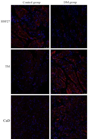
Detection of the expression and distribution of HSP27, CaD and TM with immuno-fluorescent staining
The immunofluorescent
re-sult showed that (See Figure 2) in the detrusor cells of both the DM models and con-trols, the protein expression of CaD and TM was up-regu-lated to some extent, while the expression of HSP27 was down-regulated. All of the three proteins were expre- ssed in cytoplasm and gath-ered on cytomembrane.
Genetic detection of HSP27, CaD and TM with QRT-PCR
[image:3.612.92.525.72.215.2]The relative expression lev-els of HSP27 mRNA of the models and controls were 2.35 and 5.13 respectively. In the model group, the mRNA expression levels of CaD and TM were 0.35 ±0.12E+07 and 0.63±0.22E +07 respectively, while in the control group, they were 0.16±0.08 E+07 and 0.29± Figure 1.Detection of the detrusor ultrastructure of the controls (A) and DM models (B) with electron microscopy.
[image:3.612.88.382.253.716.2]0.15E+07 respectively. The result showed that (See Table 2) compared with the controls, the expression of CaD and TM was up-regulated, while the HSP27 expression was
down-regulat-ed, with significant difference (P<0.05).
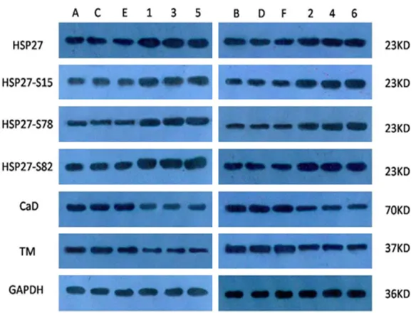
Detection of the expression of HSP27, specific phosphorylated HSP27, CaD and TM with WB
The SDS-PAGE result is shown in Figure 3. Compared with the controls, the bands of HSP27 and activated HSP27 (hsp27-ser15, hsp27-ser78 and hsp27-ser82) in the DM model group were light-colored, while the bands of CaD and TM were dark-colored. The gray value of each band was obtained via the gel processing system, and then the ratio of each test item to the internal reference was calcu-lated. The result showed that compared with the controls, in the DM model group, the
expres-sion levels of CaD and TM proteins were signifi -cantly up-regulated, while the expression levels of both HSP27 and phosphorylated HSP27 were down-regulated (Figure 3).
pivotal role in the regulation on the contraction of smooth muscles. The expression and activa-tion of HSP27 at the stress state can show a compensatory increase, which contributes to the maintenance and reestablishment of mus-cle cell contraction. HSP27 has three phos-phorylation sites [19]: Ser15, Ser78 and Ser82. Phosphorylated HSP27 can stabilize the cyto-skeleton of smooth muscle cells and enhance cellular contraction via inhibiting the depoly-merization of F-actin and promoting polymer-ization of G-actin [20]. Proteins related to thin
filament contraction, represented by CaD and
TM, undertake a critical role in regulating smooth muscle contraction [21].
A recent research showed that [22] in the rab-bit DBD model, the expression of multiple
pro-teins related to thin filament contraction, such
[image:4.612.89.526.88.143.2]as CaD and TM, was up-regulated in the blad-der detrusor, with decreased contraction of the detrusor. Likewise, this study also showed an up-regulated expression of CaD and TM and a down-regulated expression of HSP27. A study
Figure 3 Detection of the
expression of HSP27, specific
phosphorylated HSP27, CaD and TM with WB (ABCDEF: DM models; 123456: con- trols).
Discussion
DBD is a common complica-tion of DM, accounting for 40-100% among DM pa- tients. The DBD morbidity reaches 25% even among patients with stably con-trolled blood glucose [15], seriously threatening life quality. The pathogenesis of DBD has not been thoroughly unveiled by far.
Studies show that [16-18] HSP27, as an important molecular chaperone, plays a Figure 3. WB detection of specific phosphorylated HSP27, HSP27, CaD and
TM proteins (A-F represent DM group; 1, 2, 3, 4, 5, 6 represent the control group).
Table 2. Genetic detection of the DM models and controls
HSP27 CaD TM β- actin
A B A B A B A
Controls 4.92±1.64 5.13 0.16±0.08 0.17 0.29±0.15 0.30 0.96±049
DM models 2.82±0.96* 2.35 0.35±0.12* 0.29 0.63±0.22* 0.53 1.20±0.41
[image:4.612.90.382.174.394.2]shows that [23] the combination of HSP27 and phosphorylated CaD can lead to the dissocia-tion of CaD and TM, which regulates the con-traction of colon smooth muscles. This mecha-nism has not been reported in DBD cases. And the role of HSP27 in the contraction of bladder smooth muscles has also not been reported so far. The functional regulation of the bladder detrusor caused by the altered expression of HSP27, CaD and TM remains to be further
stud-ied. Our study is the first to report the
down-regulated HSP27 expression in the DM rat model, laying the foundation for further investi-gating the mechanism of functional regulation of the bladder detrusor, revealing the patho-genesis of DBD and exploring protective mea-sures as well as therapeutic targets for detru-sor contractive dysfunction.
Acknowledgements
The study was supported by the National Natural Sciences Foundation of China (No. 81270845).
Disclosure of conflict of interest
None.
Address correspondence to: Yongquan Wang and Gensheng Lu, Center of Urology, Southwest Hospital, Third Military Medical University, 35 Gaotanyan Zhengjie Street, Shapingba District, Chongqing, China. Tel: +86-2988766653; Fax: +86-2988- 766653; E-mail: [email protected] (YQW); [email protected] (GSL)
References
[1] Liu G, Daneshgari F. Diabetic bladder dysfunc-tion. Chin Med J 2014; 127: 1357-1364. [2] Wang YH, Yang J. Advances in diabetes
blad-der disease. J Mod Clin Med 2012; 2: 83-84. [3] Labib J. Diabetic cystopathy in children and
adolescents with Type 1 diabetes mellitus. J Diabetes Mell 2014; 4: 19-25.
[4] Shi XX, Wang YB, Wang RX. Pathogenesis and diagnosis of diabetic bladder disease. Zhong Guo Zong He Lin Chuang 2015; 24: 110-117. [5] Han D, Chen SZ, Shi BK. Progress and
func-tional changes in patients with urinary bladder. Shandong Yi Yao 2013; 53: 101-103.
[6] Gong Y, Song B, Jin XY. Experimental study of diabetic bladder compliance and detrusor sys-tolic and diassys-tolic function. Zhong Hua Wa Ke Za Zhi 2000; 11: 865-867.
[7] Wang DW, Shuang WB. Pathology and patho-genesis of diabetic detrusor. Lin Chuang Mi Niao Wai Ke Za Zhi 2005; 20: 649-651.
[8] Li MZ, Chu GL. Diabetic rat bladder can α1 ad -renergic receptor expression change. Jie Pou Xue Yan Jiu 2012; 3: 189-193.
[9] Zhao AJ, Huang SM, Ou ST, et al. Real-time PCR was used to detect diabetic nephropathy rat BMP-2 and MGP. Gui Yang Yi Xue Yuan Xue Bao 2011; 5: 460-464.
[10] Uoshima K, Handelman B, Cooper LF. Isolation and Characterization of a Rat HSP 27 Gene. Biochem Biophys Res Communicat 1993; 197: 1388-1395.
[11] Baik J, Ok SH, Cho H, Yu J, Kim W, Nam IK, Choi MJ, Lee HK, Sohn JT. Dexmedetomidine-induced Contraction Involves Phosphorylation of Caldesmon by JNK in Endothelium-denuded Rat Aortas. Int J Biol Sci 2014; 10: 1108-15. [12] Chen CD, Kobayashi R, Helfman DM. Binding
of hnRNP H to an exonic splicing silencer is in-volved in the regulation of alternative splicing of the rat β-tropomyosin gene. Genes Dev 1999; 13: 593-606.
[13] Nudel U, Zakut R, Shani M, Neuman S, Levy Z, Yaffe D. The nucleotide sequence of the rat cy-toplasmic beta-actin gene. Nucleic Acids Res 1983; 11: 1759-1771.
[14] Hu CY, Wang DW, Zhang XH. Changes in type 2 diabetic rats detrusor diastolic function and β receptors. Chinese Remedies & Clinics 2013; 1: 12-14.
[15] Sasaki K, Yoshimura N, Chancellor MB. Implications of diabetes mellitus in urology. Uro lClin North Am 2003; 30: 1-12.
[16] Chen HF, Xie LD and Xu CS. The signal trans-duction pathways of heat shock protein 27 phosphorylation in vascular smooth muscle cells. Mol Cell Biochem 2010; 333: 49-56. [17] MacIntyre DA, Tyson EK, Read MR. Contraction
in human myometrium is associated with changes in small heat shock protein. Endo- crinology 2008; 149: 245-252.
[18] Chaudhuri S, Smith PG. Cyclic strain-induced HSP27 phosphorylation modulates actin fila -ments in airway smooth muscle cells. Am J Respir Cell Mol Biol 2008; 39: 270-278. [19] Trott D, McManus CA, Martin JL, Brennan B,
Dunn MJ and Rose ML. Effect of phosphory-lated hsp27 on proliferation of human endo-thelial and smooth muscle cells. Proteomics 2009; 9: 3383-3394.
[20] Kostenko S, Johannessen M, Moens U. PKA-induced F-actin rearrangement requires phos-phorylation of Hsp27 by the MAPKAP kinase MK5. Cell Signal 2009; 21: 712-8.
[22] Mannikarottu AS, Changolkar AK, Disanto ME, Wein AJ, Chacko S. Over expression of smooth muscle thin filament associated proteins in the bladder wall of diabetics. J Urol 2005; 174: 360-364.