Endothelial NOS, estrogen receptor
β
,
and HIFs cooperate in the activation of
a prognostic transcriptional pattern in
aggressive human prostate cancer
Simona Nanni,1,2,3 Valentina Benvenuti,1,2 Annalisa Grasselli,1,2 Carmen Priolo,2,4
Aurora Aiello,1,3 Stefania Mattiussi,5 Claudia Colussi,6 Vittoria Lirangi,1 Barbara Illi,6
Manuela D’Eletto,1 Anna Maria Cianciulli,7 Michele Gallucci,8 Piero De Carli,8 Steno Sentinelli,9
Marcella Mottolese,9 Paolo Carlini,10 Lidia Strigari,11 Stephen Finn,4 Elke Mueller,4
Giorgio Arcangeli,12 Carlo Gaetano,5 Maurizio C. Capogrossi,5 Raffaele Perrone Donnorso,9
Silvia Bacchetti,1 Ada Sacchi,1 Alfredo Pontecorvi,2,13 Massimo Loda,4 and Antonella Farsetti1,3,13
1Department of Experimental Oncology, Regina Elena Cancer Institute, Rome, Italy. 2Department of Endocrinology, Catholic University, Rome, Italy. 3Institute of Neurobiology and Molecular Medicine, National Research Council (CNR), Rome, Italy.
4Department of Medical Oncology, Dana-Farber Cancer Institute, and Department of Pathology, Brigham and Women’s Hospital, Harvard Medical School, Boston, Massachusetts, USA. 5Laboratorio di Patologia Vascolare, Istituto Dermopatico dell’Immacolata (IDI),
Rome, Italy. 6Centro Cardiologico Monzino, Milan, Italy. 7Department of Clinical Pathology, 8Department of Urology, 9Department of Pathology, 10Department of Medical Oncology, 11Laboratory of Medical Physics and Expert Systems,
12Department of Radiotherapy, and 13Rome Oncogenomic Center, Regina Elena Cancer Institute, Rome, Italy.
The identification of biomarkers that distinguish between aggressive and indolent forms of prostate cancer
(PCa) is crucial for diagnosis and treatment. In this study, we used cultured cells derived from prostate tissue
from patients with PCa to define a molecular mechanism underlying the most aggressive form of PCa that
involves the functional activation of eNOS and HIFs in association with estrogen receptor
β
(ER
β
). Cells from
patients with poor prognosis exhibited a constitutively hypoxic phenotype and increased NO production.
Upon estrogen treatment, formation of ER
β
/eNOS, ER
β
/HIF-1
α
, or ER
β
/HIF-2
α
combinatorial complexes
led to chromatin remodeling and transcriptional induction of prognostic genes. Tissue microarray analysis,
using an independent cohort of patients, established a hierarchical predictive power for these proteins, with
expression of eNOS plus ER
β
and nuclear eNOS plus HIF-2
α
being the most relevant indicators of adverse
clinical outcome. Genetic or pharmacologic modulation of eNOS expression and activity resulted in
recipro-cal conversion of the transcriptional signature in cells from patients with bad or good outcome, respectively,
highlighting the relevance of eNOS in PCa progression. Our work has considerable clinical relevance, since
it may enable the earlier diagnosis of aggressive PCa through routine biopsy assessment of eNOS, ER
β
, and
HIF-2
α
expression. Furthermore, proposing eNOS as a therapeutic target fosters innovative therapies for PCa
with NO inhibitors, which are employed in preclinical trials in non-oncological diseases.
Introduction
In the clinical management of prostate cancer (PCa), the second most common neoplasia in men worldwide (1), the ability to dis-tinguish between aggressive and indolent forms of the disease is critical. Thus, therapeutic approaches would be substantially improved by the identification of the molecular mechanisms involved in tumor progression and the key biomarkers capable of improving patients’ stratification at diagnosis, by discriminating between those at risk for relapse and those with indolent tumors not requiring further intervention beyond surgery.
Recently, we and others (2, 3) reported on the induction of genes involved in the cell response to hypoxia in prostate, breast, and ovarian cancers and on the relevance of this phenomenon as predictor of adverse clinical outcome, suggesting that HIFs, beside their well-established role in the biology of solid tumors, represent key transcription factors specifically in endocrine tumors. High expression of the hypoxia response signature in breast cancers has a predictive power greater than parameters such as response to chemotherapy, estrogen receptors (ERs), tumor size and grade, angiogenic invasion, or age (3). In partic-ular, HIF-1α appears to promote early invasive lesions (4) and, indeed, in PCa is expressed at early stages (5, 6), supporting its specific role as predictor of poor prognosis. The more aggressive prostate tumors, in fact, are characterized by increased expression of HIF-1α, HIF-2α, and HIF-1β and their gene targets. Moreover, cells from these tumors exhibit a constitutive “hypoxic” pheno-type, even in normoxic conditions (see below), suggesting that hypoxia may confer a significant growth advantage (7), thus pro-moting and shaping cancer evolution (4, 8).
Authorship note: Simona Nanni, Valentina Benvenuti, and Annalisa Grasselli con-tributed equally to this work.
Conflict of interest: Massimo Loda is a consultant for Novartis and Director of the Center for Molecular Oncologic Pathology, Dana-Farber Cancer Institute.
Nonstandard abbreviations used: AR, androgen receptor; DSS, disease-specific survival; E2, 17β-estradiol; ER, estrogen receptor; ERE, estrogen-responsive element; HRE, hypoxia-responsive element; hTERT, human telomerase catalytic subunit; PCa, prostate cancer; PSA, prostate-specific antigen; TMA, tissue microarray.
research article
Another key molecule, on which a number of studies on PCa etiopathogenesis have been focused in the last years, is the ER. Although androgens have been traditionally considered the major hormonal regulators of the prostate gland, increasing experimen-tal evidence has recently attributed an equally important function to estrogens (9). The first ER expressed in the fetal prostate, and the predominant form in its epithelium is ERβ, which, together with the androgen receptor (AR), appears to mediate the initial stages of gland development (10, 11). Discrepancies in the litera-ture make it difficult to define the precise biological role of the 2 ER subtypes, ERβ and ERα, in PCa (9, 12, 13); however, the main function of ERβ appears to be associated with cell survival (14). Specifically, the retained expression of ERβ in the percentage of recurrent PCa associated with increased mortality (15) and in all metastatic lesions (16) is strongly suggestive of a critical involve-ment of this receptor in PCa progression. Along the hypoxia and ER pathways lays the eNOS, whose expres-sion, although abundant in endothelial cells, is widely distributed among different tissue and cell types and in tumors, including PCa (see below). The eNOS gene promoter harbors hypoxia and ER response elements, and, in fact, eNOS activity is regulated by hypox-ia and/or estrogen (17–20). In addition, the product of eNOS, NO, affects HIF-1α synthesis and accumulation in normoxia, indicating the existence of a regulatory loop between these molecules (21–24). Finally, eNOS and NO also play an important role in tumorigenesis and tumor maintenance (25–27). Remarkably, in human endothelial cells, eNOS and ER form a nuclear complex that regulates transcrip- tion of the human telomerase catalytic subunit (hTERT) (18), a mol-ecule that is an early marker of PCa development (28, 29).Since hTERT and several other genes belonging to a prognostic transcriptional signature (2) respond to both estrogens or hypoxia, we queried whether ERs, HIFs and eNOS may cooperate in PCa by co-regulating their transcription. To investigate this potential mechanism in vitro, we have used our ex vivo experimental model, consisting of immortalized cell populations derived from a cohort of patients with PCa, characterized by a prognostic gene profile, discriminating cells from tumors with poor versus good prognosis (G1 versus G2) (2). We have then validated our findings in vivo on tissue microarrays (TMAs) of primary tumors from an indepen-dent cohort of patients with known clinical outcome. Through these approaches, we have been able to demonstrate the existence of a functional cooperation among the estrogen-, hypoxia-, and NO-dependent signaling pathways in the activation of a transcrip-tional program specifically associated with aggressive PCa. Results
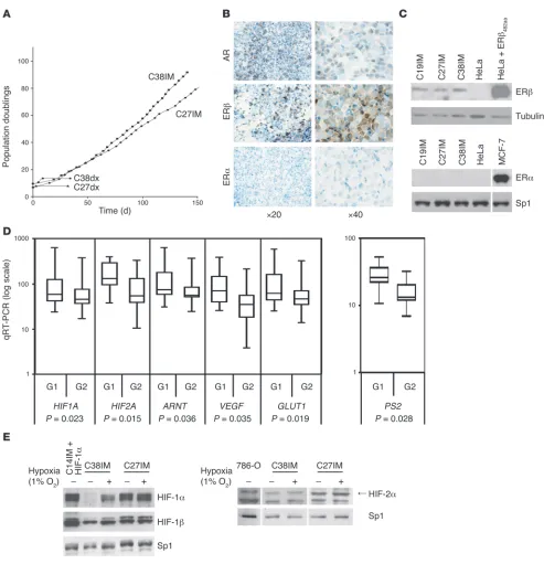
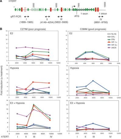
Generation and characterization of immortalized prostate tumor cells. On the basis of a defined transcriptional signature with prognostic rele-vance in PCa (2), we set out to gather interrelated information aimed at identifying molecular targets and mechanisms potentially rele-vant to PCa progression and at determining the expression pattern of genes essential to individual patient prognosis. To this end, we previously derived epithelial cell strains from prostate tissue freshly explanted from patients with benign prostate hyperplasia or clinical-ly localized carcinoma (2). These cells, even when from tumor tissue, had a limited lifespan in culture (at most 25 population doublings; Figure 1A). Telomerase was expressed at variable levels in these populations (2) but waned over time (data not shown). Telomerase reconstitution by transduction of hTERT did not affect lifespan, and transduction of SV40 large T antigen extended lifespan to about 60
population doublings (data not shown). Cotransduction of hTERT and T antigen, however, resulted in unlimited proliferation (immor-talization) of nearly all cell strains (25 of 29; Figure 1A). Immortal populations retained the markers of the epithelial luminal phe-notype of parental cells (2), as assessed by immunocytochemistry (see Supplemental Methods), being positive for AR, cytokeratin 8, and prostate-specific antigen (PSA) but negative for high-molecu-lar-weight cytokeratin, p63, vimentin, and α-smooth muscle actin (Figure 1B and data not shown). All cells expressed ERβ but not ERα (Figure 1, B and C) and showed functional responsiveness of AR to dihydrotestosterone (data not shown).
Maintenance of a prognostic transcriptional signature upon cell immor-talization . All immortalized cell lines maintained the mRNA expres-sion pattern seen in parental cells for genes of the transcriptional signature discriminating between poor (G1 group) and good (G2 group) prognosis (2). Expression of the hypoxia response fam-ily genes, their targets (30), and other genes, such as the classical estrogen target PS2 or AKT (assessed by quantitative RT-PCR; see Supplemental Methods), was significantly different between G1 and G2 cells (P = 0.028 and P = 0.007, respectively; Figure 1D and data not shown). Protein levels were determined by Western blot in C27IM and C38IM cells, representing the G1 and G2 groups, respectively. Expression of HIF-1α and HIF-2α (Figure 1E) was detected already in normoxia in C27IM cells, indicating that they have a constitutively hypoxic phenotype. In C38IM cells, on the other hand, HIF-1α appeared to be absent and HIF-2α was barely detectable. Finally, hypoxia enhanced, albeit slightly, both HIF-1α and HIF-2α in C27IM cells, while in C38IM cells, it strongly enhanced HIF-1α and had no apparent effect on HIF-2α. A more in depth analysis of this response is presented below. The substan-tial difference in ARNT mRNA could be confirmed at the protein level only in a subset of the cell lines (Figure 1E).
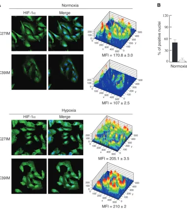
Expression of eNOS, a target of HIFs and estrogens, was exam-ined by confocal microscopy (see Supplemental Methods) in cells of both groups. Expression of the protein (Figure 2A), in terms of both amount and number of positive cells, was significantly high-er in G1 than G2 cells (P < 0.005). Most interestingly, in both cell types the protein was localized almost exclusively in the nucleus even, in the absence of the hypoxic stimulus. Nuclear relocaliza-tion of eNOS was observed in our recent study on endothelial cells upon exposure to estrogen (18). Thus, eNOS translocation to the nucleus in PCa cells may be dependent upon constitutive deregula-tion of hormone signaling in this endocrine tumor. In agreement with their higher levels of eNOS expression, G1 cells produced significantly higher amounts of NO than either benign prostate hyperplasia or G2 cells (Figure 2B), indicating that downstream effectors of the estrogen and hypoxia-inducible pathways are con-stitutively active only in G1 cells and even under normoxia.
Figure 1
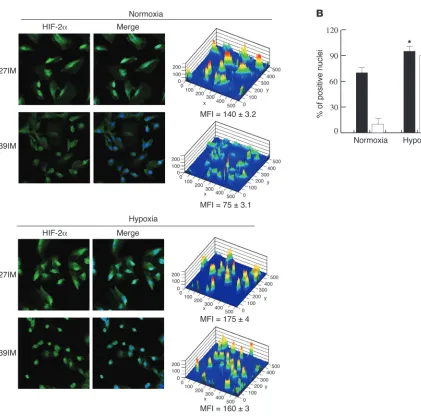
in G2 cells (C38IM) in normoxia as detected by Western blot (Fig-ure 1E) and the present results (up to 10% of nuclei were positive in basal conditions) to the higher sensitivity of detection by confo-cal microscopy. The hypoxic phenotype of C27IM cells is shared by most of the cell lines belonging to the poor prognosis group (G1) and involves both HIF-1α and HIF-2α (data not shown). This phe-notype, however, did not prevent a further significant increase in HIF-1α/HIF-2α expression, reaching almost 100% of nuclei, when cells were exposed to hypoxia (Figures 3 and 4). Indeed, confocal microscopy demonstrated a comparable response to hypoxia of both proteins in G1 and G2 cells (Figures 3 and 4).
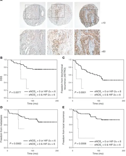
Validation in vivo of the transcriptional prognostic signature. In order to validate in vivo the prognostic power of the gene expression profile of cultured PCa cells, TMAs were constructed from an independent ret-rospective cohort of patients with PCa (n = 88), using paraffin-embed-ded tissue samples from the archive of the Pathology Department of the Regina Elena Cancer Institute. Samples were selected based on the longest follow-up period (12–14 years) and well-defined clinical outcome. The clinical and histopathological patient information is detailed in Supplemental Table 1. The poor prognosis group exclu- sively included patients deceased because of PCa or in clinical pro-gression, defined by the presence of biochemical, local, or metastatic recurrence. TMA sections were stained with antibodies to HIF-1α, HIF-2α, or eNOS (17, 27) (Figure 5A). ERβ was included in the analy- sis because of its known role in PCa. The best results in terms of inten-sity and consistency were obtained with HIF-2α, eNOS, and ERβ anti-bodies. The HIF-1α signal, instead, was mostly cytoplasmic and also only very weakly nuclear in clear cell renal carcinoma sections, which were used as positive controls. The quantitative statistical assessment of all scoring data, independently evaluated by 2 pathologists, dem-onstrated an association of HIFs, eNOS, and ERβ expression pre-dominantly in tumors with poor prognosis. There was no association between expression of these markers and Gleason score.
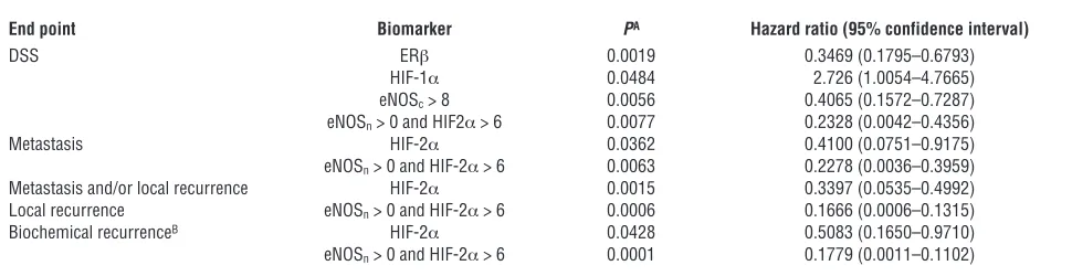
The TMA analysis has revealed at least 3 major and interesting findings. (a) A highly significant correlation between expression of HIF-1α, eNOS (nuclear and cytoplasmic), and ERβ with disease- specific survival (DSS) (Figure 5, B, D and E) was found. In par- ticular, stronger staining for each of these molecules was indepen-dently associated with decreased survival (ERβ, P = 0.0019; HIF-1α,
P = 0.0484; and eNOS, P = 0.0056) as shown by Kaplan-Meier curves. (b) While no correlation was found between HIF-2α expression and DSS, significant association between increased HIF-2α nuclear staining and decrease of recurrence-free survival (local or metastatic,
P = 0.0015) was observed (Figure 5C). Further, high expression of HIF-2α was significantly associated with increased biochemical recurrence (P = 0.0428 and P = 0.0459, respectively, for the Ameri-can Society for Therapeutic Radiology and Oncology [ASTRO] consensus and the Phoenix definition; data not shown), suggest-ing a differential but critical role of HIF-1α and HIF-2α in clinical outcome. (c) Importantly, univariate analysis of ERβ, HIF-1α, and eNOS (Figure 5F) revealed a direct correlation between increased expression of all 3 predictor variables and decreased DSS, with a much higher significance (P = 0.0001) than the individual variables. Moreover, combined expression of only ERβ and eNOS had suf- ficient predictive power on its own and even predicted patient sur-vival with a higher significance than the Gleason score (P < 0.0001 versus P = 0.0115; Figure 5G and data not shown). The power of combined ERβ/eNOS expression as an independent prognostic factor emerged also in a multivariate analysis, comprising most of the classical markers of tumor aggressiveness, e.g., Gleason score, PSA, and pathological stage, albeit with reduced significance com-pared with the univariate analysis (P = 0.0066 versus P = 0.0002 for Gleason score and P = 0.0001 for PSA). The pathological stage had no predictive power (P = 0.4195), most likely because of the relative homogeneity of the cohort (pathological stage T2 and T3).
Overall these findings confirm the strong prognostic relevance of the combined expression of eNOS, the hypoxia response, and ERβ. Given the unexpected detection of eNOS in the nucleus of PCa cells in vitro (Figure 2), localization of the protein was examined also in the TMAs (Figure 6A). Nuclear eNOS was clearly detected in a large subset (40%) of patients. Multivariate analysis revealed a very strong correlation between combined positivity for nuclear eNOS (independently of its expression levels) and HIF-2α and all clinical end points (DSS, P = 0.0077; biochemical recurrence,
P = 0.0001; metastatic recurrence, P = 0.0063; and local recurrence free survival, P = 0.0006) (Figure 6, B–E).
With the hazard ratios relative to the evaluated end points, Table 1 summarizes the contribution of each predictor variable examined in Figures 4 and 5. In conclusion, the data from the scoring of TMAs derived from tis-sues from an independent cohort of patients are fully consistent with those obtained from the analysis of tumor cell populations, confirm-ing the validity of the in vitro model that we have developed.
Chromatin remodelling upon estrogen and hypoxia stimuli at endogenous regulatory sequences of prognostic genes. Using ChIP, we previously observed a ligand-dependent involvement of both ERs in the tran-scriptional control of the hTERT promoter in the human PCa meta- static cell line LNCaP (2). Moreover, within the transcriptional pro-file of PCa-derived cells in vitro, we detected enhanced expression of genes involved in the hypoxia response (HIF1A, HIF2A, and ARNT) only in cells from patients with poor prognosis (2). Since hTERT is a direct target of both ER (2, 31) and hypoxia signaling (32), we hypothesized that HIFs might partner with ERs in PCa, through co-regulation of hTERT. Given the role of eNOS in PCa, as evidenced by the TMA analysis, we also postulated a potential contribution of this protein to the ERs and HIFs functional partnership.
To investigate these hypotheses, we performed ChIP (see Sup-plemental Methods) under normoxia (20% O2) or hypoxia (1%
O2), in the presence or absence of 17β-estradiol (E2) (10–7 M),
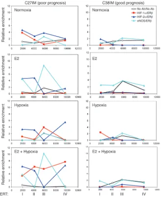
using selected PCa cells from the G1 and G2 groups. Chromatins were immunoprecipitated by antibodies to ERβ, ERα, HIF-1α, HIF-2α, HIF-1β, or eNOS, and DNA sequences proximal to or encompassing estrogen and hypoxia response elements within Figure 2
research article
a 10-kb region of the endogenous hTERT regulatory sequences were amplified (Figure 7A).
Optimal hypoxic conditions were defined by treating cells over time with E2, hypoxia, or both and evaluating induction of the HIF-1α protein at times known to be optimal for the ER cyclic occupancy in vivoof target promoters (refs. 33–35 and data not shown). Western blots on nuclear extracts from most G1 and G2 cell lines showed that the HIF-1α signal generally peaked at 135 minutes of exposure to 1% O2, a concentration within the range
observed in solid tumors (refs. 36 and 37 and data not shown). Therefore, this condition was used in further ChIP.
Substantial differences emerged in the behavior of the proteins in G1 (C27IM) and G2 (C38IM) cells (Figure 7B). Regardless of the
stimulus (E2 or hypoxia, alone or combined), eNOS was recruited to all analyzed sites of the hTERT promoter only in G1 cells. Recruit-ment upon E2 treatment (about a 6-fold increase over control) was higher than under hypoxia (3- to 4-fold increase over control), and maximum recruitment occurred upon the combined stimuli (7- to 10-fold increase with a peak on site III). In comparison, recruitment in G2 cells was, at most, 2-fold increase over control.
[image:6.585.58.438.78.507.2]Occupancy of the hTERT promoter sites by ERβ or HIFs, on the other hand, was more strongly dependent upon the stimulus and the cell context. Upon treatment with only estrogen, we observed ERβ but not ERα occupancy (about a 4-fold increase over control) of the hTERT sites III and IV in C27IM (G1) cells and II and III in C38IM (G2) cells. Unexpectedly, estrogen treatment also induced Figure 3
recruitment of HIFs, primarily HIF-1α in C27IM cells and HIF-2α in C38IM cells (3- and 4-fold increase, respectively) with the same pattern as ERβ. Hypoxia caused enrichment of HIF-1α in C27IM cells and HIF-2α in C38IM cells, with maximum enrichment (up to a 6-fold increase) at site III in both cell lines. Notably, combined E2 and hypoxia markedly increased (to about a 12-fold increase) recruitment of both HIF-1α and ERβ at site III exclusively in C27IM cells. Moreover, while HIF-2α recruitment in C27IM cells was con-stant at site III (about a 3-fold increase) upon single or combined treatments, in C38IM cells, maximum recruitment (7-fold increase) occurred under hypoxia, and combined treatment reduced it to lev-els observed with E2 alone (4-fold increase). Occupancy by HIF-1α in C38IM cells or by ERβ or HIF-1α in both cell lines was absent or minimal under all conditions (Figure 7B and data not shown).
In keeping with these results, substantial recruitment of HIF-1α, ERβ , and eNOS was also found, exclusively in G1 cells, on pro-moters of other prognostic genes, e.g., the MSH2 gene involved in mismatch repair and the cyclin B1 (CCNB1) and the PS2 genes (Supplemental Figure 1).
To assess the specificity of our findings, ChIPs were performed in von Hippel–Lindau–deficient renal carcinoma cells (786-0) lack-ing HIF-1α expression but overexpressing HIF-2α. As expected, no recruitment of HIF-1α or endogenous ERβ was observed at site III of the hTERT promoter upon combined treatment (data not shown).
[image:7.585.55.476.75.493.2]Formation of combinatorial complexes between ERβ and eNOS or HIFs. We next asked whether ERβ, HIF-1α, or HIF-2α and eNOS form combinatorial complexes on chromatin. Serial ChIPs were per-formed in PCa cells exposed to E2 or hypoxia, alone or combined. Figure 4
Cross-linked chromatin was immunoprecipitated with antibod-ies to HIF-1α, HIF-2α, or eNOS, then reimmunoprecipitated with antibody against ERβ (or vice versa). eNOS/ERβ and HIFs/ERβ complexes were all strongly enriched along the hTERT regulatory region, but with distinct dynamics, exclusively in C27IM cells (Fig-ure 8). Specifically, recruitment of the eNOS/ERβ complex, which occurred only at site III and in similar amounts upon normoxia or hypoxia, was enhanced at this site and present also at site I upon E2 treatment or treatment with E2 plus hypoxia. On the other hand, the HIF-1α/ER complex was recruited only at site III upon hypoxia but at all sites upon combined treatment, while the HIF-2α/ERβ complex was present at all sites in normoxia but was removed from site III by every treatment. Therefore, occupancy of this ER binding site by HIF-1α/ERβ or HIF-2α/ERβ complexes appears mutually exclusive, suggesting that ERβ can interact on chromatin with both HIFs, but that, upon combined treatment, the HIF-1α/ERβ complex is highly favored for site III occupancy. The absence of complexes at any site in C38IM cells underlines again the difference between G1 and G2 cell lines.
Finally, coimmunoprecipitation of soluble proteins revealed that the eNOS/HIF-1α and HIF-1α/ERβ complexes also form in the absence of DNA (data not shown). We were, however, unable to determine whether this was the case for the eNOS/ERβ interac-tion. In addition to technical reasons related to performance of available antibodies, this negative result may be due to the fact that interaction between these proteins is inherently weak and is stabilized by contacts with the cognate DNA binding sites (estro-gen-responsive elements [EREs]).
Function of combinatorial complexes on estrogen and/or hypoxia-respon-sive elements. To address whether recruitment of eNOS, as assessed by ChIP assays, is capable of transactivating estrogen- and/or hypoxia-responsive gene promoters (and requires intact ERE and hypoxia-responsive element [HRE] binding sites), we assessed the effects of eNOS on luciferase activity driven by the hTERT promot- er with intact (P-1009) or mutated ERE (P-1009Mut), in the pres-ence of ligand-activated ERβ or driven by the hypoxia-dependent erythropoietin (EPO) gene promoter (with intact or mutated HRE) upon HIF-1α/HIF-2α activation. The results (Supplemental Figure 3, A and B) clearly indicate that overexpression of eNOS, caused by cotransfection of a constitutively active eNOS (S1177D),induced P-1009 luciferase by approximately an 2-fold increase over basal activity and strongly potentiated it (~ 5-fold increase) upon overex-pression of ERβ. Conversely, both effects were virtually absent on the hTERT promoter with mutated ERE (P-1009Mut). In addition, complete abrogation of luciferase activity (basal and induced) was
brought about by pharmacological inhibition of PI3K/AKT using an AKT inhibitor, demonstrating the requirement for AKT-mediat- ed phosphorylation of eNOS in the hTERT promoter activity. Over-expression of eNOS alsosignificantly (P < 0.05) induced the EPO reporter, a 5-fold increase, over basal activity and strongly enhanced it by a 10- or 24-fold increase upon cotransfection of HIF-1α or HIF-2α, respectively. Again the effect was virtually abolished by using the HRE-mutated EPO reporter (EPOMut), confirming that synergism between eNOS and HIFs requires an intact HRE.
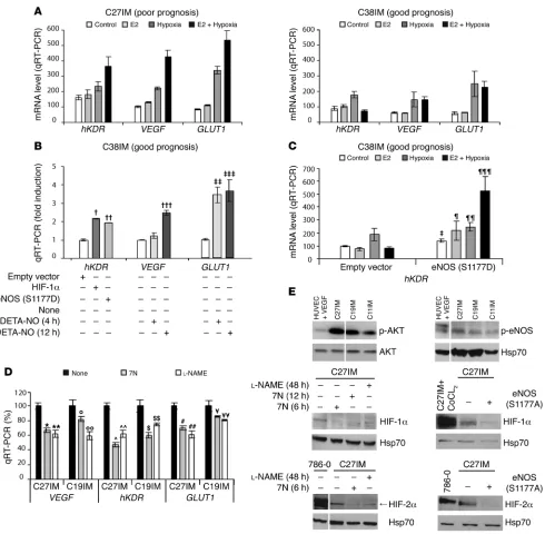
Modulation of target gene expression. We next evaluated mRNA expression of classical gene targets of HIFs or ERs (e.g., VEGF, human VEGF type 2 receptor [hKDR], and GLUT1). As shown in Figure 9A, under basal conditions, all mRNA levels were signifi-cantly higher in G1 (C27IM) than in G2 (C38IM) cells (P = 0.029 for hKDR; P = 0.014 for VEGF; and P = 0.013 for GLUT1 ) in agree-ment with results from primary (2) and immortalized cells (Figure 1D). Expression was enhanced by hypoxia to a comparable extent in both cell lines, demonstrating that the response to low oxygen tension is intact even in G2 cells. Most interestingly, and in keep-ing with the ChIP data, we observed strong induction of these mRNAs upon treatment with E2 plus hypoxia only in G1 cells. A similar transcriptional pattern was detected also in a different cell type, human umbilical vein endothelial cells, a well-character-ized model for the hypoxia-response expressing endogenous ER (38), which is responsive to estrogen (data not shown). Separate E2 or hypoxia stimuli substantially increased mRNAs of hKDR,
GLUT1, MMP9, and hTERT by 4 hours after treatment (Supple-mental Figure 2, A and B). Notably, combined stimuli accelerated this effect to within 2 hours after treatment, and synergistically enhanced recruitment of HIF-1α to the hTERT and VEGF promot-ers (Supplemental Figure 2C). No HIF-1α occupancy of the con-trol cJun promoter was detected (data not shown).
Reciprocal conversion of the transcriptional profile of G1 and G2 cells. The striking differences in the estrogen- and hypoxia-dependent chromatin remodeling by combinatorial complexes among eNOS, HIFs, and ERβ observed in G1 and G2 cells, prompted us to inves- tigate whether modulation of these factors by genetic or pharma-cological approaches could alter the cell molecular phenotype, i.e., the transcription profile of prognostic genes.
G2 cells, derived from patients with good prognoses, were trans-fected with the constitutively active forms of HIF-1α or eNOS or treated with the NO donor DETA-NO, and transcription of the tar-get genes hKDR, GLUT1, and VEGF was measured by quantitative RT-PCR (Figure 9, B and C). Basal levels of gene expression (Fig- ure 9B) were significantly increased by all 3 approaches. In addi-tion, upon transfection of active eNOS (Figure 9C), cells acquired responsiveness to E2, hypoxia, or their combination. In all respects, therefore, G2 cells mimicked the transcription profile of G1 cells. Reciprocal conversion of the G1 to the G2 transcriptional profile was achieved by treating G1 cells with the NOS inhibitors 7-nitro-indazole (7N) or N(G)-nitro-l-arginine methyl ester (l-NAME), both of which resulted in significant transcriptional downregula-tion of the target genes (Figure 9D). Most interestingly, in G1 cells exhibiting constitutive phosphorylation of eNOS and AKT, phar-macological interference with NO production or overexpression of a dominant-negative mutant of eNOS (eNOS S1177A) caused a marked reversal of the constitutive hypoxic phenotype, as shown by decreased HIF-1α and HIF-2α expression (Figure 9E), demon-strating the existence of a regulatory loop between NO production and the hypoxia signaling.
Figure 5
research article
Discussion
The results presented here support the hypothesis that the more aggressive prostate tumors are characterized by concomitant
[image:10.585.86.502.89.601.2]abnormal activation of multiple signaling pathways, particu-larly those mediated by the ER, the HIFs, and NOS. Specifically, our results reveal the formation and prognostic power for PCa of combinatorial complexes of ERβ with eNOS and/or HIF-1α and HIF-2α, and their enhanced co-recruitment on chromatin upon Figure 6
combined estrogen and hypoxia stimuli. These results are validat-ed by the data obtained in vivo on TMAs, demonstrating the very high predictive power of (a) the combined enhanced expression of ERβ and eNOS in terms of DSS and (b) the association between nuclear localization of eNOS and enhanced expression of HIF-2α for all clinical end points. These findings represent a substantial advance toward anticipation of diagnosis and better refinement of prognosis. Indeed, routine assessment of these biomarkers, particularly the combination of ERβ and eNOS and of nuclear eNOS and HIF-2α, by traditional immunohistochemistry, as early as at the time of biopsy, should contribute to increase the accuracy of PCa patient stratification.
The finding, which we believe to be novel, of a functional crosstalk among ERβ, HIFs, and eNOS provides an intriguing alternative interpretation of PCa pathogenesis and progres-sion. Tumors with a constitutively hypoxic phenotype and with enhanced production of NO and eNOS nuclear localization (as is the case of G1 cells), may be more sensitive to changes in the prostate microenvironment, such as those determined by estro-gen signaling or estrogen/androgen metabolites. Thus, upon exposure to estrogen they may acquire a more malignant pheno-type, enabling colonization at distant sites. Our previous study (34) has indeed outlined the relevance of intracellular conversion of androgens to estrogens in the etiopathogenesis of PCa, by dem-onstrating that aromatase inhibitors reduce telomerase activity, an early marker of prostate carcinogenesis. Of relevance to this line of arguing is that ERβ is the first ER expressed in the fetal prostate and the only ER present in epithelial and stromal cells during early ductal morphogenesis (10, 11). We can therefore assume that, with the AR, ERβ mediates the very early stage of fetal prostate development, suggesting its key role in hormone-based chemoprevention strategies for PCa. Our observation that ERβ forms functional complexes with eNOS on chromatin and that combined overexpression of the 2 proteins strongly potenti-ates hTERT promoter activity opens new perspectives about the role of estrogens and NO in PCa. The presence of eNOS in the tumor cell nuclei may in fact influence the specificity of estrogen action on selected genes that in turn may contribute to tumor maintenance and progression (18, 25). Interestingly, binding to DNA, indeed the integrity of the ERE binding site, appears to be a
conditio sine qua non for the formation of the complex and there- fore, for the execution of eNOS nuclear function (see Supplemen-
tal Figure 3). This requirement may explain our failure to coim-munoprecipitate the eNOS/ERβ complex in the absence of DNA (data not shown).Stabilization of complexes between the ER and other transcription factors by contacts with the cognate DNA response elements (EREs) has been previously reported (39). In addition to its functional interaction with ER, eNOS acts as downstream effector of the hypoxia-activated response (17). Numerous reports have assigned to HIFs an essential role in the progression of solid tumors (4, 30, 40), including a role in the lethal phenotype of endocrine cancers (41). Our in vitro and in vivo results confirm and reinforce this role. Within the transcrip-tional signature with prognostic value we previously described (2), HIFs emerged as a predominant functional class of genes upregulated in cells from patients with poor prognosis (G1). These cells exhibit a constitutively hypoxic phenotype but retain responsiveness to both estrogen and hypoxia. Indeed, upon the combined stimuli, we observed chromatin remodeling by HIFs/ ERβ and eNOS/ERβ complexes, accompanied by enhanced transcription of prognostic estrogen and hypoxia target genes. Interestingly, this transcription pattern mimics that observed in human endothelial cells (Supplemental Figure 2) in response to estrogen and hypoxia, in agreement with the more undifferenti-ated phenotype of G1 as compared with G2 cells.
In vivo, high expression of HIF-1α or HIF-2α on their own is predictive of poor outcome. However, this predictive power is sig-nificantly potentiated by the concomitant expression of ERβ and eNOS in the case of HIF-1α and especially of nuclear eNOS in the case of HIF-2α (Figures 5 and 6 and Table 1). In this regard, it should be emphasized that, rather than the recruitment of a single protein, the crucial event in determining the tumor phe-notype and ultimately affecting clinical outcome appears to be the dynamic assembly on chromatin of at least 2 proteins with prognostic value. Specifically, formation of the ERβ/eNOS, ERβ/ HIF-1α, and ERβ/HIF-2α combinatorial complexes, as demon-strated by serial ChIP (Figure 8), is virtually negligible in C38IM cells, under any treatment evaluated, as compared with C27IM cells. These findings reiterate the importance of eNOS, specifi-cally the eNOS-associated complexes and particularly eNOS/ERβ and eNOS/HIF-2α, in the progression of PCa.
The relevant role of eNOS is further supported by our findings that modulation of eNOS expression and activity is capable of reverting the transcriptional pattern of tumor cells with opposite phenotype (G1 and G2 cells; Figure 9, B–E). Specifically, overex- pression of eNOS or treatment with a NO donor confers to con-Table 1
Significance and hazard ratio of Kaplan-Meier analysis of TMA data
End point Biomarker PA Hazard ratio (95% confidence interval)
DSS ERβ 0.0019 0.3469 (0.1795–0.6793)
HIF-1α 0.0484 2.726 (1.0054–4.7665)
eNOSc > 8 0.0056 0.4065 (0.1572–0.7287)
eNOSn > 0 and HIF2α > 6 0.0077 0.2328 (0.0042–0.4356)
Metastasis HIF-2α 0.0362 0.4100 (0.0751–0.9175)
eNOSn > 0 and HIF-2α > 6 0.0063 0.2278 (0.0036–0.3959)
Metastasis and/or local recurrence HIF-2α 0.0015 0.3397 (0.0535–0.4992)
Local recurrence eNOSn > 0 and HIF-2α > 6 0.0006 0.1666 (0.0006–0.1315)
Biochemical recurrenceB HIF-2α 0.0428 0.5083 (0.1650–0.9710)
eNOSn > 0 and HIF-2α > 6 0.0001 0.1779 (0.0011–0.1102)
[image:11.585.50.535.109.234.2]research article
stitutively unresponsive G2 cells the responsiveness to estrogen and hypoxia typical of G1 cells (Figure 9, B and C). Conversely, interference with NO synthesis by pharmacological or genetic inhibition of eNOS activity downregulates expression of the HIFs protein and concurrently abrogates the constitutively hypoxic phenotype of G1 cells (Figure 9E).
[image:12.585.94.492.85.542.2]Overall, the molecular data reported here assign a master role to eNOS in the coordinated activation of a transcriptional program in response to estrogen and hypoxia. In this resides the innova-tive concept of eNOS as an essential cofactor of ERβ and HIFs in transcriptional regulation. The evidence of dynamic assembly on chromatin of functional combinatorial complexes among ERβ Figure 7
and eNOS or HIFs, highly sensitive to local changes in the hor- monal and oxygen tension levels, substantially deepens our under-standing of the molecular mechanisms underlying aggressiveness in hormone-dependent tumors. In particular, NO production and the constitutive phosphorylation and nuclear localization of eNOS appear to mark tumor cells more prone to progression. Consistent with this model, we recently demonstrated in endothelial cells that activated eNOS can translocate into the nucleus, in which it regu-lates gene transcription (18). The in vivo detection of association between nuclear eNOS and enhanced expression of HIF-2α rein-forces this concept and is in agreement with the observation that HIF-2α seems to preferentially promote cell de-differentiation and proliferation (42). In addition, our accurate and systematic assess-ment of the prevalence and localization of eNOS within the cancer tissue contributes to the deciphering of the role of NO in tumor progression by overcoming the lack of such in vivo data.
eNOS is an important downstream target of the PI3K/AKT sig- naling pathway, which is strongly deregulated in our experimen-tal system. In fact, in the original PCa primary cell populations, we found an extremely elevated expression of the serine/threo-nine kinase AKT/protein kinase B (AKT1) mRNA by microarray
as compared to the normal/hyperplastic cells (2). More interest-ingly, mRNA levels of AKT1, a key regulator of cell survival, were significantly higher in G1 than in G2 cells (P < 0.05; data not shown) and the AKT1 protein was constitutively phosphorylated (Figure 9E), reiterating the phenotypic difference of cells derived from patients with adverse prognosis. In line with this conclu-sion is our finding of the abrogation of hTERT-driven luciferase activity, basal and induced, upon pharmacological inhibition of the PI3K/AKT (Supplemental Figure 3), underscoring the requirement for AKT-mediated phosphorylation of eNOS in the regulation of hTERT promoter activity.
[image:13.585.57.388.82.488.2]Resistance to apoptosis is another feature linked to the activa-tion of PI3K/AKT and greatly enhanced by eNOS function (43), suggesting that reduced susceptibility to cell death may represent an important element toward acquisition of a more aggressive PCa phenotype. Although E2 appears to protect both G1 and G2 cells from apoptosis (data not shown), treatment with the testosterone metabolite 5α-androstane-3β,17β-diol, a specific ERβ ligand (44), prevented apoptosis in G1 cells while enhancing it in G2 cells. This suggests that this metabolite may play agonist or antagonist roles in different cell environments, substantiating our hypothesis of a
Figure 8
research article
deregulated estrogen signaling, exclusively in cells from tumors with more aggressive phenotype and resistance to apoptosis (2). This con-
[image:14.585.51.541.78.558.2]clusion is also supported by the observations that the classical estro-gen target gene PS2 is overexpressed in G1 cells (Figure 1D), and that ERβ and eNOS are both recruited to the PS2 promoter in response to estrogen and hypoxia stimuli (Supplemental Figure 1).
Figure 9
Altogether, these data lead us to postulate that activation of eNOS is a crucial requirement for tumor progression in the prostate microenvironment, which is highly sensitive to abnormal estrogen level and hypoxia. ERs and the hypoxia-response family members are already exploited in clinical oncology as therapeutic targets in endocrine cancers. We contribute to this approach by adding a key molecule, eNOS, which together with abnormal ER and hypoxia-response signaling, drives PCa toward a more aggressive phenotype. These findings facilitate optimization of patient stratification for predicting clinical outcome and foster innovative therapies aimed at targeting the combined NO, estrogen, and hypoxia responses. Methods
Generation of immortalized cell lines. Immortalized PCa cellswere derived from primary cell cultures obtained from fresh tissue explants as described previously (2). Informed consent forms were signed by patients prior to surgery and experiments were approved by the Ethical Committee of the Regina Elena Cancer Institute. The cDNAs for hTERT and SV40 large T antigen were cloned into the retrovirus vectors pLXSP (45) and pBabepuro, respectively, both of which carry the puromycin resistance gene. Produc-tion of the retroviruses pLhTERTSP and pBabepuroTwt and infection of target cells were as described previously (45).
TMA construction, immunohistochemistry, and scoring. Paraffin-embedded tissues from 88 patients that had undergone prostatectomy for clini- cally localized PCa at the Urology Division, Regina Elena Cancer Insti-tute, between 1990 and 1998, were used for TMA construction. None of the cancer patients had received neoadjuvant hormonal treatment or radiotherapy before surgery. The use of archival formalin-fixed, paraffin-embedded prostate tumors from the Department of Pathology human tissue repository was approved by the Ethical Committee of the Regina Elena Cancer Institute. The characteristics of the study group are detailed in Supplemental Table 1. The tissue blocks were selected, sectioned, and stained with H&E and then graded (all histopathological data were systematically reevaluated by the pathologist) in accordance with the Union Internationale Contre le Cancer TNM system (UICC) (TNM Classification of Malignant Tumors. 6th edition. Wiley-Liss. 2002), prior to TMA construction. Representative areas of PCa were marked on both slide and matching paraffin tissue block for TMA construction. TMAs were prepared at the Medical Oncology Depart-ment of the Dana-Farber Cancer Institute, Harvard Medical School, Boston, USA. Briefly, 1 tissue cylinder with a diameter of 0.6 mm was punched from morphologically representative tissue areas of each “donor” tissue block and brought into 1 recipient paraffin block, using a homemade tissue arrayer. Two or three 0.6-mm diameter tissue cores (from tumoral and normal area) were taken from each PCa case. Four composite high-density TMA blocks were designed, and serial 5-μm sections were cut with a Leica microtome and transferred to adhesive-coated slides. Immunohistochemistry was performed as described (2). The nuclear and cytoplasmic compartments were scored separately for all markers except for HIF-1α, which was mostly cytoplasmic. TMA sections were scored semiquantitatively based upon the proportion of tumor cells stained (quantity [Q]) and the staining intensity (I) to obtain the final score as the product of I × Q (46, 47). The scoring system was as follows for Q: 0, negative; 1, 1%–9% positive cells; 2, 10%–39% positive cells; 3, 40%–69% positive cells; 4, 70%–100% positive cells. The scoring system was
as follows for I: 0, negative; 1, low; 2, moderate; 3, high. Immunostaining was assessed by a histopathologist and reviewed independently by a second his-topathologist, both blinded to patients outcome. Only representative tissue cores containing at least 20% of tumor cells were scored.
Outcome analysis. The outcome for the patients (survival, recurrence rate, biochemical recurrence) was followed up to June 2008 (observation period, January 1990 to June 2008). A complete database with all clinical and his-topathological information was generated from the medical records of the Regina Elena Cancer Institute. Patients with PCa were divided in 2 groups based on clinical outcome: unfavorable prognosis was defined by presence of biochemical/local recurrence, metastasis, or disease-specific mortality, and favorable prognosis was defined by complete remission with surgery alone. The cause of death was classified as PCa, other disease, or undefined cause. The survival average was 12.9 years after surgery (95% confidence interval, 12.0–13.8 years) and the median observation period was 9.9 years (25th–75th percentile, 6.2–11.2 years). Biochemical recurrence was defined according to the American Society for Therapeutic Radiology and Oncol- ogy (ASTRO) consensus definition (3 consecutive rises), with time of recur-rence backdated to the midpoint between the PSA nadir and the first PSA rise (48), and the Phoenix definition (PSA nadir + 2 ng/ml) (49). Statistics. Statistical analysis was performed using Prism 2.01 statistical software (GraphPad). Differences among subject groups were assessed by 2-tailed Mann-Whitney U test. A 95% confidence interval (P < 0.05) was considered significant. For TMA data analysis, the DSS, local or distant relapse-free survival, or biochemical recurrence rates were calculated using the Kaplan-Meier method from the time of prostatectomy (50). The statistical significance of differences in survival rates between groups was established by the log-rank test. Hazards ratios and 95% confidence intervals were calculated for each biomarker as predictors of different end points in the Kaplan-Meier analysis. Multivariate analysis of prognostic factors was performed using the Cox proportional hazard model (51). The data analysis was performed with an SPSS version 10 (SPSS Inc.). Acknowledgments We thank F. Graham and S. Soddu for critical reading of the manuscript, D. Del Bufalo for reagents and helpful suggestions, and all members of the Division of Radiotherapy, Regina Elena Cancer Institute for constructive discussions. This work was sup-ported by research grants from Associazione Italiana Ricerca sul Cancro and Ministero del Lavoro, della Salute e delle Politiche Sociali (to A. Farsetti); Ministero dell’Istruzione, dell’Università e della Ricerca; grants from NIH (Specialized Programs of Research Excellence 5P50CA90381, 5R01CA131945) and the Prostate Can-cer Foundation (to M. Loda). Received for publication January 18, 2008, and accepted in revised form February 11, 2009. Address correspondence to: Antonella Farsetti, Department of Experimental Oncology, Molecular Oncogenesis Laboratory, Regi-na Elena Cancer Institute — Experimental Research Center (CRS), Via delle Messi d’Oro 156, 00158 Rome, Italy. Phone: 39-06-5266-2531; Fax: 39-06-41805-26; E-mail: afarsetti@gmail.com. 1. Ferlay, J., Bray, F., Pisani, P., and Parkin, D.M. 2004. GLOBOCAN 2002: Cancer incidence, mortality and prevalence worldwide. IARC CancerBase No. 5, version 2.0. IARC Press. Lyon, France. http://www-dep.iarc.fr/. 2. Nanni, S., et al. 2006. Epithelial-restricted gene profile of primary cultures from human prostate tumors: a molecular approach to predict clini-cal behavior of prostate cancer. Mol. Cancer Res.
4:79–92.
3. Chi, J.T., et al. 2006. Gene expression programs in
response to hypoxia: cell type specificity and prog-nostic significance in human cancers. PLoS Med.
3:e47.
5. Zhong, H., et al. 1999. Overexpression of hypoxia-research article
inducible factor 1alpha in common human cancers and their metastases. Cancer Res. 59:5830–5835.
6. Dales, J.P., et al. 2005. Overexpression of hypoxia-inducible factor HIF-1alpha predicts early relapse in breast cancer: retrospective study in a series of 745 patients. Int. J. Cancer. 116:734–739.
7. Gatenby, R.A., and Gillies, R.J. 2004. Why do can-cers have high aerobic glycolysis? Nat. Rev. Cancer.
4:891–899.
8. Keith, B., and Simon, M.C. 2007. Hypoxia-inducible factors, stem cells, and cancer. Cell. 129:465–472.
9. Ho, S.M., Leung, Y.K., and Chung, I. 2006. Estro-gens and antiestrogens as etiological factors and therapeutics for prostate cancer. Ann. N. Y. Acad. Sci. 1089:177–193.
10. Adams, J.Y., Leav, I., Lau, K.M., Ho, S.M., and Pflueger, S.M. 2002. Expression of estrogen recep-tor beta in the fetal, neonatal, and prepubertal human prostate. Prostate. 52:69–81.
11. Shapiro, E., et al. 2005. Immunolocalization of estrogen receptor alpha and beta in human fetal prostate. J. Urol. 174:2051–2053.
12. Signoretti, S., and Loda, M. 2001. Estrogen receptor beta in prostate cancer: brake pedal or accelerator?
Am. J. Pathol. 159:13–16.
13. Imamov, O., Shim, G.J., Warner, M., and Gustafs-son, J.A. 2005. Estrogen receptor beta in health and disease. Biol. Reprod. 73:866–871.
14. Zhu, X., et al. 2004. Dynamic regulation of estrogen receptor-beta expression by DNA methylation dur-ing prostate cancer development and metastasis.
Am. J. Pathol. 164:2003–2012.
15. Horvath, L.G., et al. 2001. Frequent loss of estrogen receptor-beta expression in prostate cancer. Cancer Res. 61:5331–5335.
16. Leav, I., et al. 2001. Comparative studies of the estrogen receptors beta and alpha and the andro- gen receptor in normal human prostate glands, dys-plasia, and in primary and metastatic carcinoma.
Am. J. Pathol. 159:79–92.
17. Coulet, F., Nadaud, S., Agrapart, M., and Soubrier, F. 2003. Identification of hypoxia-response element in the human endothelial nitric-oxide synthase gene promoter. J. Biol. Chem. 278:46230–46240. 18. Grasselli, A., et al. 2008. Estrogen receptor-alpha
and endothelial nitric oxide synthase nuclear com-plex regulates transcription of human telomerase.
Circ. Res. 103:34–42.
19. Zeng, L., and Xu, Q. 2008. eNOS-ERalpha complex goes to telomerase. Circ. Res. 103:10–12.
20. Chambliss, K.L., and Shaul, P.W. 2002. Estrogen modulation of endothelial nitric oxide synthase.
Endocr. Rev. 23:665–686.
21. Brune, B., and Zhou, J. 2007. Nitric oxide and
superoxide: interference with hypoxic signaling.
Cardiovasc. Res. 75:275–282.
22. Sandau, K.B., Fandrey, J., and Brune, B. 2001. Accu-mulation of HIF-1alpha under the influence of nitric oxide. Blood. 97:1009–1015.
23. Kasuno, K., et al. 2004. Nitric oxide induces hypox-ia-inducible factor 1 activation that is dependent on MAPK and phosphatidylinositol 3-kinase signaling.
J. Biol. Chem. 279:2550–2558.
24. Thomas, D.D., et al. 2004. Hypoxic inducible factor 1alpha, extracellular signal-regulated kinase, and p53 are regulated by distinct threshold concentra-tions of nitric oxide. Proc. Natl. Acad. Sci. U. S. A.
101:8894–8899.
25. Lim, K.H., Ancrile, B.B., Kashatus, D.F., and Coun-ter, C.M. 2008. Tumour maintenance is mediated by eNOS. Nature. 452:646–649.
26. Fukumura, D., Kashiwagi, S., and Jain, R.K. 2006. The role of nitric oxide in tumour progression. Nat. Rev. Cancer. 6:521–534.
27. Ying, L., and Hofseth, L.J. 2007. An emerging role for endothelial nitric oxide synthase in chronic inflam-mation and cancer. Cancer Res. 67:1407–1410. 28. Zhang, W., Kapusta, L.R., Slingerland, J.M., and
Klotz, L.H. 1998. Telomerase activity in prostate can-cer, prostatic intraepithelial neoplasia, and benign prostatic epithelium. Cancer Res. 58:619–621. 29. Sommerfeld, H.J., et al. 1996. Telomerase activity:
a prevalent marker of malignant human prostate tissue. Cancer Res. 56:218–222.
30. Semenza, G.L. 2003. Targeting HIF-1 for cancer therapy. Nat. Rev. Cancer. 3:721–732.
31. Kyo, S., et al. 1999. Estrogen activates telomerase.
Cancer Res. 59:5917–5921.
32. Nishi, H., et al. 2004. Hypoxia-inducible factor 1 mediates upregulation of telomerase (hTERT). Mol. Cell. Biol. 24:6076–6083.
33. Carroll, J.S., et al. 2005. Chromosome-wide map- ping of estrogen receptor binding reveals long-range regulation requiring the forkhead protein FoxA1. Cell. 122:33–43.
34. Nanni, S., et al. 2002. Signaling through estrogen receptors modulates telomerase activity in human prostate cancer. J. Clin. Invest. 110:219–227. 35. Shang, Y., Hu, X., DiRenzo, J., Lazar, M.A., and
Brown, M. 2000. Cofactor dynamics and sufficien-cy in estrogen receptor-regulated transcription.
Cell. 103:843–852.
36. Jiang, B.H., Semenza, G.L., Bauer, C., and Marti, H.H. 1996. Hypoxia-inducible factor 1 levels vary exponentially over a physiologically relevant range of O2 tension. Am. J. Physiol. 271:C1172–C1180. 37. Seifeddine, R., et al. 2007. Hypoxia and estrogen
co-operate to regulate gene expression in T-47D
human breast cancer cells. J. Steroid Biochem. Mol. Biol. 104:169–179.
38. Venkov, C.D., Rankin, A.B., and Vaughan, D.E. 1996. Identification of authentic estrogen receptor in cultured endothelial cells. A potential mecha-nism for steroid hormone regulation of endothelial function. Circulation. 94:727–733.
39. Klinge, C.M. 1999. Role of estrogen receptor ligand and estrogen response element sequence on inter-action with chicken ovalbumin upstream promoter transcription factor (COUP-TF). J. Steroid Biochem. Mol. Biol. 71:1–19.
40. Krishnamachary, B., and Semenza, G.L. 2007. Anal-ysis of hypoxia-inducible factor 1alpha expression and its effects on invasion and metastasis. Methods Enzymol. 435:347–354.
41. Kimbro, K.S., and Simons, J.W. 2006. Hypoxia-inducible factor-1 in human breast and prostate cancer. Endocr. Relat. Cancer. 13:739–749. 42. Gordan, J.D., and Simon, M.C. 2007.
Hypoxia-inducible factors: central regulators of the tumor phenotype. Curr. Opin. Genet. Dev. 17:71–77. 43. Mattiussi, S., et al. 2006. Papilloma protein E6
abrogates shear stress-dependent survival in human endothelial cells: evidence for specialized functions of paxillin. Cardiovasc. Res. 70:578–588. 44. Weihua, Z., et al. 2001. A role for estrogen receptor beta
in the regulation of growth of the ventral prostate.
Proc. Natl. Acad. Sci. U. S. A. 98:6330–6335. 45. Bossi, G., et al. 2004. Wild-type p53 gene transfer is
not detrimental to normal cells in vivo: implications for tumor gene therapy. Oncogene. 23:418–425. 46. Signoretti, S., et al. 2002. Oncogenic role of the
ubiquitin ligase subunit Skp2 in human breast cancer. J. Clin. Invest. 110:633–641.
47. Ogino, S., et al. 2008. Cohort study of fatty acid synthase expression and patient survival in colon cancer. J. Clin. Oncol. 26:5713–5720.
48. [No authors listed]. 1997. Consensus statement: guidelines for PSA following radiation therapy. American Society for Therapeutic Radiology and Oncology Consensus Panel. Int. J. Radiat. Oncol. Biol. Phys. 37:1035–1041.
49. Roach, M., 3rd, et al. 2006. Defining biochemical failure following radiotherapy with or without hormonal therapy in men with clinically localized prostate cancer: recommendations of the RTOG-ASTRO Phoenix Consensus Conference. Int. J. Radiat. Oncol. Biol. Phys. 65:965–974.
50. Kaplan, E.L., and Meier, P. 1958. Nonparametric estimation from incomplete observations. J. Am. Stat. Assoc. 53:457–481.
51. Cox, D.R. 1975. Partial likelihood. Biometrika.