Original Article
Dedifferentiation patterns in DTC: is PDTC
an intermediate state between DTC and ATC?
Duo Wen1,2*, Jia-Qian Hu1,2*, Wen-Jun Wei1,2*, Ben Ma1,2, Zhong-Wu Lu1,2, Yu-Long Wang1,2, Yu Wang1,2,
Qing-Hai Ji1,2
1Department of Head and Neck Surgery, Fudan University Shanghai Cancer Center, Shanghai 200032, China; 2Department of Oncology, Shanghai Medical College, Fudan University, Shanghai 200032, China. *Equal
contribu-tors.
Received September 30, 2018; Accepted November 20, 2018; Epub January 1, 2019; Published January 15, 2019
Abstract: Purpose: The majority of poorly differentiated thyroid cancer (PDTC) and anaplastic thyroid cancer (ATC) are thought to dedifferentiated from differentiated thyroid cancer (DTC). PDTC is regarded as an intermediate form, through which DTC dedifferentiates into ATC. Although accumulation of gene mutations has been observed in PDTC and ATC, evidence of a direct link between DTC, PDTC, and ATC is still lacking. This work aims to illustrate if PDTC is the intermediate state between DTC and ATC. Patients and methods: Whole-exome sequencing was performed in a single patient presenting with primary papillary thyroid cancer (PTC) with simultaneous PTC, PDTC, and ATC in metastatic lymph nodes. Results: 167 nonsynonymous mutations were identified, and 24 of them were exclusive to metastatic loci. Comparison of mutational spectra revealed following results: 1) PDTC and ATC were associated with PTC-LN2 but not PTC-LN1; 2) the majority (5/6) of non-ubiquitous mutations in ATC were also present in PDTC; and 3) despite one mutation observed simultaneously in PTC-LN2, PDTC and ATC, no other associations were found between the ATC and PTC samples (primary or metastatic). Additionally, we identified five mutations (four mutations present in both PDTC and ATC, and one exclusive to ATC), including TP53 p.N107_S108delinsX, possibly related with DTC dedifferentiation. Conclusions: We observed an apparent potential, stepwise dedifferentiation process linking DTC, PDTC, and ATC, with PDTC serving as an intermediate state between DTC and ATC in one certain patient. One key mutation, combined with several low-frequency mutations, appears to underlie this dedifferentiation pathway.
Keywords: PDTC, ATC, pattern of dedifferentiation, whole-exome sequencing, intratumor heterogeneity
Introduction
Thyroid cancer is the most common malignancy of the endocrine system, with follicular cancer being the most common form [1, 2]. Follicular-derived thyroid tumors can be broadly divided into three types: differentiated thyroid cancer (DTC), poorly differentiated thyroid cancer (PDTC), and anaplastic thyroid cancer (ATC): DTC accounts for > 90% of all cancers with a relatively benign clinical presentation and good prognosis [3]. In comparison, PDTC and ATC are relatively rare, accounting for only 5-10% of all follicular-derived thyroid tumor cases, but are much more aggressive and likely to be associ-ated with lymph node and distant metastases [4]. The median survival time of PDTC and ATC patients is 3.2 and 0.5 years, respectively;
a result of accumulating gene mutations over time, although a direct link remained elusive [8].
Here, we report a rare clinical case with well dif-ferentiated papillary thyroid cancer (PTC) diag-nosed in primary tumor, but presenting with PTC, PDTC, and ATC in metastatic lymph nodes simultaneously. The biological behavior and clinical outcome of this case was more typical of ATC rather than DTC. We hypothesize that changes in the tumor microenvironment result in a stepwise, evolutional-like dedifferentiation cascade in certain tumor cells during the pro-cess of lymph node metastasis. To test this hypothesis, we examined a series of tissue
Final pathological report confirmed bilateral PTC. On the right side, 19 lymph nodes were examined, of which 7 were positive: 5 for DTC, 1 for PDTC, and 1 for ATC. Five DTC metastatic lymph nodes were found in the left side. The primary tumors, after a thorough pathological investigation, were diagnosed as PTC solely. There were no traces of PDTC or ATC in other PTC metastatic lymph nodes, either. All of these results were reviewed by two local pathologists. The histological types of tumor were classified according to World Health Organization criteria. PDTC was defined on the basis of the Turin pro-posal: 1) Presence of a solid/trabecular/insular pattern of growth. 2) Absence of the
conven-Figure 1. General information of the PTC sample. A. Schematic information of sample positions in the neck and medical history of this patient. B. Hematoxylin and eosin stain of primary tumor and lymph node metastasis.
samples isolated from the patient. By performing wh- ole-exome sequencing, we evaluated the basic pat- tern and possible key mol-ecules involved with the process of DTC dedifferen- tiation.
Material and methods Case description
[image:2.612.91.385.73.487.2]tional nuclear features of papillary carcinoma. 3) Presence of at least one of the following fea-tures: convoluted nuclei; mitotic activity ≥ 3 × 10 HPF, and tumor necrosis.
Two months after surgery (April 2016), radioac-tive iodine therapy (RAI) was started at a dose of 100 mci and whole body scan showed no signs of uptake outside the neck at that time. On follow-up examination in June 2016, medi-astinal lymphadenopathy and pulmonary me- tastases were found. Unfortunately, due to her poor general condition, the second RAI could not be performed. This patient died in October 2016, 8 months after the operation (Figure 1). For the publication of this case, written informed consent was provided by the patient’s husband.
DNA extraction and quality control
Samples of the primary lesion (one of the two PTC primary tumors), para-cancerous normal tissue, and ATC metastatic lesion were collect-ed from fresh frozen tissue, and DNA was ex- tracted using InvitrogenTM genomic DNA
extrac-tion kits (Life Technologies, USA). For two PTC
for validation by polymerase chain reaction (PCR) amplification and Sanger sequencing. Whole-exome sequencing and bioinformatics analysis
The NimbleGen 2.1M chip (NimbleGen Sys- tems, Madison, WI, USA) captures exome se- quences and allows for performance of DNA quality control on the Agilent 2100 Bioanalyzer (Agilent Technologies, Santa Clara, CA, USA) to ensure sufficient DNA quality for sequencing on the Genome Analyzer IIx sequencing platform (Illumina, San Diego, CA, USA).
[image:3.612.99.383.74.348.2]Sequencing data quality assessment: The base mass value is expressed as the Q value, and the sequencing error rate is expressed by E; the relationship between E and the base quality can therefore be expressed as Q = -10log10 (E), with Q = 40 deemed acceptable. The GC con-tent distribution was checked to evaluate the presence of A/T, G/C separation phenomenon. Sequencing data were aligned by BWA-MEM (http://bio-bwa.sourceforge.net) against the human standard genome. Sequencing reads with a ≥ 95% match to the target sequence were included in all downstream analyses.
Figure 2. Detected mutations in the PTC sample. Portions of different types
of mutations illustrated in the upper figure. Exact number of different types of
mutations among samples is listed in the lower table.
and one PDTC metastatic lymph nodes, DNA was extracted from formalin-fixed and paraffin-embed-ded (FFPE) samples by using QIAamp® DNA FFPE Tissue kit (Qiagen, USA) and punches performed specifically in the tumor regions of the nodal me- tastases.
Bioinformatics analysis: Image recognition of the sequencing results were obtained, followed by removal of contaminated and linker sequenc-es, and sequence length, number, and data yield were determined. Data were then com-pared against a reference genome (GRCh38/ hg38) for detection of single nucleotide varia-tions. The mutation frequency and statistical significance of identified mutation sites were validated using a combination of Exgroup (Exome Aggregation Consortium), the National Heart, Lung, and Blood Institute Exome Sequ- encing Project (NHLBI-ESP) exome database, (4) and Clinvar, a database for predicting the pathogenicity of gene mutations and analyzing whether mutations are likely to be pathogenic, and whether they are associated with a particu-lar causative agent.
Results
Identification and validation of somatic muta-tions
For this patient, we performed exon-capture sequencing on DNA from samples of primary tumor, adjacent normal thyroid tissue, two PTC metastatic lymph nodes (PTC-LN1, PTC-LN2), one PDTC metastatic lymph node (PDTC-LN), and one ATC metastatic lymph node (ATC-LN). The average read length was 150 bp, with an average depth of 146.12, 95.07, 108.85, 146.97, 116.6, and 116.59-fold coverage, res- pectively. Nonsynonymous somatic point mu- tations and insertions and deletions (indels)
identified in one type of metastatic sample but not in any other samples including the primary tumor; and 3. Shared mutations (n = 7), defined as mutations that were identified in at least two different metastatic samples, but were not found in the primary tumor. As shown in Figure 2, we mapped the regional distribution of muta-tions across different samples. All of 24 non-ubiquitous mutations were listed in Supple- mentary Table 1. From this analysis, 36 muta-tions were selected for validation by PCR ampli-fication and Sanger sequencing, of which, 32 (88.9%) were specifically validated in samples in which they were initially identified.
Molecular evidence of a stepwise transforma-tion of DTC to ATC via PDTC
[image:4.612.91.383.73.234.2]To compare the mutational spectra between different metastatic lesions, we focused on those 24 non-ubiquitous mutations in this study. Data analyzing revealed four important results (Figures 2, 3): 1. PTC-LN1 exhibited five unique mutations, along with one mutations also observed in PTC-LN2. In contrast, PTC-LN1 shared no mutations with the PDTC or ATC sam-ples. 2. In addition to the one mutation shared with PTC-LN1, PTC-LN2 harbored four unique mutations, along with one mutation shared with PDTC, and other one mutation simultane-ously shared with PDTC and ATC. 3. The PDTC sample exhibited seven unique mutations, four PDTC/ATC shared mutations and one PTC-LN2/ PDTC/ATC shared mutation. 4. The ATC sample contained one unique mutations and five sh-
Figure 3. Map of 24 non-ubiquitous mutations. Twenty-four non-ubiquitous mu-tations were mapped into all of metastatic samples. Horizontal axis presents gene names that mutations were located within; Vertical axis presents different
tissue samples; Grey: mutation identified; Blue: no mutation identified.
ared mutations as mentioned above. All of these five shared mutations were also seen in the PDTC sample.
Identification of PTC dedifferentiation-related mutations
In our sequencing data, we found a total of four mutations shared between PDTC and ATC, along with one mutation unique to ATC (Table 1). Among these mutations, the role of the tu- mor suppressor gene TP53 is well-established, with newer estimates suggesting a mutation frequency up to 50% higher than that of earlier reports [9, 10]. The p.N107_S108delinsX muta-tion of TP53 identified in this study is a stop-gain mutation, in which deletion of the stop codon results in a decrease in the efficiency of transcriptional translation. We also identified mutations in PDE10A, a gene that has been linked to tumorigenesis in non-small cell lung cancer [11].
All of mutations identified in this study were fur-ther compared against the two thyroid databas-es of the The Cancer Genome Atlas (TCGA). With the exception of TP53, all of the mutations identified in this study were low-frequency mutations. Examples include ARMC3 and CRP,
for which mutations were observed in only one PTC sample. No mutations were detected in either PDE10A or RBMXL1.
Discussion
Dedifferentiation of DTC is an important factor in PTC diagnosis, having a significant impact on both therapeutic intervention strategy and clini-cal outcome [12]. Tumor dedifferentiation, as is widely acknowledged, can result in advanc- ed invasion and metastasis, limiting surgical options for certain patients. RAI is another ther-apy in the treatment of DTC apart from surgery, whose success depends on the presence of sodium-iodide symporters (NIS) on the surface of thyroid follicular cells [13]. Decreased NIS activity caused by tumor dedifferentiation is likely to result in a diminished response to RAI, reducing the possible chance of a successful outcome [14]. Dedifferentiation is a major cause of RAI failure [4, 15]. For those who are diagnosed with RAI refractory DTC, we now have targeted drugs indicated for their situa-tion. Unfortunately, there are currently no tar-geted drugs approved for use against either ATC or PDTC [16, 17]. Better methods for identi-fying tumor dedifferentiation in early stage patients, in combination with targeted thera-Table 1.Potential PTC dedifferentiation-related mutations
Gene Chrd Pose Reff Altg ExonicFunc AA changeh Primary
a PDTC-LNb ATC-LNc APi Covj AP Cov AP Cov RBMXL1 chr1 89449509 T - Frameshift deletion p.G71A 0 121 0.11 55 0.28 67 CRP chr1 159683661 G A Nonsynonymous p.A110V 0 110 0.31 134 0.18 178 ARMC3 chr10 23326291 GA - Frameshift deletion p.R834fs 0 41 0.21 42 0.18 45
p.R571fs p.R827fs
TP53 chr17 7577566 - A Stopgain - 0 68 0.44 52 0.34 50
PDE10A chr6 166075488 G T Nonsynonymous p.P5H 0 15 0 11 0.33 12
[image:5.612.91.521.85.197.2]a. Primary: primary tumor; b. PDTC-LN: lymph node with poorly differentiated cancer metastases; c. ATC-LN: lymph node with anaplastic thyroid carcinoma metastases; d. Chr: chromosome; e. Pos: position; f. Ref: reference Genomic sequences; g. Alt: Alternative genomic bases; h. AA change, Amino acid changes; i. AP: Alternative allelic portion; j. Cov, coverages.
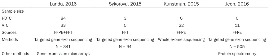
Table 2. Reported second generation sequencing results of PDTC/ATC
Landa, 2016 Sykorova, 2015 Kunstman, 2015 Jeon, 2016
Sample size
PDTC 84 3 0 0
ATC 33 5 22 11
Sources FFPE+FFT FFT FFPE FFPE
Methods Targeted gene exon sequencing Targeted gene exon sequencing Whole exome sequencing Targeted gene exon sequencing
N = 341 N = 94 N = 505
[image:5.612.90.524.261.352.2]pies effectively against dedifferentiated DTC, represent important unmet goals in thyroid cancer research.
A better understanding of dedifferentiation is the first step to treat these patients. To date, two basic models have been proposed to this end: stepwise transformation from DTC to ATC via PDTC, and the de novo model of indepen-dent transformation from DTC to ATC and PDTC. [18] Unfortunately, both models lack sufficient evidence, yielding further research in this field [19]. Our hospital has treated 60 patients with PDTC or ATC between 2006 and 2015, includ-ing 5 cases of ATC in a DTC background and 18 cases of DTC and PDTC in the same tumor. However, no clear coexistence of DTC, PDTC, and ATC within a single primary lesion has been observed, so we cannot confirm direct transfor-mation from PDTC to ATC.
With recent advances in next-generation sequ- encing, significant advances have been made in terms of quantitative data analysis. Xu et al. assessed the role of known mutations in PTC. A total of 87 PDTC and 71 ATC cases, drawn from four publications (Table 2): the analysis showed that both PDTC and ATC are the results of DTC cells gradually accumulating mutations over time [20-22]. Delving deeper into these data, ATC lesions exhibited higher mutational fre-quencies in TERT, as well as in the PIK3CA-PTEN pathway, tumor suppressors, and DNA repair genes [23, 24]. However, none of these studies provided direct supporting evidence of transformation of DTC to ATC via PDTC. Furthermore, most of the studies used targeted
and compare the mutational spectra among the different samples. Results showed three important findings. First, the majority (5/6; 77.8%) of the non-ubiquitous mutations in the ATC sample were also present in the PDTC sa- mple. Second, with the exception of one PTC-LN2/PDTC/ATC shared mutation, there was no other common mutation existing between ATC and PTC samples (primary or metastatic). Finally, the PTC-LN2/PDTC/ATC shared muta-tion represent the only mutamuta-tional link between the PTC and dedifferentiated samples, mean-ing that PTC-LN1 was unrelated to the PDTC and ATC lesions. On observation of the close relationship between PDTC and ATC, we specu-lated that the dedifferentiation from PDTC to ATC occurred more likely during the metastatic phase of the disease, rather than progression from different primary tumor subclones sepa-rately. It suggests a relatively straightforward stepwise transformation of DTC to ATC via PDTC, which acts as an essential intermediate state. A phylogenetic overview of this mutation-al linkage is shown in Figure 4.
Mutations shared between PDTC and ATC are the most likely drivers of the PTC dedifferentia-tion process, while unique ATC mutadedifferentia-tions may also play a role in this transformation. Our study identified the p.N107_S108delinsX mutation of TP53. This mutation may act as a switch in PTC dedifferentiation. Mutations in the PDE10A ge- ne were also revealed by sequencing. Notably, this gene has been reported to be related with tumorigenesis in non-small cell lung cancer [11]. Other mutations identified in this study have not been linked to tumorigenesis, and
Figure 4. Phylogenetic overview of mu-tational linkages between primary and metastatic lesions. Thickness of lines and arrow symbols represents number of
mu-tations identified in different samples.
gene exome sequencing, which cannot evaluate the whole genome.
their role in PTC dedifferentiation and tumor progression are unclear. More studies need to be done to discover their biological functions. However, it is the most likely explanation but not the only one. Actually, we could not rule out the possibility that PDTC and ATC metastatic lesions were arise from the same subclone in the primary tumor and developed in different lymph node with a parallel way. The reason, that those unique and shared mutations identi-fied in PDTC and ATC samples have not been detected in the primary tumor, may be caused by tumor purity and heterogeneity. This possi-bility is relatively low, because the locations of PDTC and ATC lymph nodes are the furthest from the primary tumor than other PTC meta-static lesions. To exclude this possibility to the maximum extent, multipoint sampling and microdissection methods are necessary during sample collection.
Since lymphatic reflux represents a self-sus-tained dynamic system, there may be internal cross-connection; theoretically, the primary cells of the ATC metastases could have ori- ginated from upstream PDTC metastases, or from PTC metastases. However, based upon our sequencing results, the non-ubiquitous mutations carried by the ATC metastases did not intersect with PTC-LN1. Meanwhile, two mutations identified in the PDTC metastases were shared with PTC-LN2, suggesting that PDTC is likely to be a necessary intermediate state between DTC and ATC. To further limit the influence of the complex lymph node cross-nection, a full exome sequencing of all con-firmed positive lymph nodes is indispensable. The last and most important limitation of this study is its single-case design. This case may not be reflective enough of the wider popula-tion, for the mutations identified could be case-sensitive, which means these genes may only mutate in this case. Accordingly, the possibility of other dedifferentiation patterns in DTC can-not be ruled out. Therefore, to address this limi-tation, more cases need to be collected and more aspects, such as RAI therapy and target-ed drug clinical trials, should also be taken into account in the future.
In conclusion, our data provide evidence of a possible link between the mutational profile of lesions and their dedifferentiation status. In
this model, the transformation process from PTC to ATC was more likely stepwise. PDTC may play an important role as an intermediary between DTC and ATC, with the transformation driven by a single key mutation in combination with several low-frequency mutations.
Acknowledgements
The abstract of this paper was presented at the 87th annual meeting of the American Thyroid Association (ATA) as a poster presentation. The poster’s abstract was published in Thyroid. Vol- ume: 27 Issue S1: October 1, 2017. http://doi. org/10.1089/thy.2017.29050.sc.abstracts. This work was supported by funds from the National Natural Science Foundation of China, 81572622 to Qing-Hai Ji; 81502317 to Wei-Wen Jun, 81472498 and 81772851 to Yu-Long Wang.
Disclosure of conflict of interest
None.
Address correspondence to: Qing-Hai Ji and Yu
Wang, Department of Head and Neck Surgery, Fudan University Shanghai Cancer Center, Shanghai 200032, China; Department of Oncology, Shanghai Medical College, Fudan University, Shanghai 2000- 32, China. Tel: 021-64175590; Fax: 0086-021-64175590; E-mail: [email protected] (QHJ); [email protected] (YW)
References
[1] Sherma SI. Thyroid carcinoma. Lancet 2003; 361: 501-511.
[2] Schlumberger MJ. Papillary and follicular thy-roid carcinoma. N Engl J Med 1998; 338: 297-306.
[3] Pacini F, Castagna M, Brilli L, Pentheroudakis G; ESMO Guidelines Working Group. Thyroid cancer: ESMO clinical practice guidelines for diagnosis, treatment and follow-up. Ann Oncol 2012; 23 Suppl 7: vii110-9.
[4] Viola D, Valerio L, Molinaro E, Agate L, Bottici V, Biagini A, Lorusso L, Cappagli V, Pieruzzi L and Giani C. Treatment of advanced thyroid cancer with targeted therapies: ten years of experi-ence. Endocr Relat Cancer 2016; 23: R185-R205.
ge-netics and advanced therapies. Nat Rev Endo-crinol 2017; 13: 644-660.
[6] Lee DY, Won JK, Lee SH, Park DJ, Jung KC, Sung MW, Wu HG, Kim KH, Park YJ and Hah JH. Changes of clinicopathologic characteris-tics and survival outcomes of anaplastic and poorly differentiated thyroid carcinoma. Thy-roid 2016; 26: 404-413.
[7] Sasanakietkul T, Murtha TD, Javid M, Korah R and Carling T. Epigenetic modifications in poorly differentiated and anaplastic thyroid cancer. Mol Cell Endocrinol 2018; 469: 23-37. [8] Landa I, Ibrahimpasic T, Boucai L, Sinha R,
Knauf JA, Shah RH, Dogan S, Ricarte-Filho JC, Krishnamoorthy GP and Xu B. Genomic and transcriptomic hallmarks of poorly differenti-ated and anaplastic thyroid cancers. J Clin In-vest 2016; 126: 1052-66.
[9] Stengel A, Kern W, Haferlach T, Meggendorfer M, Fasan A and Haferlach C. The impact of TP53 mutations and TP53 deletions on sur-vival varies between AML, ALL, MDS and CLL: an analysis of 3307 cases. Leukemia 2017; 31: 705-711.
[10] Forbes SA, Beare D, Bindal N, Bamford S, Ward S, Cole C, Jia M, Kok C, Boutselakis H and De T. COSMIC: high-resolution cancer ge-netics using the catalogue of somatic muta-tions in cancer. Curr Protoc Hum Genet 2016; 91: 10.11.1-10.11.37.
[11] Zhu B, Lindsey A, Li N, Lee K, Ramirez-Alcanta-ra V, Canzoneri JC, Fajardo A, da Silva LM, Thomas M and Piazza JT. Phosphodiesterase 10A is overexpressed in lung tumor cells and inhibitors selectively suppress growth by block-ing β-catenin and MAPK signalblock-ing. Oncotarget 2017; 8: 69264-69280.
[12] Haugen BR, Alexander EK, Bible KC, Doherty GM, Mandel SJ, Nikiforov YE, Pacini F, Ran-dolph GW, Sawka AM and Schlumberger M. 2015 American Thyroid Association manage-ment guidelines for adult patients with thyroid nodules and differentiated thyroid cancer: the American Thyroid Association guidelines task force on thyroid nodules and differentiated thyroid cancer. Thyroid 2016; 26: 1-133. [13] Seregni E, Mallia A, Chiesa C, Scaramellini G,
Massimino M and Bombardieri E. Radioiodine therapy of differentiated thyroid cancer. In: editors. Nuclear medicine therapy. Springer; 2013. pp. 133-153.
[14] Lakshmanan A, Scarberry D, Shen DH and Jhi-ang SM. Modulation of sodium iodide symport-er in thyroid cancsymport-er. Horm Cancsymport-er 2014; 5: 363-373.
[15] Fröhlich E and Wahl R. The current role of tar-geted therapies to induce radioiodine uptake in thyroid cancer. Cancer Treat Rev 2014; 40: 665-674.
[16] Gilliland FD, Hunt WC, Morris DM and Key CR. Prognostic factors for thyroid carcinoma. A population-based study of 15,698 cases from the Surveillance, Epidemiology and End Re-sults (SEER) program 1973-1991. Cancer 1997; 79: 564-573.
[17] Mazzaferri EL and Kloos RT. Clinical review 128: current approaches to primary therapy for papillary and follicular thyroid cancer. J Clin Endocrinol Metab 2001; 86: 1447-1463. [18] Dellaire G, Berman JN and Arceci RJ. Cancer
genomics: from bench to personalized medi-cine. Academic Press; 2013.
[19] Xing M. Molecular pathogenesis and mecha-nisms of thyroid cancer. Nat Rev Cancer 2013; 13: 184-199.
[20] Sykorova V, Dvorakova S, Vcelak J, Vaclavikova E, Halkova T, Kodetova D, Lastuvka P, Betka J, Vlcek P and Reboun M. Search for new genetic biomarkers in poorly differentiated and ana-plastic thyroid carcinomas using next genera-tion sequencing. Anticancer Res 2015; 35: 2029-2036.
[21] Kunstman JW, Juhlin CC, Goh G, Brown TC, Stenman A, Healy JM, Rubinstein JC, Choi M, Kiss N and Nelson-Williams C. Characteriza-tion of the mutaCharacteriza-tional landscape of anaplastic thyroid cancer via whole-exome sequencing. Hum Mol Genet 2015; 24: 2318-2329. [22] Jeon MJ, Chun SM, Kim D, Kwon H, Jang EK,
Kim TY, Kim WB, Shong YK, Jang SJ and Song DE. Genomic alterations of anaplastic thyroid carcinoma detected by targeted massive paral-lel sequencing in a BRAFV600E mutation-prev-alent area. Thyroid 2016; 26: 683-690. [23] Xu B and Ghossein R. Genomic landscape of
poorly differentiated and anaplastic thyroid carcinoma. Endocr Pathol 2016; 27: 205-212. [24] Rosove MH, Peddi PF and Glaspy JA. BRAF
Supplementary Table 1. Unique and shared mutations in all of lymph node metastasis specimens
Gene Chr Pos Ref Alt Alternation type AA Change PRIMARY PTC-LN1 PTC-LN2 PDTC ATC
AP Cov AP Cov AP Cov AP Cov AP Cov
Unique mutations of PTC-LN1
PLBD1 chr12 14720590 T G Nonsynonymous SNV p.Q14P 0 19 0.22 9 0 28 0 20 0 20
SPOPL chr2 139326516 G A Nonsynonymous SNV p.D349N 0 65 0.21 47 0 47 0 34 0 35
YTHDF1 chr20 61834858 G T Nonsynonymous SNV p.S145Y 0 44 0.23 35 0 53 0 25 0 45
FGFRL1 chr4 1019055 CA - Frameshift deletion p.H479fs 0 66 0.23 40 0 100 0 54 0 54
RNF145 chr5 158630629 - TT Frameshift insertion p.N29fs; p.N16fs; p.N13fs;
0 115 0.17 47 0 91 0 152 0 62
Mutations shared by PTC-LN1 and PTC-LN2
CASP5 chr11 104878040 - T Frameshift insertion p.T10fs; p.T81fs; p.T68fs
0 86 0.16 64 0.14 65 0 73 0 70
Unique mutations of PTC-LN2
LRRIQ3 chr1 74575212 - T Frameshift insertion p.Q245fs 0 183 0 101 0.13 191 0 117 0 115
TRIM77 chr11 89444610 - A Frameshift insertion p.W148fs 0 165 0 81 0.15 151 0 104 0 141
TNS2 chr12 53451822 G A Nonsynonymous SNV p.G354D;
p.G344D; p.G220D
0 6 0 5 0.29 7 0 7 0 8
FEV chr2 219846580 A G Nonsynonymous SNV p.S176P 0 6 0 6 0.29 14 0 7 0 8
Mutations shared by PTC-LN2 and PDTC
LENG9 chr19 54974774 A T Nonsynonymous SNV p.M1K 0 7 0 4 0.62 8 0.73 11 0 5
Mutations shared by PDTC-LN2, PDTC and ATC
LNX1 chr4 54327211 - A Splice - 0 49 0 35 0.16 31 0.19 31 0.22 27
Mutations shared by PDTC and ATC
RBMXL1 chr1 89449509 T - Frameshift deletion p.G71A 0 121 0 54 0 82 0.11 55 0.28 67
CRP chr1 159683661 G A Nonsynonymous SNV p.A110V 0 110 0 94 0 97 0.31 134 0.18 178
ARMC3 chr10 23326291 GA - Frameshift deletion p.R571fs; p.R827fs; p.R834fs
0 41 0 42 0 44 0.21 42 0.0.9 45
TP53 chr17 7577566 - A Stopgain - 0 68 0 56 0 59 0.44 52 0.34 50
Unique mutations of PDTC
LAMA1 chr18 7117665 G A Stopgain p.Q19X 0 44 0 7 0 36 0.33 24 0 30
PRX chr19 40901323 C T Nonsynonymous SNV p.G979E 0 46 0 22 0 50 0.28 18 0 31
AIRE chr21 45712283 C T Nonsynonymous SNV p.P365L 0 20 0 9 0 19 0.67 6 0 13
BOC chr3 112998209 T A Nonsynonymous SNV p.Y644N;
p.Y643N 0 38 0 42 0 67 0.25 28 0 38
CYP39A1 chr6 46620316 C - Frameshift deletion p.E2fs 0 76 0 49 0 65 0.23 30 0 67
SNAP91 chr6 84269884 A C Nonsynonymous SNV p.V550G; p.V827G; p.V733G; p.V857G; p.V766G
0 55 0 30 0 67 0.19 48 0 43
SVEP1 chr9 113173407 G A Nonsynonymous SNV p.P2195L 0 41 0 55 0 39 0.33 46 0 60
Unique mutations of ATC
PDE10A chr6 166075488 G T Nonsynonymous SNV p.P5H 0 15 0 9 0 4 0 11 0.33 12