Original Article
Knockdown of eukaryotic translation initiation factor
3 subunit B inhibits cell proliferation and migration
and promotes apoptosis by downregulating WNT
signaling pathway in acute myeloid leukemia
Yonghuai Feng1, Liusong Wu2
1Department of Hematology, Affiliated Hospital of Zunyi Medical University, Guizhou, China; 2The Second Depart-ment of Pediatrics, Affiliated Hospital of Zunyi Medical University, Guizhou, China
Received April 26, 2019; Accepted September 24, 2019; Epub January 1, 2020; Published January 15, 2020
Abstract: The study aimed to investigate the effect of eukaryotic translation initiation factor 3 subunit B (EIF3B) on cell proliferation, migration, and apoptosis as well as the underlying mechanism in acute myeloid leukemia (AML). EIF3B expression was detected in AML-193, HL-60, OCI-AML2, and KG-1 cell lines and human primary bone mar-row mononuclear cells (BMMC). EIF3B knockdown was realized by transfecting EIF3B ShRNA plasmids, and EIF3B knockdown and WNT2 overexpression were established by transfecting EIF3B ShRNA plasmids and WNT2 overex-pression plasmids into KG-1 cells. The effect of EIF3B knockdown, and EIF3B knockdown plus WNT2 overexpres-sion on cell proliferation, apoptosis, migration, glycogen synthase kinase 3B (GSK3B) and catenin beta 1 (CTNNB1) was assessed. EIF3B mRNA and protein expression were higher in AML-193, OCL-AML2 and KG-1 cell lines, but unchanged in the HL-60 cell line compared with human primary BMMC. The expression of WNT2 was decreased by EIF3B downregulation, while it had no effect on EIF3B expression. As for cell activities, EIF3B knockdown inhib-ited the cell proliferation and migration but promoted apoptosis by inhibiting WNT2 expression. In addition, EIF3B knockdown downregulated the expression of CTNNB1 but upregulated the expression of GSK3B by blocking WNT2 expression in AML, implying an inhibitory effect of EIF3B downregulation on WNT signaling pathway. EIF3B is up-regulated and its knockdown inhibits cell proliferation, and migration, while promoting apoptosis by downregulating the WNT signaling pathway in AML.
Keywords: EIF3B, AML, proliferation, migration, apoptosis, WNT signaling pathway
Introduction
According to a 2018 global statistics report, the incidence of leukemia accounts for 2.4% of all cancers globally, and acute myeloid mia (AML) is a major pathologic type of
leuke-mia, that is identified as the malignant
trans-formation of clonal hematopoietic stem cells in the bone marrow and peripheral blood [1-3]. Although treatments such as intensive induc-tion chemotherapy and stem cell transplanta-tion contribute to the improvement of the sur-vival in AML patients, long-term mortality and recurrence remain a challenge, which lead to a poor prognosis in AML patients and a heavy burden to social and medical resources [1, 4]. Therefore, it is essential to explore novel thera-peutic targets for alternative AML therapies to
improve treatment outcomes and prognosis in AML patients.
aberrant self-renewal ability of hematopoietic cells [8-12]. The regulatory proteins within the WNT signaling pathway, including catenin beta 1 (CTNNB1), glycogen synthase kinase 3B (GSK3B), cyclin D1 (CCND1) and C-Myc, are dysregulated under the induction of AML-re- lated translocation, accelerating the progres-sion of AML [11-13]. Based on the aforemen-tioned evidence and considering that there is still limited information about the function of EIF3B in AML, we speculated that EIF3B might be dysregulated in AML and it might affect AML cell function by regualting the WNT sig- naling pathway. In the present study we per-formed cellular experiments to explore the underlying mechanism of EIF3B in AML.
Materials and methods
Cell culture
Human AML cell lines AML-193, HL-60, OCI-AML2 and KG-1 were purchased from Leibniz Institute DSMZ-German Collection of Micro- organisms and Cell Cultures (Braunschweig, Germany), AML-193 cells were cultured in 90%
Iscove’s Modified Dulbecco’s Medium (Gibco,
USA) and 10% fetal bovine serum (FBS) (Gibco, USA), and HL-60, OCI-AML2 and KG-1 cells were cultured in 90% Roswell Park Memorial Institute 1640 Medium (Gibco, USA) and 10% FBS (Gibco, USA) under 95% air and 5% CO2 at 37°C.
EIF3B expression in AML cell lines
EIF3B mRNA and protein expressions in AML-193, HL-60, OCI-AML2 and KG-1 cell lines were detected using real-time quantitative poly-merase chain reaction (RT-qPCR) and western blot (WB). EIF3B mRNA and protein expressi- ons in human primary bone marrow mononu-clear cells (BMMC) were also detected as con-trols. Human primary BMMC were bought from American Type Culture Collection (Manassas, USA).
Transfections
Control short hairpin RNA (ShRNA), control overexpression EIF3B ShRNA, and WNT2 over-expression plasmids were constructed using pEX (GenePharma, China) or pGPH1 (GenePhar- ma, China). Control ShRNA plasmids and con-trol overexpression plasmids were transfected
into KG-1 cells as a Control group; EIF3B Sh- RNA plasmids and control overexpression plas-mids were transfected into KG-1 cells as Sh-EIF3B group; lastly, EIF3B ShRNA plasmids and WNT2 overexpression plasmids were tr- ansfected into KG-1 cells as Sh-EIF3B & WNT2 group.
EIF3B and WNT2 expressions after transfec-tion
Protein and mRNA expressions of EIF3B and WNT2 were detected by RT-qPCR and WB re- spectively at 48 h post transfection.
Counting Kit-8 (CCK-8) assay after transfection
Proliferation of KG-1 cells was detected by CCK-8 assay at 0 h, 24 h, 48 h, and 72 h post transfection using CCK-8 Kit (Dojindo, Japan).
In brief, 10 μl CCK-8 and 90 μl serum-free
medium were added to each group of KG-1 cells, then the cells were incubated in mixed gas with 95% air and 5% CO2 at 37°C. Optical density (OD) value was measured by microplate reader (BioTek, USA).
Annexin V/Propidium Iodide (AV/PI) assay after transfection
Apoptosis rate of KG-1 cells was detected by AV/PI assay at 48 h post transfection using FITC Annexin V Apoptosis Detection Kit II (BD, USA). In brief, KG-1 cells in each group were digested by trypsin (Thermo, USA) and washed with phosphate buffer solution (PBS), then
sus-pended in 100 μl binding buffer. Then 5 μl Annexin V (AV) and 5 μl propidium iodide (PI)
were added to the cells and then cells were left
in dark for 15 min. Subsequently, 400 μl bind -ing buffer was added and apoptosis rate was
analyzed using flow cytometry (FCM) (Beckman,
USA).
Apoptotic markers expression after transfec-tion
Cleaved Caspase 3 (C-Caspase 3) and B-cell lymphoma-2 (Bcl-2) protein expressions in KG- 1 cells were detected at 48 h post transfection by WB assay.
Transwell assay after transfection
hydrating the Chamber (Costar, USA) with 8 μm Polycarbonate film, 200 μl of serum-free medi -um containing 5×10⁵ cells were seeded in the upper chamber. Then the lower chamber was
filled with 500 μul 10% FBS medium. The whole
chamber was incubated at 37°C for 12 h. The cells were collected from the medium in the lower chamber and the number of cells was
counted by flow cytometry (Beckman, USA).
GSK3B and CTNNB1 expressions after trans-fection
Genes downstream of WNT signaling, including GSK3B and CTNNB1 expressions were detect-ed at 48 h post transfection by RT-qPCR and WB respectively.
RT-qPCR
Total RNA was extracted from cells using TRIzol™ Reagent (Invitrogen, USA) and then reversely transcribed to cDNA using Prime- Script™ RT Master Mix (Takara, Japan). Fol- lowing that, RT-qPCR was performed using TB Green™ Fast qPCR Mix (Takara, Japan) to qu- antify EIF3B, WNT2, GSK3B and CTNNB1 expressions. The result was calculated using 2-ΔΔCt method with GAPDH as an internal
refer-ence. The primers used in RT-qPCR are listed in Table 1.
Western blot analysis
Total protein was extracted with RIPA Lysis and Extraction Buffer (Thermo, USA). The protein concen-tration in each sample was mea-sured using the Pierce™ Rapid Go- ld BCA Protein Assay Kit (Thermo,
USA). 20 μg protein samples were
added to NuPAGE™ 4-20% Tris-Acetate Midi Protein Gels (Thermo, USA) and transferred onto Polyvin- ylidene Fluoride membrane (Milli- pore, USA). After blocking with BSA (Thermo, USA) for 2 h, the mem-branes were incubated with the pri-mary antibodies overnight at 4°C. Then, the membranes were incu-bated with the secondary antibody for 90 min at 37°C. Pierce™ ECL Plus Western Blotting Substrate (Thermo, USA) was used to illumi-nize the bands and X-ray film
(Ko-Table 1. Primers for qPCR
[image:3.612.90.355.83.165.2]Gene Forward Primer (5’-3’) Reverse Primer (5’-3’) EIF3B CGTATGTGCGTTGGTCTCCTAA CCTTGGTGGCTGAATCTCTGAA WNT2 TCTCGGTGGAATCTGGCTCTG CCTGGCACATTATCGCACATCA GSK3B GCACTCTTCAACTTCACCACTC CCACGGTCTCCAGTATTAGCAT CTNNB1 TGACCAGCCGACACCAAGA GCACGAACAAGCAACTGAACT GAPDH GACCACAGTCCATGCCATCAC ACGCCTGCTTCACCACCTT
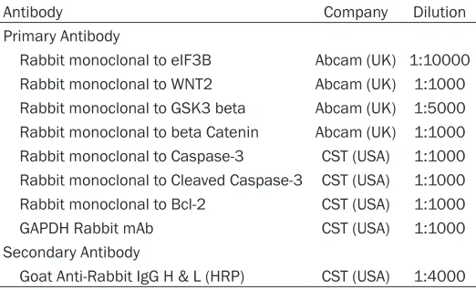
Table 2. Antibodies for western blot
Antibody Company Dilution Primary Antibody
Rabbit monoclonal to eIF3B Abcam (UK) 1:10000 Rabbit monoclonal to WNT2 Abcam (UK) 1:1000 Rabbit monoclonal to GSK3 beta Abcam (UK) 1:5000 Rabbit monoclonal to beta Catenin Abcam (UK) 1:1000 Rabbit monoclonal to Caspase-3 CST (USA) 1:1000 Rabbit monoclonal to Cleaved Caspase-3 CST (USA) 1:1000 Rabbit monoclonal to Bcl-2 CST (USA) 1:1000 GAPDH Rabbit mAb CST (USA) 1:1000 Secondary Antibody
Goat Anti-Rabbit IgG H & L (HRP) CST (USA) 1:4000
dak, USA) was used to visualize the result. The antibodies used in this study are summarized in Table 2.
Statistics
Data are shown as mean value ± standard de- viation, and comparison among groups was detected by One-Way ANOVA followed by Dun- nett’s multiple comparisons test, while com-parison between two groups was detected by t
test. The statistical software used in this study was SPSS 21.0 Software (IBM, USA), and the graph-making software used in this study was GraphPad Prism 6.01 (GraphPad Int, USA). P <
0.05 was considered significant.
Results
EIF3B expression in AML
[image:3.612.91.352.200.359.2]Interaction between EIF3B and WNT2 in KG-1 cells
In order to examine the effect of EIF3B on re- gulating WNT2, the following experiments were carried out in KG-1 cells. EIF3B mRNA was downregulated in Sh-EIF3B group compared with the Control group (P < 0.001), and EIF3B mRNA expression was similar in the Sh-EIF3B group and Sh-EIF3B & WNT2 group (P > 0.05) (Figure 2A). WNT2 mRNA was downregulated in the Sh-EIF3B group compared with Control group (P < 0.01). In addition, WNT2 mRNA was upregulated in Sh-EIF3B & WNT2 group com-pared with Sh-EIF3B group (P < 0.01) (Figure 2B). WB assay indicated that EIF3B protein expression was reduced in Sh-EIF3B group compared with Control group but similar bet- ween the Sh-EIF3B & WNT2 group and Sh-EIF3B group. WNT2 protein expression was decreas- ed in the Sh-EIF3B group compared with Con- trol group but elevated in the Sh-EIF3B & WNT2 group compared with Sh-EIF3B (Figure 2C).
The above findings indicated that knockdown
of EIF3B downregulated the WNT2 expression, while WNT2 had no effect on EIF3B expression in AML.
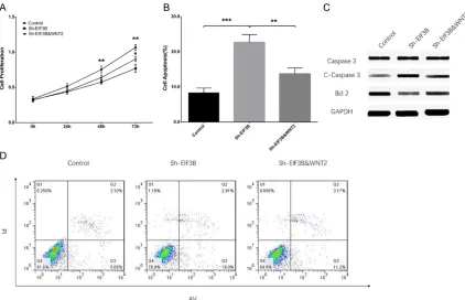
EIF3B knockdown reduced cell proliferation while promoting apoptosis by downregulating WNT2 in KG-1 cells
In order to investigate the effect of EIF3B on regulating AML cell function and its interaction
with WNT2, cell proliferation and apoptosis were detected after tr- ansfection. The cell proliferation ability was unchanged at 24 h (P > 0.05) but reduced at 48 h (P < 0.01) and 72 h (P < 0.01) after transfection in Sh-EIF3B gr- oup compared with the control group (Figure 3A). Cell prolifera-tion ability was similar at 24 h and 48 h after transfection between the Sh-EIF3B & WNT2 group and Sh-EIF3B group (both P > 0.05) but increased at 72 h after trans-fection in the Sh-EIF3B & WNT2 group compared with the ShEIF3B group (P < 0.05). Cell apoptosis rate was increased after transfec-tion in the Sh-EIF3B group com-pared with Control group (P < 0.001), while reduced in Sh-EIF3B & WNT2 group compared with Sh-EIF3B group (P < 0.01) (Figure 3B, 3D). WB assay visualiz- ed that C-Caspase 3 was upregulated in the Sh-EIF3B group compared with controls but downregulated in the Sh-EIF3B & WNT2 group compared with the ShEIF3B group (Figure 3C). Conversely, Bcl-2 was downregulated in the Sh-EIF3B group compared with controls, while upregulated in the Sh-EIF3B & WNT2 group compared with the ShEIF3B group. The above data suggested that knockdown of EIF3B re- duced cell proliferation but promoted cell apop-tosis by downregulating WNT2 in AML.
EIF3B knockdown reduced cell migration by downregulating WNT2 in KG-1 cells
The number of migrated cells was decreased in Sh-EIF3B group compared with the control group (P < 0.01) but increased in the Sh- EIF3B & WNT2 group compared with the Sh- EIF3B group (P < 0.05) (Figure 4). The above data show that knockdown of EIF3B decreased cell migration by inhibiting WNT2 in AML.
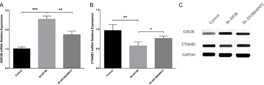
EIF3B knockdown suppressed WNT signaling pathway in KG-1 cells
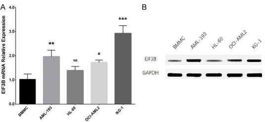
[image:4.612.92.360.70.193.2]In addition, the effect of EIF3B on regulating genes downstream of WNT signaling, includ- ing GSK3B and CTNNB1 were evaluated. GS- K3B mRNA relative expression was increased in the Sh-EIF3B group compared with the con-trol group (P < 0.001) but reduced in the Sh- Figure 1. EIF3B relative expression in human AML cell lines. EIF3B
mRNA relative expression (A) and protein expression (B) were increased in AML-193, OCI-AML2, and KG-1 cell lines compared to human primary BMMC. The comparison of EIF3B mRNA relative expression between human AML cell lines and human primary BMMC was conducted using one-way ANOVA followed by Dunnett’s multiple comparisons test. P <
0.05 was considered significant. NS, non-significant; *P < 0.05, **P
Figure 2. EIF3B and WNT relative expression in KG-1 after transfection. Sh-EIF3B decreased EIF3B mRNA
expres-sion and protein expresexpres-sion, while Sh-EIF3B & WNT2 had no influence on EIF3B mRNA expresexpres-sion or protein expres -sion (A, C). Sh-EIF3B reduced WNT2 mRNA expres-sion and protein expres-sion, while Sh-EIF3B & WNT2 rescued the effect of Sh-EIF3B on WNT2 expression (B, C). Comparisons of EIF3B mRNA relative expression between the Sh-EIF3B group and control, Sh-EIF3B & WNT2 group, and Sh-EIF3B group were made by t test. P < 0.05 was
con-sidered significant. NS, non-significant, **P < 0.01, ***P < 0.001. EIF3B, eukaryotic translation initiation factor 3 subunit B; AML, acute myeloid leukemia; Sh-EIF3B, eukaryotic translation initiation factor 3 subunit B short hairpin RNA plasmids and control overexpression plasmids were transfected into KG-1; Sh-EIF3B & WNT2, eukaryotic trans-lation initiation factor 3 subunit B short hairpin RNA plasmids and WNT2 overexpression plasmids were transfected into KG-1 cells.
Figure 3. Sh-EIF3B increased cell proliferation but inhibited apoptosis by blocking WNT signaling pathway in AML. Sh-EIF3B had no effect on cell proliferation at 24 h but reduced cell proliferation at 48 h and 72 h; Sh-EIF3B & WNT2 rescued the effect of Sh-EIF3B on proliferation at 72 h, but not at 24 h or 48 h (A). Sh-EIF3B increased apoptosis
rate, while Sh-EIF3B & WNT2 rescued the influence of Sh-EIF3B on cell apoptosis (B, D). Sh-EIF3B increased the
expression of C-Caspase 3, while Sh-EIF3B & WNT2 compensated for the effect of Sh-EIF3B on C-Caspase 3
expres-sion (C). Sh-EIF3B decreased the expresexpres-sion of Bcl-2, while Sh-EIF3B & WNT2 rescued the influence of Sh-EIF3B
[image:5.612.93.515.342.614.2]and KG-1 cell lines compared with human pri-mary BMMC. (2) Knockdown of EIF3B inhibited cell proliferation and migration but promoted apoptosis in KG-1 cells through blocking the WNT signaling pathway.
EIF3B, as a subunit of the EIF3 family, plays a crucial role in stimulation of cell growth-regulat-ing protein synthesis, which contributes to the oncogenic progression of several cancers [14]. Existing studies show that EIF3B is overex-pressed in some malignancies and is a poten-tial oncogene [5-7, 15]. For example, overex-pression of EIF3B is observed in SW116 cells of human colon cancer as well as SKOV3 and HO-8910 cells of human ovarian cancer [6, 15]. Another study indicates that EIF3B is upregu-lated in bladder and prostate cancer compared with normal tissues [5]. According to the pre- vious studies, EIF3B is upregulated in several solid tumors and plays an important role in the process of tumorigenesis; however, the regula-tory role of EIF3B in hematologic malignancies including AML remained unknown. In the pres-ent study, we observed that EIF3B was over- expressed in AML-193, OCL-AML2, and KG-1 AML cell lines compared with human primary BMMC. The possible explanations were (1) con-sistent with previous studies, EIF3B might act as an oncogene in AML and accelerate the pro-gression of AML by initiating oncogenic cell activities, which was validated in our cellular experiments. (2) ELB3B might serve as a promoter of some oncogenes in AML, such as CTNNB1, CCND1 and C-Myc, which are related to the self-renewal of leukemic cells and accel-erated AML metabolism [13].
Existing evidence suggests that ELB3B is in- volved in tumorigenesis and affects cancer cell activities in some carcinomas [5-7, 14]. For example, knockdown of EIF3B inhibits cell pro-liferation and clonability but elevates the cell apoptosis rate in colon cancer SW1116 cells [6]. Another study disclosed that the knock-down of EIF3B reduces cell proliferation and invasion but stimulates cell apoptosis by down-regulating the WNT/CTNNB1 signaling pathway in breast cancer [16]. Overexpression of EIF3B
is found in immortal fibroblast cells and EIF3B
[image:6.612.91.284.71.241.2]participates in the complex mechanism of tu- mor formation, which leads to stimulation of tumor growth in human breast carcinoma [14]. In addition, emerging evidence exhibits that the WNT signaling pathway is implicated in the Figure 4. Sh-EIF3B decreased cell migration by
down-regulating WNT signaling pathway in AML. Sh-EIF3B decreased the number of migrated cells, while Sh-EIF3B & WNT2 rescued the effect of Sh-Sh-EIF3B on the number of migrated cells. P < 0.05 was considered
significant. NS, non-significant, **P < 0.01. EIF3B, eukaryotic translation initiation factor 3 subunit B; AML, acute myeloid leukemia; Sh-EIF3B, eukaryotic translation initiation factor 3 subunit B short hairpin RNA plasmids and control overexpression plasmids were transfected into KG-1; Sh-EIF3B & WNT2, eu-karyotic translation initiation factor 3 subunit B sh- ort hairpin RNA plasmids and WNT2 overexpression plasmids were transfected into KG-1 cells.
EIF3B&WNT2 group compared with Sh-EIF3B group (P < 0.01) (Figure 5A). To the contrary, CTNNB1 mRNA relative expression was de- creased in the Sh-EIF3B group compared with the control group (P < 0.01) while elevated in the Sh-EIF3B & WNT2 group compared with the Sh-EIF3B group (P < 0.05) (Figure 5B). WB assay displayed that the protein expression of GSK3B was higher in the Sh-EIF3B group com-pared with controls but was lower in the Sh- EIF3B & WNT2 group compared with Sh-EIF3B group (Figure 5C). Conversely, protein expres-sion of CTNNB1 was decreased in the Sh-EIF- 3B group compared with controls while incre- ased in the Sh-EIF3B & WNT2 group compared with the Sh-EIF3B group. These indicated that knockdown of EIF3B decreased the WNT sig- naling pathway in AML. Collectively, the afore-mentioned data suggested knockdown of EI- F3B decreased cell proliferation andmigration while increasing cell apoptosis by downregulat-ing the WNT signaldownregulat-ing pathway in AML.
Discussion
AML pathology by enhancing proliferation of hematopoietic stem and progenitor cells [9-11, 17]. Based on previous studies, we speculat- ed that EIF3B might affect the AML cell activi-ties by activating the WNT signaling pathway. We performed experiments that suggested th- at knockdown of EIF3B inhibited proliferation and migration but promoted apoptosis in AML by blocking the WNT signaling pathway. Possi- ble explanations included: (1) Knockdown of EIF3B decreased WNT2 and other WNT signal-ing related genes and proteins by regulatsignal-ing protein synthesis, thus its knockdown could inhibit AML cell proliferation and migration whi- le promoting apoptosis by blocking the WNT signaling pathway; (2) EIF3B might affect other carcinogenic pathways by regulating related pr- otein synthesis; thus its knockdown inhibited AML progression, but further studies are need-ed for validation.
To assess the regulatory effect of EIF3B on the WNT signaling pathway in AML, we selected three genes in the WNT pathway (WNT2, GSK3B and CTNNB1). In detail, WNT2 is overexpressed in AML and it promotes AML development and progression by inducing aberrant gene promot-er methylation and subsequently promoting cell growth and reducing cell apoptosis in AML
[18]. GSK3B is identified as an anti-cancer
gene and the knockdown of GSK3B promot- es the progression of AML [17]. Regarding CTN- NB1, it contributes to cancer pathology and enhances metastasis in AML [11-13]. In
addi-tion, the high expression of CTNNB1 impairs bone marrow hematopoiesis and is related to the elevated self-renewing ability of leukemic
cells in AML [13]. Regarding these findings, we
propose that EIF3B might regulate the expres-sion of GSK3B and CTNNB1 in AML cells as well, and discovered that knockdown EIF3B upregulated GSK3B but downregulated CTN- NB1 by blocking WNT2. The possible reasons were that knockdown of EIF3B might activate the leukemia inhibitory factor through acting synergistically with the WNT antagonists GS- K3B and WNT producer CTNNB1. CTNNB1 en- hanced the expression of GSK3B and inhibit- ed renewal of hematopoietic stem cells depen- ding on CTNBB1, attenuating the progression of AML [17]. According to our study, knockdown of EIF3B inhibited cell proliferation and migra-tion but elevated apoptosis through blocking the WNT signaling pathway in AML, which sug-gested that EIF3B might be an innovative thera-peutic target for AML treatment.
In conclusion, EIF3B is upregulated, and its knockdown inhibits cell proliferation and migra-tion, while promoting apoptosis by downregu-lating the WNT signaling pathway in AML.
Acknowledgements
This study was supported by National Natural Science Foundation of China (No. 81270572).
Disclosure of conflict of interest
[image:7.612.93.519.70.208.2]None.
Figure 5. Sh-EIF3B elevated GSK3B expression but decreased CTNNB1 expression by downregulating the WNT sig-naling pathway in AML. Sh-EIF3B increased GSK3B mRNA and protein expression, while Sh-EIF3B & WNT2 rescued the effect of Sh-EIF3B on GSK3B expression (A, C). Sh-EIF3B reduced CTNNB1 mRNA and protein expression, while
[9] Mikesch JH, Steffen B, Berdel WE, Serve H and Muller-Tidow C. The emerging role of Wnt sig-naling in the pathogenesis of acute myeloid leukemia. Leukemia 2007; 21: 1638-1647. [10] Taskesen E, Staal FJ and Reinders MJ. An
inte-grated approach of gene expression and
DNA-methylation profiles of WNT signaling genes
uncovers novel prognostic markers in acute myeloid leukemia. BMC Bioinformatics 2015; 16 Suppl 4: S4.
[11] Minke KS, Staib P, Puetter A, Gehrke I, Gandhi-rajan RK, Schlosser A, Schmitt EK, Hallek M and Kreuzer KA. Small molecule inhibitors of WNT signaling effectively induce apoptosis in acute myeloid leukemia cells. Eur J Haematol 2009; 82: 165-175.
[12] Serinsoz E, Neusch M, Busche G, Wasielewski R, Kreipe H and Bock O. Aberrant expression of beta-catenin discriminates acute myeloid leu-kaemia from acute lymphoblastic leuleu-kaemia. Br J Haematol 2004; 126: 313-319.
[13] Bhavanasi D, Wen KW, Liu X, Vergez F, Danet-Desnoyers G, Carroll M, Huang J and Klein PS. Signaling mechanisms that regulate ex vivo survival of human acute myeloid leukemia ini-tiating cells. Blood Cancer J 2017; 7: 636. [14] Zhang L, Pan X and Hershey JW. Individual
overexpression of five subunits of human
translation initiation factor eIF3 promotes
ma-lignant transformation of immortal fibroblast
cells. J Biol Chem 2007; 282: 5790-5800. [15] Wang L and Ouyang L. Effects of EIF3B gene
downregulation on apoptosis and proliferation of human ovarian cancer SKOV3 and HO-8910 cells. Biomed Pharmacother 2019; 109: 831-837.
[16] Fan Y and Guo Y. Knockdown of eIF3D inhibits breast cancer cell proliferation and invasion through suppressing the Wnt/beta-catenin sig-naling pathway. Int J Clin Exp Pathol 2015; 8: 10420-10427.
[17] Bhavanasi D and Klein PS. Wnt signaling in normal and malignant stem cells. Curr Stem Cell Rep 2016; 2: 379-387.
[18] Mochmann LH, Bock J, Ortiz-Tanchez J, Schlee C, Bohne A, Neumann K, Hofmann WK, Thiel E and Baldus CD. Genome-wide screen reveals WNT11, a non-canonical WNT gene, as a direct target of ETS transcription factor ERG. Onco-gene 2011; 30: 2044-2056.
Address correspondence to: Yonghuai Feng, De-
partment of Hematology, Affiliated Hospital of Zunyi
Medical University, 149 Dalian Road, Zunyi 563000, China. Tel: +86-10-88324958; E-mail: fengyong-huai@163.com
References
[1] Jongen-Lavrencic M, Grob T, Hanekamp D, Ka-velaars FG, Al Hinai A, Zeilemaker A, Erpelinck-Verschueren CAJ, Gradowska PL, Meijer R, Cloos J, Biemond BJ, Graux C, van Marwijk Kooy M, Manz MG, Pabst T, Passweg JR, Have-lange V, Ossenkoppele GJ, Sanders MA, Schuurhuis GJ, Lowenberg B and Valk PJM. Molecular minimal residual disease in acute myeloid leukemia. N Engl J Med 2018; 378: 1189-1199.
[2] Chen WQ, Li H, Sun KX, Zheng RS, Zhang SW, Zeng HM, Zou XN, Gu XY and He J. Report of cancer incidence and mortality in China, 2014. Zhonghua Zhong Liu Za Zhi 2018; 40: 5-13. [3] Bray F, Ferlay J, Soerjomataram I, Siegel RL,
Torre LA and Jemal A. Global cancer statistics 2018: GLOBOCAN estimates of incidence and mortality worldwide for 36 cancers in 185 countries. CA Cancer J Clin 2018; 68: 394-424.
[4] Koreth J, Schlenk R, Kopecky KJ, Honda S, Si-erra J, Djulbegovic BJ, Wadleigh M, DeAngelo DJ, Stone RM, Sakamaki H, Appelbaum FR, Dohner H, Antin JH, Soiffer RJ and Cutler C. Al-logeneic stem cell transplantation for acute
myeloid leukemia in first complete remission:
systematic review and meta-analysis of pro-spective clinical trials. JAMA 2009; 301: 2349-2361.
[5] Wang H, Ru Y, Sanchez-Carbayo M, Wang X, Kieft JS and Theodorescu D. Translation initia-tion factor eIF3b expression in human cancer and its role in tumor growth and lung coloniza-tion. Clin Cancer Res 2013; 19: 2850-2860. [6] Wang Z, Chen J, Sun J, Cui Z and Wu H. RNA
interference-mediated silencing of eukaryotic translation initiation factor 3, subunit B (EI-F3B) gene expression inhibits proliferation of colon cancer cells. World J Surg Oncol 2012; 10: 119.
[7] Liang H, Ding X, Zhou C, Zhang Y, Xu M, Zhang C and Xu L. Knockdown of eukaryotic transla-tion initiatransla-tion factors 3B (EIF3B) inhibits prolif-eration and promotes apoptosis in glioblasto-ma cells. Neurol Sci 2012; 33: 1057-1062. [8] Xu F, Xu CZ, Gu J, Liu X, Liu R, Huang E, Yuan Y,