Original Article
SPOP mutation in prostate cancers in Korean
population: variation in its mutation
frequency among ethnic groups
Nara Yoon1, Ji-Youn Sung2, So Young Kang3, Ghee Young Kwon3, Yoon-La Choi4,5
1Department of Pathology, The Catholic University of Korea Incheon St. Mary’s Hospital, The Catholic University of
Korea, Incheon, Korea; 2Department of Pathology, Kyunghee University Medical Center, Kyunghee University
Col-lege of Medicine, Seoul, Korea; 3Department of Pathology and Translational Genomics, Samsung Medical Center,
Sungkyunkwan University College of Medicine, Seoul, Korea; 4Department of Pathology and Translational
Genom-ics, Samsung Medical Center, Sungkyunkwan University College of Medicine, Seoul, Korea; 5Department of Health
Sciences and Technology, SAIHST, Sungkyunkwan University, Seoul, Korea
Received December 15, 2015; Accepted February 25, 2016; Epub March 1, 2016; Published March 15, 2016
Abstract: Speckle-type POZ protein, SPOP, mediates SRC-3 oncogene ubiquitination and proteolysis in human can-cer. Frequent SPOP mutation is observed in exome sequencing of prostate cancer (Pca) in western population. We performed SPOP mutation analysis and investigated its protein expression of PCa in Korean population. We selected 108 cases of prostatectomy specimen for PCa at Samsung Medical Center in Seoul, Korea from 1995 to 2006. All cases were sequenced by Sanger sequencing to analyze SPOP somatic mutations in paraffin-embedded tissue.
In addition, we also applied SPOP immunohistochemistry (IHC) on the tissue microarray blocks for 108 cases. All cases were successfully sequenced. Three missense mutations, p.Phe102Cys in one case and double mutation
with p.Phe125Val and p.Glu145 Lys in another case, were identified (1.85%). p.Phe102Cys and p.Phe125Val have
been previously reported in the literature; however, p.Glu145 Lys was newly discovered in this study. All but one case
were successfully stained with IHC. Thirty two (32.7%) out of 107 cases showed SPOP expression loss and loss of
SPOP expression was not correlated with mutation status. In conclusion, we identified three missense mutations including double mutation in 108 PCa in Korean population. The incidence (2/108, 1.85%) is lower than previous
studies which had done in western population.
Keywords: SPOP protein, mutation, prostate cancer
Introduction
Prostate cancer (PCa) is the second most fre-quent cancer among men worldwide and has been increasing in nearly all countries including Korean due to westernization [1-3]. Given the increasing incidence of PCa and technological developments in genomics, there has been increased interest in molecular diagnosis and
classification of PCa. There are new attempts at PCa classification according to genomic
alteration. The main division of molecular
clas-sification is gene fusion involving ETS family
member accounting for up to 80% of PCa [4, 5].
Whole exome sequencing in PCa which had performed in western population has revealed a number of somatic mutations including Speckle-type POZ protein (SPOP) gene [6-8]. Analysis SPOP mutations in Korean population
4.4% (2/45) and 6.9% (6/87) [9, 10]. We
performed SPOP mutation analysis on more expanded cohort, 108 surgically excised PCa specimens and determined its protein expre- ssion by immunohistochemistry (IHC).
Materials and methods
Patients and samples
One hundred eight cases of radical prostatec-tomy for PCa at Samsung Medical Center in Seoul, Korea from 1995 to 2006 were select-ed. Cases with incomplete resection or neoad-juvant treatment (radiotherapy or hormonal therapy) were excluded. Clinical data were reviewed based on electronic medical records, and pathologic diagnoses were reviewed by three pathologists (N. Yoon, Y. Choi and G.
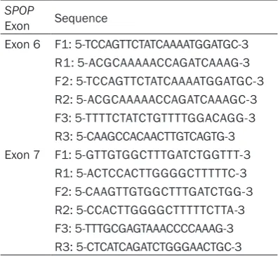
Table 2. Primer sequences of SPOP gene SPOP
Exon Sequence
Exon 6 F1: 5-TCCAGTTCTATCAAAATGGATGC-3 R1: 5-ACGCAAAAACCAGATCAAAG-3
F2: 5-TCCAGTTCTATCAAAATGGATGC-3 R2: 5-ACGCAAAAACCAGATCAAAGC-3 F3: 5-TTTTCTATCTGTTTTGGACAGG-3 R3: 5-CAAGCCACAACTTGTCAGTG-3 Exon 7 F1: 5-GTTGTGGCTTTGATCTGGTTT-3
[image:2.629.98.298.272.455.2]R1: 5-ACTCCACTTGGGGCTTTTTC-3 F2: 5-CAAGTTGTGGCTTTGATCTGG-3 R2: 5-CCACTTGGGGCTTTTTCTTA-3 F3: 5-TTTGCGAGTAAACCCCAAAG-3 R3: 5-CTCATCAGATCTGGGAACTGC-3
Table 1. Clinicopathologic parameters of the 108 patients with prostate carcinoma
Clinicopathologic parameters Mean (range) Age 65 (44-78)
No (%) of patients
Gleason score 6 9 (8.3%)
7 77 (71.3%)
8-10 22 (20.4%)
T stage 2 75 (69.4%)
3a 22 (20.4%)
3b 8 (7.4%)
4 3 (2.8%)
Gleason grading. Tumor stage was determined according to 7th Edition of the AJCC Cancer Staging Manual. Clinicopathological data in- cluding age, Gleason score and pathologic stage are summarized in Table 1. The study protocol was approved by the Samsung Me- dical Center Institutional Review Board.
Tissue microarray construction
We selected representative formalin-fixed, par
-affin-embedded (FFPE) blocks for TMA from archived pathology files of Samsung Medical
Center preparation by reviewing hematoxylin and eosin (H&E)-stained slides. Using a manual tissue microarrayer (Accumax, ISU Abxis, Seoul, Korea), two representative tissue cores (0.2 cm in diameter) were taken from each tumor and
placed into two recipient paraffin blocks.
Immunohistochemistry
The SPOP rabbit monoclonal antibody (ERG- 3864, Epitomics, Burlingame, CA, USA, dilution
1:100) was used for IHC, which was performed using a BenchMark XT (Roche/Ventana Medi- cal Systems, Tucson, AZ, USA) according to the
manufacturer’s instructions. In brief, 4-μm sec
-tions of FFPE tissues were deparaffinized and
were immersed in Tris/Borate/EDTA buffer (pH 8.0-8.5) for 8 minutes at room temperature to retrieve the antigen. Endogenous peroxidase was quenched via incubation with hydrogen peroxide for 5 minutes at room temperature. The sections were incubated for 32 minutes at room temperature with primary antibodies. The secondary antibody (OmniMap anti-Rabbit HRP, Tucson, AZ, USA) and chromogenic substrate Diamiobenzidine (ChromoMap DAB; Tucson, AZ, USA) were applied for 16 and 8 minutes, respectively, at room temperature. Normal prostate glands in each core were used as a positive internal control. The SPOP protein was expressed in the cytoplasm of normal prostate epithelium or tumor cells. If the intensity of tumor cells is weaker than normal epithelium, the case was judged as presence of SPOP ex- pression loss.
Extraction of DNA
Two 5-μm-thick unstained slides were obtained
from representative blocks of FFPE samples. Original H&E slides were reviewed to identify the region of tumor and tumor tissues were microdissected from corresponding areas of unstained slides. Additional H&E slides were prepared after obtaining the slides for DNA
extraction and reviewed to confirm the pres -ence of tumor. Genomic DNA was isolated from FFPE tumor samples using a ReliaPrep™ FFPE gDNA extraction kit (Promega, Madison, WI, USA). The concentration of DNA was deter-mined by a NanoDrop ND-1000 spectropho-tometer (NanoDrop Technologies).
Sequencing of SPOP
PCR was performed in 20-µl reactions contain-ing 100 ng of template DNA, 2 µl×10 buffer, 0.25 mmol/L deoxynucleoside triphosphate (dNTP), 10 pmol primers and 1.25 U Taq DNA polymerase (iNtRON, Seoul, Korea). Primer pairs were used to amplify the coding sequen- ces of SPOP exons 6 and 7 (Table 2). The ther-mal cycling conditions were 5 min at 95°C, fol-lowed by 40 cycles of denaturation at 94°C for 30 s, annealing at 58°C for 30 s, and extension at 72°C for 30 s. PCR products were subjected
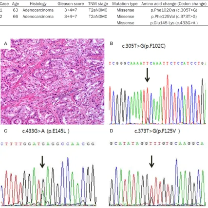
Table 3. Features of cases with SPOP missense mutation
Case Age Histology Gleason score TNM stage Mutation type Amino acid change (Codon change) 1 63 Adenocarcinoma 3+4=7 T2aN0M0 Missense p.Phe102Cys (c.305T>G) 2 66 Adenocarcinoma 3+4=7 T2aN0M0 Missense p.Phe125Val (c.373T>G) Missense p.Glu145 Lys (c.433G>A )
sequencing was performed using the BigDye Terminator v1.1 kit (Applied Biosystems) on the ABI 3130XL genetic analyzer (Applied Biosys- tems). The results were regarded as mutation positive if a mutation was detected in both the forward and reverse Sequencher version 4.10.1 (Gene Codes Corporation, Ann Arbor, MI, USA) along with a manual review of chromatograms.
Statistical analysis
Statistical analysis was performed using SPSS 20.0 statistical software (SPSS, Chicago, IL,
USA). Correlation analyses between the SPOP
mutation and clinicopathologic parameters were done using the x2-test or Fisher’s exact test. A p-value of < 0.05 was regarded as
significant.
Results
SPOP mutation
The tumor content was approximately 70% on
average after microdissection. All cases are successfully sequenced demonstrating clear Figure 1. The pathologic features (A) and results from Sanger sequencing (B) of the first case with SPOP mutation. On microscopic analysis, the tumor showed well-formed atypical glands with some fused structures. SPOP DNA
se-quence revealed a missense mutation (p.Phe102Cys) in the first case. Chromatogram showed the double missense
signal peaks. The results from sequencing the
SPOP gene are summarized in Table 3.
Three missense mutations, p.Phe102Cys (p. F102C) in one case, and double mutation with p.Phe125Val (p.F125V) and p.Glu145 Lys (p.
E145K) in another case, were identified (1.85%)
(Figure 1). The patient with the p.F102C muta-tion was 63 years old, with a PSA level of 12.51 ng/ml at the time of initial diagnosis. The patient’s primary Gleason grade was 3 and sec-ondary Gleason grade was 4, giving a total score of 7. The pathologic stage was T2c with no lymph node metastasis. The patient who had double mutation with the p.F125V and p.E145K mutations was 66 years old, with a PSA level of 7.93 ng/ml at the time of initial diagnosis. The primary Gleason grade was 3 and the secondary Gleason grade was 4, giving a total score of 7. The pathologic stage was T2a, with no lymph node metastasis. There was no metastasis or recurrence during the follow-up periods of 80 and 98 months, respectively. As for clinicopathologic parameters, there was
no significant correlation with the SPOP mu- tations.
SPOP IHC
All but one case were successfully stained with
IHC. Thirty two (32.7%) out of 107 cases showed
expression loss in tumor cells compared to nor-mal epithelium (Figure 2). No association was
noted between SPOP mutation and protein expression. Two cases with SPOP mutation was well expressed SPOP IHC.
Discussion
SPOP is Bric-a-brac/Tramtrack/Broad complex (BTB) protein that is a constituent of Cul3-based ubiquitination [10, 11]. The SPOP gene consists of two domains, the N-terminal MATH domain, which directly binds to steroid receptor coactivator-3 (SRC-3), and the C-terminal BTB/ POZ domain, which directly binds to Cul3 [12]. Overexpression and overactivation of SRC-3 occur in many human cancers, and SPOP pro-tein regulates SRC-3-mediated oncogenic sig-naling and tumorigenesis by promoting its deg-radation [12, 13]. It is speculated that SPOP in human cancers, including PCa, functions as a tumor suppressor [10, 12, 13]. SPOP mutation contribute to PCa development by altering the steady-state levels of key component in the androgen-signaling pathway [14].
We screened for somatic mutations in exons six and seven of the SPOP gene in 108 PCa because all of the SPOP mutations previously reported occurred within these two exons [6, 9, 10]. Three somatic missense mutations were found in two cases: p.F102C in one case, and double mutation with p.F125V and p.E145K in another case. These two cases have no dis- tinctive clinicopathologic characteristics com-Figure 2. The SPOP immunohistochemistry result. A. SPOP is well expressed in cytoplasm of cancer cells. B. SPOP
[image:4.629.98.534.78.299.2]pared with the case without SPOP mutation. Although p.F102C and p.F125V have been pre-viously reported in the literature, p.E145K was newly discovered in this study [6, 12]. Especially, p.F102C and p.F125V SPOP mu- tants are known to exert a gain-of-function oncogenic effect by increasing SRC-3 protein levels and AR transcriptional activity above baseline [12]. p.F102 and p.F125 are revealed to be frequently mutated site in previous study [9]. Three mutations of this study are gathered in MATH domain in coincidence with previous works [6, 9, 10]. SPOP mutants in PCA weaken the interaction between SPOP and SRC-3 and the coactivator function of SRC-3 on AR tran-scriptional activity, thus inhibiting ubiquitina-tion and degradaubiquitina-tion of SRC-3 [12].
In our study, SPOP mutation in PCa is identifi-ed in about 1.85% of all cases, which is lower than the 3.5-15% reported previously in studies
including multi-organ cohorts [6, 7, 9]. This result can add a new data to mutation inci-dence rate of SPOP gene especially in Asian population. In several Korean series, the fre-quencies of SPOP mutation in PCa are similar or relatively lower than western group [9, 10]. PCa exhibits different molecular characteris- tics across ethnic group [15]. Recent studi- es assessed the difference in prevalence of ERG rearrangements and revealed that Asian and African American showed lower fre-quency than Caucasian Men [16, 17]. More
specifically, ERG rearrangements have been revealed lower frequency in Korean, Japanese and Chinese than in Caucasian men [18-20]. Khani et al compared prevalence of molecular alteration in PCa between African American and Caucasian men revealed that there are dis-tinctive differences in frequency of SPINK1 overexpression, ERG rearrangement and PTEN
deletion between two ethnic groups. They also found that SPOP mutation was less frequent in
African American (4.5%) than in Caucasian (10.3%) [15]. In addition, the study that we
investigated in same cohort [22] revealed no mutation in MED12 exon 26 which were fre-quently observed in western population with
5.4% [6].
Aside from genetic heterogeneity among ethnic groups, the technical difference may contribute to difference of mutation prevalence. Exome sequencing which performed by Barbieri et al is known to be more sensitive method compared to Sanger sequencing performed in the study by Kim MS et al and ours [6, 21].
In summary, Sanger sequencing for SPOP
mutations in FFPE of 108PCa specimens in Korean population reveals lower frequency
than western population. Two (1.85%) cases
had missense mutation and one out of two
had double mutation. In addition, 32.7% cases
showed loss of expression of SPOP protein in IHC but not correlated with SPOP mutation. Acknowledgements
This work was supported by the R&D Program for Society of the National Research Foundation (NRF) funded by the Ministry of Science, ICT & Future Planning (NRF-2013M3C8A1078501). Address correspondence to: Dr. Yoon-La Choi, De- partment of Pathology and Translational Genomics, Samsung Medical Center, Sungkyunkwan University School of Medicine, #50 Ilwon-Dong, Kangnam-Gu, Seoul, Korea; Department of Health Sciences and Technology, SAIHST, Sungkyunkwan University, Se- oul, Korea. Tel: 2797; Fax: +82-2-3410-0025; E-mail: [email protected]; Dr. Ghee Young Kwon, Department of Pathology and Translational Genomics, Samsung Medical Center, Sungkyunkwan University School of Medicine, #50 Ilwon-Dong, Kangnam-Gu, Seoul, Korea. Tel: +82-2-3410-2770; Fax: +82-2-3410-0025; E-mail: [email protected]
References
[1] Siegel R, Ma J, Zou Z and Jemal A. Cancer sta-tistics, 2014. CA Cancer J Clin 2014; 64: 9-29. [2] Jung KW, Park S, Kong HJ, Won YJ, Lee JY, Seo
HG and Lee JS. Cancer statistics in Korea: inci-dence, mortality, survival, and prevalence in 2009. Cancer Res Treat 2012; 44: 11-24. [3] Center MM, Jemal A, Lortet-Tieulent J, Ward E,
Ferlay J, Brawley O and Bray F. International variation in prostate cancer incidence and mortality rates. Eur Urol 2012; 61: 1079-1092. [4] Barbieri CE and Tomlins SA. The prostate can-cer genome: perspectives and potential. Urol Oncol 2014; 32: 53, e15-22.
[5] Shaikhibrahim Z, Braun M, Nikolov P, Boehm D, Scheble V, Menon R, Fend F, Kristiansen G, Perner S and Wernert N. Rearrangement of the ETS genes ETV-1, ETV-4, ETV-5, and ELK-4 is a clonal event during prostate cancer progres-sion. Hum Pathol 2012; 43: 1910-1916. [6] Barbieri CE, Baca SC, Lawrence MS, Demichelis
AK, Mosquera JM, Rupp N, Wild PJ, Moch H, Morrissey C, Nelson PS, Kantoff PW, Gabriel SB, Golub TR, Meyerson M, Lander ES, Getz G, Rubin MA and Garraway LA. Exome
sequenc-ing identifies recurrent SPOP, FOXA1 and
MED12 mutations in prostate cancer. Nat Genet 2012; 44: 685-689.
[7] Berger MF, Lawrence MS, Demichelis F, Drier Y, Cibulskis K, Sivachenko AY, Sboner A, Esgueva
R, Pflueger D, Sougnez C, Onofrio R, Carter SL,
Park K, Habegger L, Ambrogio L, Fennell T, Parkin M, Saksena G, Voet D, Ramos AH, Pugh TJ, Wilkinson J, Fisher S, Winckler W, Mahan S, Ardlie K, Baldwin J, Simons JW, Kitabayashi N, MacDonald TY, Kantoff PW, Chin L, Gabriel SB, Gerstein MB, Golub TR, Meyerson M, Tewari A, Lander ES, Getz G, Rubin MA and Garraway LA. The genomic complexity of primary human prostate cancer. Nature 2011; 470: 214-220. [8] Grasso CS, Wu YM, Robinson DR, Cao X,
Dhanasekaran SM, Khan AP, Quist MJ, Jing X,
Lonigro RJ, Brenner JC, Asangani IA, Ateeq B, Chun SY, Siddiqui J, Sam L, Anstett M, Mehra R, Prensner JR, Palanisamy N, Ryslik GA, Vandin F, Raphael BJ, Kunju LP, Rhodes DR, Pienta KJ, Chinnaiyan AM and Tomlins SA. The mutational landscape of lethal castration-re-sistant prostate cancer. Nature 2012; 487: 239-243.
[9] Blattner M, Lee DJ, O’Reilly C, Park K, Macdonald TY, Khani F, Turner KR, Chiu YL, Wild PJ, Dolgalev I, Heguy A, Sboner A, Ramazangolu S, Hieronymus H, Sawyers C, Tewari AK, Moch H, Yoon GS, Known YC, Andren O, Fall K, Demichelis F, Mosquera JM, Robinson BD, Barbieri CE and Rubin MA. SPOP mutations in prostate cancer across demo-graphically diverse patient cohorts. Neoplasia 2014; 16: 14-20.
[10] Kim MS, Je EM, Oh JE, Yoo NJ and Lee SH. Mutational and expressional analyses of SPOP, a candidate tumor suppressor gene, in pros-tate, gastric and colorectal cancers. APMIS 2013; 121: 626-33.
[11] Kwon JE, La M, Oh KH, Oh YM, Kim GR, Seol JH, Baek SH, Chiba T, Tanaka K, Bang OS, Joe CO and Chung CH. BTB domain-containing speckle-type POZ protein (SPOP) serves as an adaptor of Daxx for ubiquitination by Cul3-based ubiquitin ligase. J Biol Chem 2006; 281: 12664-12672.
[12] Geng C, He B, Xu L, Barbieri CE, Eedunuri VK, Chew SA, Zimmermann M, Bond R, Shou J, Li C, Blattner M, Lonard DM, Demichelis F, Coarfa C, Rubin MA, Zhou P, O’Malley BW and Mitsiades N. Prostate cancer-associated muta-tions in speckle-type POZ protein (SPOP) regu-late steroid receptor coactivator 3 protein turn-over. Proc Natl Acad Sci U S A 2013; 110: 6997-7002.
[13] Li C, Ao J, Fu J, Lee DF, Xu J, Lonard D and O’Malley BW. Tumor-suppressor role for the SPOP ubiquitin ligase in signal-dependent pro-teolysis of the oncogenic co-activator SRC-3/ AIB1. Oncogene 2011; 30: 4350-4364. [14] Mani RS. The emerging role of speckle-type
POZ protein (SPOP) in cancer development. Drug Discov Today 2014; 19: 1498-1502. [15] Khani F, Mosquera JM, Park K, Blattner M,
O’Reilly C, MacDonald TY, Chen Z, Srivastava A, Tewari AK, Barbieri CE, Rubin MA and Robinson BD. Evidence for molecular differ-ences in prostate cancer between African American and Caucasian men. Clin Cancer Res 2014; 20: 4925-4934.
[16] Magi-Galluzzi C, Tsusuki T, Elson P, Simmerman K, LaFargue C, Esgueva R, Klein E, Rubin MA and Zhou M. TMPRSS2-ERG gene fusion
prev-alence and class are significantly different in
prostate cancer of Caucasian, African-American and Japanese patients. Prostate 2011; 71: 489-497.
[17] Rosen P, Pfister D, Young D, Petrovics G, Chen
Y, Cullen J, Bohm D, Perner S, Dobi A, McLeod DG, Sesterhenn IA and Srivastava S. Differences in frequency of ERG oncoprotein expression between index tumors of Caucasian and African American patients with prostate cancer. Urology 2012; 80: 749-753.
[18] Miyagi Y, Sasaki T, Fujinami K, Sano J, Senga Y, Miura T, Kameda Y, Sakuma Y, Nakamura Y, Harada M and Tsuchiya E. ETS family-associat-ed gene fusions in Japanese prostate cancer: analysis of 194 radical prostatectomy sam-ples. Mod Pathol 2010; 23: 1492-1498. [19] Mao X, Yu Y, Boyd LK, Ren G, Lin D, Chaplin T,
Kudahetti SC, Stankiewicz E, Xue L, Beltran L, Gupta M, Oliver RT, Lemoine NR, Berney DM, Young BD and Lu YJ. Distinct genomic altera-tions in prostate cancers in Chinese and Western populations suggest alternative path-ways of prostate carcinogenesis. Cancer Res 2010; 70: 5207-5212.
[20] Lee K, Chae JY, Kwak C, Ku JH and Moon KC. TMPRSS2-ERG gene fusion and clinicopatho-logic characteristics of Korean prostate cancer patients. Urology 2010; 76: 1268, e1267-1213.
[21] Kim MS, Je EM, Oh JE, Yoo NJ and Lee SH. Mutational and expressional analyses of SPOP, a candidate tumor suppressor gene, in pros-tate, gastric and colorectal cancers. Apmis 2013; 121: 626-633.