Original Article
Expression of PTEN and TGF-β in prostate cancer tissues
and correlation with clinical pathological features
Lijun Li, Jing Liu, Mingxing Qiu, Baisheng Gong
Department of Urology, Sichuan Academy of Medical Sciences and Sichuan Provincial People’s Hospital, Cheng-du, Sichuan, China
Received November 24, 2015; Accepted January 25, 2016; Epub March 1, 2016; Published March 15, 2016
Abstract: Prostate cancer has higher mortality rate with complicated mechanisms. PTEN is one tumor-suppressor gene that codes for dual-specific phosphatase. TGF-β is one polypeptide cytokine that can regulate cell growth and differentiation. Recent study has revealed the importance of PTEN and TGF-β in the pathogenesis and progression of prostate cancer. This study thus investigated the expression of those two genes in prostate cancer and benign prostate tissues, in addition to the correlation between gene expression and clinical features. A total of 60 prostate cancer and 20 benign prostate hypertrophy patients were collected for tissue samples during the surgery. The expressions of PTEN and TGF-β in both tissues were detected examined by immunohistochemical (IHC) staining. The mRNA level of PTEN was measured by in situ hybridization (ISH). PTEN gene expression was significantly higher in benign prostate tissues compared to tumors. With advanced grade of tumors, the expression level of PTEN was further down-regulated. IHC results showed significantly elevated expression of TGF-β in prostate cancer tissues compared to hypertrophy tissues. PTEN gene expression was down-regulated in prostate cancer tissues compared to hypertrophy tissues, while TGF-β expression level was potentiated. There was a negative correlation between PTEN and TGF-β levels, both of which may reflect the malignancy of prostate cancer to some extents.
Keywords: Prostate cancer, PTEN, TGF-β, immunohistochemistry, in situ hybridization
Introduction
As one malignant tumor occurred in epithelial cells of male prostate tissue, prostate cancer can be divided into adenoma, ductal adenocinoma, urothelium caradenocinoma, squamous car-cinoma and adenosquamous carcar-cinoma based on pathological features [1, 2]. Prostate cancer has a relatively higher incidence in Western countries. In China, however, the occurrence rate is now 9.92 per 100, 000 people and is continuously rising [3]. Prostate cancer has a complicated pathogenesis mechanism involv-ing multiple factors includinvolv-ing genetic, age, diet habit and sex activity. With recent progresses regarding the molecular biological mechanism of prostate cancer, the occurrence of tumor has been found to be closely correlated with various oncogenes and tumor-suppressor genes [4].
3234 Int J Clin Exp Pathol 2016;9(3):3233-3238 of PTEN and TGF-β and clinical features of pros
-tate cancer.
Materials and methods
Clinical samples and patients
A total of 60 prostate cancer patients (aging between 58 and 84 years old, average = 71.8 ± 8.3 years) who received surgical resection in Sichuan Academy of Medical Sciences and Sichuan Provincial People’s Hospital from January 2014 to January 2015 were recruited in this study. All patients had not received any chemo-, radio- or immune therapy before the surgery. Prostate adenoma has been confirmed on all patients by pathological examinations. According to WHO classification, there were 11 cases at class I, 12 patients at class II and 37 cases of class III patients. Using Whitmore-Jewett grading scale, there were 14 patients at stage A or stage B, 15 cases of stage C, plus 31 stage D patients. Another cohort of 20 benign prostatic hyperplasia (BPH) patients (aging between 55 and 86 years old, average = 69.6 ± 8.8 years) who received surgical treatment at the same period were recruited as the control group. Tissue samples were collected from both groups during the surgery, and were fixed in 10% neutral buffered formalin (NBF). Tissues were embedded in paraffin block, which was sectioned into 4-μm thickness slices for hema -toxylin-eosin (HE) staining, pathological grad -ing, immunohistochemistry (IHC) stain-ing, and in situ hybridization (ISH). This study has been pre-approved by the ethical committee of Sichuan Academy of Medical Sciences and Sichuan Provincial People’s Hospital, and has obtained written consents from all partici- pants.
Reagents
Ready-to-use mouse anti-human PTEN or TGF-β monoclonal antibody was purchased from Shengxing Biotech (China). DAB development kit was a product of Boxin Reagent (China). SP staining kit for IHC was obtained from RD (US). ISH probes were synthesized by Sangon (China). IHC staining
The expression of PTEN or TGF-β was detected by IHC staining using previously established method [9]. In brief, paraffin tissues slices were de-waxed in xylene, and were re-hydrated in gradient ethanol and distilled water. Endo-
genous peroxidase activity was quenched by 3% H2O2 for 20 min. After PBS rinsing twice, 10% normal goat serum was applied for 10-min blocking at room temperature. Primary anti -body was then added for 2-hour incubation at 37°C. Secondary antibody with biotin labelled was then added for 30-min incubation. After further PBS rinsing, horseradish peroxidase working solution was added for further 30-min incubation. DAB substrates were added to develop tissue slices. Hematoxylin was then added for counter-staining. After dehydration, tissues slices were observed under a micro-scope. Five high-magnification (400×) fields were randomly selected to count the total num-ber of cells and positive stained cells. Positive staining was identified as brown-yellow gran -ules in the cytoplasm. The overall positive staining was determined as more than 40% of positive stained cells; otherwise cells were deduced as having negative staining.
ISH method
The mRNA level of PTEN was detected by ISH as previously reported [10]. In brief, paraffin-based tissues slices were dew axed and re-hydrated using routine procedures. After 3% H2O2 quenching for 10 min, proteinase K was added for incubation. The tissue slices were rinsed in 0.5 M PBS (pH 7.4) for three times, and were immersed in pre-hybridization buffer under 65°C for 4 hours. The hybridization buf -fer containing specific RNA probes was then used for hybridization at 65°C overnight. After hybridization, tissues were incubated sequen-tially by 2XSSC and 0.2XSSC (10 min, for three times each). Rabbit anti-DIG working solution was then added for 60-min incubation at 37°C, followed by PBS washing. Alkaline phosphatase buffer was added for 5-min incubation, with the addition of chromogenic substrates for devel-oping in dark. 4% paraformaldehyde was used to fix tissue slices, which were then observed under a microscope after mounting coverslips. Positive staining was deduced as blue-violet granules in cytoplasm. Overall positive staining was identified when more than 40% of positive stained cells existed. Parallel negative control slices were employed using 0.5 M PBS instead of hybridization probes [11].
Statistical methods
while enumeration data were presented as ratios. Between-group comparison was per-formed by rank-sum test. Spearman analysis was used for correlation analysis. A statistical significance was define when P<0.05.
tumor tissues with advanced grade, the stain-ing intensity was even higher (Figure 2). When examining the staining pattern in each tumor tissue, we found higher expression of TGF-β in peripheral region of tumor tissues, while
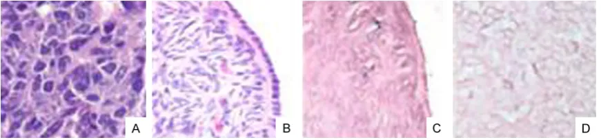
[image:3.629.97.535.79.186.2]cen-Figure 1. PTEN staining images. A. BPH tissues; B. Grade I prostate cancer; C. Grade II prostate cancer; D. Grade III prostate cancer.
Figure 2. TGF-β staining images. A. Grade III prostate cancer; B. Grade II prostate cancer; C. Grade I prostate cancer; D. BPH tissues.
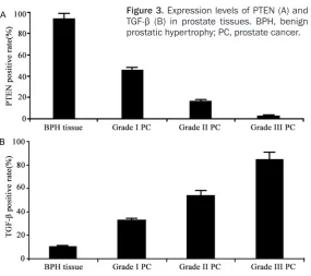
Figure 3. Expression levels of PTEN (A) and TGF-β (B) in prostate tissues. BPH, benign prostatic hypertrophy; PC, prostate cancer.
Results
PTEN IHC staining
The staining intensity of PTEN was significantly higher in BPH tissues compared to prostate cancer tissues (Fig
-ure 1). When comparing stain-ing patterns across different grades, we found higher PTEN-positive rate in grade I and II tissues of prostate can-cer than grade III.
TGF-β staining
[image:3.629.99.532.235.340.2] [image:3.629.102.388.384.637.2]3236 Int J Clin Exp Pathol 2016;9(3):3233-3238 tral and necrotic regions had relatively lower
expression (Figure 2). Expression level analysis
After quantification of all samples by IHC stain -ing, we found the highest positive rate of PTEN as 85% (17/20) in BPH tissues. With advanced grade of tumor tissues, the expression level of PTEN was significantly decreased, as grade III prostate cancer tissue had PTEN-positive rate at 2.7% (1/37) only (P<0.05, Figure 3A). In con-trast to PTEN, TGF-β had significantly lower positive expression rate in BPH tissues at only 10.0% (2/20), while grade III prostate cancer had 86.5% (32/37) positive rate (P<0.05,
Figure 3B).
We further collect all results of IHC and ISH staining, and compared them with clinical fea-tures of prostate cancer. As shown in Table 1, PTEN expression level was decreased while TGF-β level was increased with advanced his -tology and clinical stages of prostate cancer. Moreover, expression levels of PTEN and TGF-β were negatively correlated (P<0.01).
Discussion
Current studies have revealed the involvement of PTEN and TGF-β gene in multiple malignant tumors including hepatocyte carcinoma, breast cancer and pulmonary carcinoma [12, 13]. This study investigated the expression of those two genes by both IHC and ISH staining in both nor
-Figure 4. ISH images. A. BPH tissues; B. Grade I prostate cancer; C. Grade II prostate cancer; D. Grade III prostate cancer.
[image:4.629.98.534.80.182.2]Figure 5. MRNA level of PTEN. BPH, benign prostatic hypertro -phy; PC, prostate cancer.
Table 1. Clinical indexes and PTEN/TGF-β expression
Clinical features N PTEN (ISH) PTEN (IHC) TGF-β (IHC)
+ - % + - % + - %
Histo-grade Grade I 11 7 4 63.6 5 6 45.5 4 7 36.4 Grade II 12 4 8 33.3 2 10 16.7 7 5 58.3 Grade III 37 2 35 5.4 1 36 2.7 32 5 86.5 Clinic stage Phase A to C 25 11 14 78.6 10 15 66.7 11 14 78.6
Phase D 35 2 33 5.7 3 32 8.6 32 3 91.4
PTEN mRNA level
We further employed ISH to detect the level of PTEN mRNA. We found similar results as those in IHC stain -ing, as BPH tissues had more intensive staining than cancer tissues (Figure 4). Further quantitative analysis revealed a 90% positive rate (18/20) in BPH tissues, which was re-markable higher than that in cancer tissue (5.4%, 2/37, P<0.05, Figure 5). These re- sults suggested negative cor-relation between PTEN mRNA contents and advanced gra- des.
Expression of PTEN and
TGF-β and clinical features of
[image:4.629.101.384.234.371.2] [image:4.629.97.388.405.499.2]mal BPH and prostate cancer tissues across different histological grades and clinical stag-es, in an attempt to analyze the relationship between gene expression and clinical features. Our results showed 85% positive rate of PTEN expression by IHC staining in BPH tissues, while ISH positive rate was as high as 90%, both of which were significantly higher than those in prostate cancer tissues (13.3% and 21.7%). Moreover, with advanced stages of prostate cancer, the positive rate of PTEN was gradually decreased. TGF-β, on the other hand, had only 10% positive ICH rate in BPH tissues, as con -trast to 71.7% in prostate tissues. With advancement of clinical stages, positive rate of TGF-β was further elevated. Our results thus showed a close relationship between expres-sion of PTEN and TGF-β and the malignancy, histological grades and clinical stages of pros-tate cancer.
As one important tumor-suppressor gene, PTEN exerts its function mainly via inhibiting phos-phatidyl inositol kinase (PI3K) activity via PI3K/ Akt signaling pathway [14]. Under stimulus by growth factors, PI3K can phosphorylate 3,4,5- triphosphate phosphatidyl inositol, which acts as the secondary messenger for activating Akt for facilitating cell proliferation and growth [15]. PTEN gene can inhibit PI3K/Akt signaling path -way. It has lipid phosphatase activity and can dephosphorylate 3,4,5-triphosphate phospha-tidyl inositol, for antagonizing PI3K activity and maintaining Akt activity at a relatively stable level, for maintaining normal metabolism, pro-liferation, differentiation and apoptosis of cells [16]. Under abnormal expression of PTEN, a cascade reaction may lead to over-activation of Akt, leading to uncontrolled cell proliferation and malignancy transformation. In multiple malignant tumors the PTEN expression disor-ders have been found [17, 18]. Our results also showed down-regulation of PTEN in prostate cancer, as the lower PTEN expression is always accompanied with higher malignancy.
As one polypeptide cytokine, TGF-β exerts criti -cal functions in both normal and tumor cells. In normal epithelial tissues, it can stimulate cells for differentiation for inhibiting cell prolifera-tion. In tumor tissues, however, it can facilitate cell proliferation and infiltration [8]. This study also observed the expression of TGF-β in nor -mal BPH tissues and prostate cancer with dif -ferent stages. We found positive relationship between TGF-β positive rate and malignancy,
and higher TGF-β expression in peripheral tumor regions, suggesting the possible involve-ment of TGF-β in the growth, infiltration and invasion of prostate cancer cells.
Our study has shown elevated PTEN expression and decreased TGF-β expression in prostate cancer tissues as compared to BPH tissues, suggesting a negative relationship between PTEN and TGF-β expression. Our results indi -cated possible correlation between PTEN and TGF-β expression. Previous study has found lowered PTEN mRNA level after over-expressing TGF-β in mice [19]. The treatment of pancreatic carcinoma cell line PANC-1 using TGF-β led to suppressed PTEN expression level [20]. These results agreed that TGF-β might inhibit PTEN expression. One recent study suggested the facilitating of cell growth and infiltration poten -cy in addition to down-regulating PTEN by TGF-β via activating NF-κB [21]. This is consistent with our results.
Currently, novel anti-tumor treatment based on TGF-β and PTEN has become one research hotspot [22]. This study provided evidences for related studies. As the early diagnosis of malig-nant tumor is one major challenge in clinics, our results regarding correlation between PTEN and TGF-β may provide new insights regarding the early diagnosis of prostate cancer.
Disclosure of conflict of interest
None.
Address correspondence to: Dr. Jing Liu, Department of Urology, Sichuan Academy of Medical Sciences and Sichuan Provincial People’s Hospital, 32 West Second Section First Ring Road, Chengdu 610072, Sichuan, China. Tel: 28-87393690; Fax: +86-028-87393690; E-mail: [email protected]
References
[1] Stanford JL and Ostrander EA. Familial pros -tate cancer. Epidemiol Rev 2001; 23: 19-23. [2] Epstein JI, Amin MB, Beltran H, Lotan TL, Mos
-quera JM, Reuter VE, Robinson BD, Troncoso P, Rubin MA. Proposed morphologic classifica -tion of prostate cancer with neuroendocrine differentiation. Am J Surg Pathol 2014; 38: 756-67.
3238 Int J Clin Exp Pathol 2016;9(3):3233-3238 [4] Grasso CS, Wu YM, Robinson DR, Cao X,
Dha-nasekaran SM, Khan AP, Quist MJ, Jing X, Loni -gro RJ, Brenner JC, Asangani IA, Ateeq B, Chun SY, Siddiqui J, Sam L, Anstett M, Mehra R, Prensner JR, Palanisamy N, Ryslik GA, Vandin F, Raphael BJ, Kunju LP, Rhodes DR, Pienta KJ, Chinnaiyan AM, Tomlins SA. The mutational landscape of lethal castration-resistant pros-tate cancer. Nature 2012; 487: 239-43. [5] Song MS, Salmena L and Pandolfi PP. The
functions and regulation of the PTEN tumour suppressor. Nat Rev Mol Cell Biol 2012; 13: 283-96.
[6] Carver BS, Chapinski C, Wongvipat J, Hierony -mus H, Chen Y, Chandarlapaty S, Arora VK, Le C, Koutcher J, Scher H, Scardino PT, Rosen N, Sawyers CL. Reciprocal feedback regulation of PI3K and androgen receptor signaling in PTEN-deficient prostate cancer. Cancer Cell 2011; 19: 575-86.
[7] Oshimori N, Oristian D and Fuchs E. TGF-beta promotes heterogeneity and drug resistance in squamous cell carcinoma. Cell 2015; 160: 963-76.
[8] Drabsch Y and ten Dijke P. TGF-beta signalling and its role in cancer progression and metas-tasis. Cancer Metastasis Rev 2012; 31: 553-68.
[9] Lotan TL, Gurel B, Sutcliffe S, Esopi D, Liu W, Xu J, Hicks JL, Park BH, Humphreys E, Partin AW, Han M, Netto GJ, Isaacs WB, De Marzo AM. PTEN protein loss by immunostaining: analytic validation and prognostic indicator for a high risk surgical cohort of prostate cancer pa -tients. Clin Cancer Res 2011; 17: 6563-73. [10] Zhang X, Lu X and Lopez-Berestein G. In situ
hybridization-based detection of microRNAs in human diseases. MicroRNA Diagnost Thera-peut 2014; 1.
[11] Burdelski C, Menan D, Tsourlakis MC, Kluth M, Hube-Magg C, Melling N, Minner S, Koop C, Graefen M, Heinzer H, Wittmer C, Sauter G, Si -mon R, Schlomm T, Steurer S, Krech T. The prognostic value of SUMO1/Sentrin specific peptidase 1 (SENP1) in prostate cancer is lim-ited to ERG-fusion positive tumors lacking PTEN deletion. BMC Cancer 2015; 15: 538. [12] Mulholland DJ, Kobayashi N, Ruscetti M, Zhi A,
Tran LM, Huang J, Gleave M, Wu H. Pten loss and RAS/MAPK activation cooperate to pro-mote EMT and metastasis initiated from pros-tate cancer stem/progenitor cells. Cancer Res 2012; 72: 1878-89.
[13] Furuta C, Miyamoto T, Takagi T, Noguchi Y, Kaneko J, Itoh S, Watanabe T, Itoh F. Trans -forming growth factor-beta signaling enhance-ment by long-term exposure to hypoxia in a tu-mor microenvironment composed of Lewis lung carcinoma cells. Cancer Sci 2015; 106: 1524-33.
[14] Dong Y, Richards JA, Gupta R, Aung PP, Emley A, Kluger Y, Dogra SK, Mahalingam M, Wajap-eyee N. PTEN functions as a melanoma tumor suppressor by promoting host immune re-sponse. Oncogene 2014; 33: 4632-42. [15] Ying J, Xu Q, Liu B, Zhang G, Chen L, Pan H. The
expression of the PI3K/AKT/mTOR pathway in gastric cancer and its role in gastric cancer prognosis. Onco Targets Ther 2015; 8: 2427-33.
[16] Ferraldeschi R, Nava Rodrigues D, Riisnaes R, Miranda S, Figueiredo I, Rescigno P, Ravi P, Pe -zaro C, Omlin A, Lorente D, Zafeiriou Z, Mateo J, Altavilla A, Sideris S, Bianchini D, Grist E, Thway K, Perez Lopez R, Tunariu N, Parker C, Dearnaley D, Reid A, Attard G, de Bono J. PTEN protein loss and clinical outcome from castra-tion-resistant prostate cancer treated with abi-raterone acetate. Eur Urol 2015; 67: 795-802. [17] Mithal P, Allott E, Gerber L, Reid J, Welbourn W,
Tikishvili E, Park J, Younus A, Sangale Z, Lanch -bury JS, Stone S, Freedland SJ. PTEN loss in biopsy tissue predicts poor clinical outcomes in prostate cancer. Int J Urol 2014; 21: 1209-14.
[18] Stern HM, Gardner H, Burzykowski T, Elatre W, O’Brien C, Lackner MR, Pestano GA, Santiago A, Villalobos I, Eiermann W, Pienkowski T, Mar -tin M, Robert N, Crown J, Nuciforo P, Bee V, Mackey J, Slamon DJ, Press MF. PTEN loss is associated with worse outcome in HER2-am -plified breast cancer patients but is not associ -ated with trastuzumab resistance. Clin Cancer Res 2015; 21: 2065-74.
[19] Lan R, Geng H, Polichnowski AJ, Singha PK, Saikumar P, McEwen DG, Griffin KA, Koesters R, Weinberg JM, Bidani AK, Kriz W, Venkatacha -lam MA. PTEN loss defines a TGF-beta-induced tubule phenotype of failed differentiation and JNK signaling during renal fibrosis. Am J Physi -ol Renal Physi-ol 2012; 302: F1210-23. [20] Singha PK, Pandeswara S, Geng H, Lan R, Ven
-katachalam MA, Saikumar P. TGF-beta induced TMEPAI/PMEPA1 inhibits canonical Smad sig-naling through R-Smad sequestration and pro-motes non-canonical PI3K/Akt signaling by re -ducing PTEN in triple negative breast cancer. Genes Cancer 2014; 5: 320-36.
[21] Chow JY, Ban M, Wu HL, Nguyen F, Huang M, Chung H, Dong H, Carethers JM. TGF-beta downregulates PTEN via activation of NF-kap -paB in pancreatic cancer cells. Am J Physiol Gastrointest Liver Physiol 2010; 298: G275-82.