Original Article
Temporal and spatial expression change of GPR124 in
mouse retinal development and in oxygen-induced
retinopathy model
Boyu Yang*, Yue Xu*, Yaguang Hu, Yi Gao, Ching Kit Tsui, Xi Lu, Lin Lu, Xiaoling Liang
State Key Laboratory of Ophthalmology, Zhongshan Ophthalmic Center, Sun Yat-sen University, Guangzhou 510000, Guangdong Province, People’s Republic of China. *Equal contributors.
Received July 15, 2016; Accepted July 22, 2016; Epub September 1, 2016; Published September 15, 2016
Abstract: G protein-coupled receptor 124 (GPR124), a member of the class II GPR family, is up-regulated in endo-thelial cells during central nervous system and tumor angiogenesis. However, the expression change of GPR124 in normal retinal development and in oxygen-induced retinopathy (OIR) model remains to be elucidated. In this study, neonatal C57BL/6J mice were randomly divided into normal group and OIR group, eyes were enucleated at
each time point, then qRT-PCR, immunohistochemistry and immunofluorescence double staining were performed
to observe the location and expression change of GPR124 in normal mice retina and in OIR retina. GPR124 was progressively up-regulated in the normal retinal development, peaked at P14, and then gradually decreased. In OIR retina, the expression of GPR124 was weak at P10 and P12, then elevated gradually and peaked at P17. GPR124 expression was located in ganglion cell layer and inner nuclear layer in a time-dependent manner in both groups, and also expressed in the abnormal pre-retinal neovascularization in OIR. Cellular localization indicated that GPR124 was expressed in endothelial cells and in retinal ganglion cells. The spatiotemporal changes of GPR124 expression demonstrated that GPR124 involved in retinal neurovascular development and neovascularization. We conclude that GPR124 might become a new target for the study of retinal development and to investigate the occur-rence of pathological retinal neovascularization, and could be used as a potential candidate to prevent the retinal neovascular diseases.
Keywords: GPR124, endothelial cells, retinal ganglion cells, retinal development, retinal neovascularization
Introduction
Vascular endothelium cells, neurons, glial cells interdependently form neurovascular unit in the retina, which are involved in the regulation of retinal angiogenesis. Retinal neovascular (RNV) diseases, including retinopathy of pre-maturity (ROP) and diabetic retinopathy (DR), are the neurovascular related diseases can lead to severe visual impairment [1]. With the improvement of living standards and the raised survival rate of premature infants, the inci-dence of the DR and ROP are significantly increased. It has been confirmed that there are many pro-angiogenic factors involved in RNV [2]. Undoubtedly, vascular endothelial growth factor (VEGF) is the most important angiogen-esis factor, playing a crucial role in the develop-ment of RNV disease and anti-VEGF agents have become the most effective treatment in
clinic [3, 4]. However, the application of these agents are limited in some ways for many patients are not respond to it and for their potential side-effects such as inflammation reaction, endophthalmitis, retinal fibrosis, glial toxicity, etc [5, 6]. Moreover, scholars speculat-ed that anti-VEGF therapy could cause retinal vascular regression, further intensify retinal ischemia and induce upregulation of placental growth factor, fibroblast growth factor or other pro-angiogenic factors, which in turn reactivate the neovascularization formation [2]. Therefore, it is urgent to explore the pathogenesis of RNV and find a more effective and economical thera-peutic target.
played a significant role in the central nervous system (CNS) angiogenesis [9-11]. Various studies have determined that GPR124 was expressed in endothelial cells and pericytes during mouse embryogenesis, most prominent-ly in the forebrain and neural tube [9]. Both global deletion and endothelial-specific dele-tion of GPR124 in mice resulted in embryonic lethality for the defective angiogenesis of the forebrain and spinal cord, characterized by the delayed vascular penetration, pathological glo-meruloid tufts formation, and hemorrhage [9- 11]. In contrast, over-expression of GPR124 in mice resulted in the CNS-specific hyperprolif-erative vascular malformations [9]. In addition, GPR124 was found abundantly elevated in endothelial cells during tumor angiogenesis [7]. As a part of the CNS, the formation and the characteristics of retina blood vessels are simi-lar with the cerebral vessels, such as the blood barrier function, the interactions among peri-cytes, neurocytes and glial cells. Previous study has shown that GPR124 was expressed in endothelial cells of adult mouse retina [9]. However, the expression changes of GPR124 in the process of retinal angiogenesis and in the oxygen-induced retinopathy (OIR) model have not been reported. In the present study, we observed the expression and distribution changes of GPR124 in normal mice retina and in OIR retina, and further explored the cellular localization of GPR124. These results may gain an insight into the possible roles of GPR124 in the normal retinal development and in the pathogenesis of RNV, further developing a new direction for the study of RNV.
Materials and methods
Experimental animals and OIR Model
C57BL/6J pregnant mice in this study were obtained from the Animal Laboratory of Zhongshan Ophthalmic Center (Guangzhou, China). The neonatal mice were randomly div- ided into normal group and OIR group. In nor-mal group, all mice were kept in room air during the whole experiment. In OIR group, the model was induced as described as our previously studies [12-14]. Briefly, the neonatal C57BL/6J mice with their nursing mothers were exposed to 75% O2 for five consecutive days from post-natal day (P) 7 to P12. At P12, the mice were
returned to room air. All procedures with ani-mals in our study were performed in accor-dance with the ARVO Statement for the Use of Animals in Ophthalmic and Vision Research, and were approved by the Institutional Animal Care and Use Committee of Zhongshan Oph- thalmic Center.
Quantitative Real-Time PCR (qRT-PCR) Analysis
GPR124 mRNA was detected by qRT-PCR and total RNA was isolated using Trizol reagent (Invitrogen; Thermo Fisher Scientific, Inc.). The purity and concentration of RNA samples were measured by violet spectrophotometry at 260 and 280 nm. Reverse-Transcriptase reaction was performed using reverse-transcriptase reagent kit with gDNA Eraser (Takara, Tokyo, JAPAN). qRT-PCR was performed using the SYBR Green qRT-PCR Master mix, according to manufacturer’s protocol (Biotool, Houston, USA). The sample in each tube was repeated in triplicate. The CT value was normalized against β-actin and the 2-ΔΔCt method was used to
cal-culate GPR124 expression. The primers for mice GPR124 and β-actin were as follows: GPR124 Forward 5’-ACC ACC GTC TAG GTC CAG AT-3’, GPR124 Reverse 5’-GGG TTA CCC TAG GGA CCG AT-3’; β-actin Forward 5’-CAT CCG TAA AGA CCT CTA TGC CAA C-3’, β-actin Reverse 5’-ATG GAG CCA CCG ATC CAC A-3’.
Immunohistochemistry
antibodies for GPR124 (1:200; Novus Biolo- gicals), and co-stained with mouse monoclonal primary antibodies for CD31 (1:200; Santa Cruz) or Brn3a (1:200; Santa Cruz). On the fol-lowing day, sections were washed in PBS for 30 min, and incubated for 2 h with secondary anti-body conjugated anti-rabbit IgG (Alexa Fluor 488, green) and anti-mouse IgG (Alexa Fluor 555, red). The nucleus was labeled with DAPI for 10 min. Images were observed under confo-cal microscopy (Zeiss 510; Carl Zeiss).
Statistical analysis
Each experiment consisted of at least three replicates per condition and the values were presented as mean ± SEM. Statistical analyses were performed by one-way ANOVA followed by Bonferroni post hoc test. In all cases, P values less than 0.05 was considered to be statisti-cally significant. All statistical analyses were performed using SPSS 20.0 software version (SPSS Inc, Chicago, IL).
Results
Changes of GPR124 mRNA expression in the normal mice retina and in the OIR mice retina
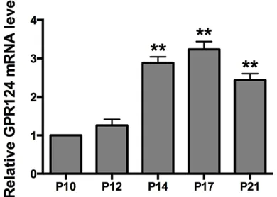
qRT-PCR analysis was performed to study the expression pattern of GPR124 mRNA level in different time points in normal retinal develop-ment and in OIR model. As shown in Figure 1, GPR124 mRNA level in normal mice retina increased from birth, peaked at P14, and then gradually decreased at P17 and P21. Besides, qRT-PCR analysis for GPR124 mRNA in OIR mice retina was performed. As shown in Figure 2, the GPR124 mRNA level was elevated gradu-ally from P10 to P17 in OIR mice retina. The most robust increase was found at P17 and declined subsequently. These results demon-strated that the expression of GPR124 mRNA was in a time-dependent manner in both nor-mal and OIR mice retina.
Expression and localization of GPR124 protein in the normal mice retina and in the OIR mice retina
To determine the spatial changes of GPR124 in the normal and OIR mice retina, Immunohis- tochemistry was performed. Similarly to other rodent, the C57BL/6J mice retina has not been fully developed at birth. Immunohistochemistry
Double labeling immunofluorescence staining
[image:3.612.91.286.76.218.2]Mice in two groups were euthanized at P17, and eyes were enucleated and fixed in 4% parafor-maldehyde, then were equilibrated in 30% sucrose and embedded in OCT. Frozen sections were made with a thickness of 6 μm. The sec-tions were permeabilized with 1% Triton X-100 for 20 min, and blocked with 10% normal goat serum for 2 h. Then, sections were incubated overnight at 4°C with rabbit polyclonal primary Figure 1. GPR124 mRNA expression in the normal mice retina. Mice retinas from normal group were harvested at P4, P7, P10, P12, P14, P17 and P21 and detected by qRT-PCR. qRT-PCR was performed at least three times and the expression of GPR124 was
normalized by β-actin. Significantly different com -pared with P4. These data were presented as means ± SEM. *P<0.01, **P<0.001.
Figure 2. GPR124 mRNA expression in the OIR mice retina. Mice retinas from OIR group were harvested at P10, P12, P14, P17 and P21 and detected by qRT-PCR. qRT-PCR was performed at least three times and the expression of GPR124 was
normal-ized by β-actin. Significantly different compared with
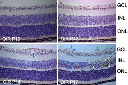
[image:3.612.91.286.339.478.2]GPR124 expression level was elevated in GCL and INL compared with the OIR mice retina at P12. On P17 in the OIR retina, the neovascular-ization and the vascular clusters grew within retina and then grew toward the retinal surface or into the vitreous (Figure 4D, green arrows), besides, there was an abundance of chromatin condensation, pyknotic nuclei (Figure 4D, yel-low arrows), and vacuoles (Figure 4D, red arrows). In addition to the localization of GPR124 in the GCL and INL in OIR mice at P17, we found that GPR124 was expressed in the new vessels either.
Immunofluorescence localization of GPR124
in the normal mice retina and in the OIR mice retina at P17
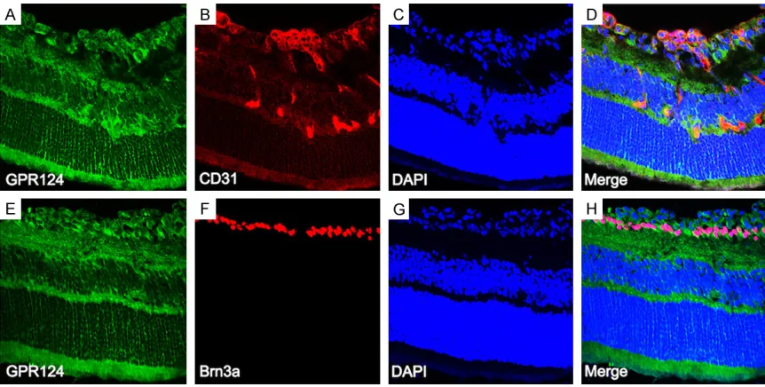
To further address the cell types expressing GPR124, double labeling immunofluorescence staining was performed on the P17 retinas from normal mice (Figure 5) and OIR mice (Figure 6) with retinal ganglion cells (RGCs) specific mark-er (Brn3a) and with vascular endothelial cells specific marker (CD31). GPR124 was expressed in the endothelial cells as previous researches reported [7, 9, 15], and also expressed in the new vessels of retina from OIR mice (Figure 6A-D). In addition, the results revealed that GPR124 expression was co-localized with Brn3a, indicating that the location of GPR124 is in the RGCs either (Figures 5A-D, 6E-H). showed that the retina was composed of three
layers in normal retina at P4 (Figure 3A): the RPE cell layer, the neuroblast layer and the gan-glion cell layer (GCL). At P7 (Figure 3B), the neu-roblast layer differentiated into inner nuclear layer (INL) and outer nuclear layer (ONL), but the boundary was not clear and the cells were irregular arranged, and the photoreceptor layer still not differentiated into inner-segment/out-er-segment (IS/OS). As time goes by, the bound-ary of INL and ONL became increasingly clear, and the short IS/OS could be observed. At P14 (Figure 3E), the mice retinal structure has been improved, manifested as the distinguished ten layers structure of the retina, and the complete-ly separated INL and ONL as the result of the appearance of outer plexiform layer (OPL). At P17 (Figure 3F), the retinal structure was more mature as the adult mice retina.
GPR124 immunolabeling signal was observed weakly in the GCL at P4 and P7, and was appeared in the INL at P7, but the expression was still weak. As the retina development, a dramatic increase in the expression of GPR124 was occurred in the GCL and INL from P10 to P17, peaked at P14, and then stabled at P17. In OIR mice retina, GPR124 was weaker at P10 (Figure 4A) and P12 (Figure 4B) compared with the corresponding time point of normal retina. On P14, vascular expansion (Figure 4C, blue arrow) could be seen at the GCL, and the
Figure 3. Immunohistochemical detection of GPR124 protein in the normal mice retina. Mice retinal paraffin sec -tions at P4 (A), P7 (B), P10 (C), P12 (D), P14 (E) and P17 (F) from normal group were immunohistochemical staining
with GPR124. Original magnification, ×400. GCL, ganglion cell layer; INL, inner nuclear layer; ONL, outer nuclear
(LRRs) domains, one LRR of the COOH-terminal domain, an immunoglobulin (Ig) domain, a hor-mone-receptor (HormR) domain containing an RGD (Arg-Gly-Asp) motif and a membrane proxi-mal GPCR proteolysis site domain [7, 15]. Research has found that the RGD motif of GPR124 was exposed by proteolytic process, and then by directly interacted with integrin αvβ3 to mediate endothelial cells adhesion and survival [15]. Previous studies have reported
Discussion
[image:5.612.92.519.72.356.2]GPR124 was originally reported as a gene over-expressed in tumor vessels of human colorec-tal cancer [7], and was classified as orphan receptor for which the ligand has not been dis-covered yet [9]. GPR124 is a seven-pass trans-membrane receptor, which NH2-terminal extra-cellular region contains several conserved sub-domains and motifs: four leucine rich repeats
Figure 4. Immunohistochemical detection of GPR124 protein in the OIR mice retina. Mice retinal paraffin sections at
P10 (A), P12 (B), P14 (C) and P17 (D) from OIR group were immunohistochemical staining with GPR124. Represen-tative images of vascular expansion (C, blue arrow), chromatin condensation and pyknotic nuclei (D, yellow arrows),
vacuoles (D, red arrows) and new vessels (D, green arrows) are shown. Original magnification, ×400. GCL, ganglion
cell layer; INL, inner nuclear layer; ONL, outer nuclear layer.
Figure 5. Immunofluorescence localization of GPR124 in the normal mice retina at P17. Mice retinal frozen sections
at P17 from normal group were immunostained with GPR124 (A, green), Brn3a (B, red), DAPI (C, blue), and Merge
[image:5.612.95.520.441.547.2]sis; however, it was not upregulated in mature blood vessels [16]. To further explore the rea-son, researchers found that GPR124 could mediate contact inhibition of proliferation in endothelial cells during angiogenesis but not to maintain it in mature vessels [17].
OIR is a widely used mouse model for the research on molecular mechanisms of RNV. In the hyperoxic stage of OIR, high oxygen arrest the development of retinal vascular, leading to the existing blood vessels degenerate and the vaso-obliterated zones formation in central ret-ina. In the hypoxic stage of OIR, the ischemic retina triggers a compensatory release of pro-angiogenic factors, leading to abnormal retinal neovascularization. In our present study, the expression of GPR124 was weak at the hyper-oxic stage, then elevated gradually and peaked at P17. These results revealed that the expres-sion of GPR124 may be influenced by the oxy-gen stress and may participate in the formation of retinal neovascularization.
In the next experiments, we investigated the localization and spatial expression change of GPR124. Immunohistochemistry results sho- wed that GPR124 expression was located in GCL and INL in both normal and OIR mice reti-na, and also expressed in the abnormal pre-retinal neovascularization in OIR. To further that GPR124 displayed elevated expression in
endothelial cells during tumor and physiologic angiogenesis [7, 9, 15]. Besides expressed in CNS angiogenesis, GPR124 was also widely expressed in non-CNS embryonic organs or tis-sues throughout the mouse embryogenesis, including the heart, liver, kidney, epithelium of lung and esophagus and in mesenchyme. In adult mouse, however, the GPR124 was specifi-cally expressed in brain endothelium and angio-genic pericytes and in pericytes of non-neural organs, including the kidney, pancreas, and cor-pus luteum [9].
As the extended part of the brain, the develop-ment of retina is highly correlated with the CNS. In consideration of the pivotal role of GPR124 in CNS and in tumor angiogenesis, we have rea-sons to speculate that GPR124 plays a role in retinal angiogenesis and in retinal neovascular-ization. Our data revealed that GPR124 was progressively up-regulated in the normal retinal development, peaked at P14, and then gradu-ally decreased. As we know, the development of mouse retinal vascular is approximately mature at P14. Thus, the high consistency of time axis between GPR124 expression and reti-nal angiogenesis indicates that GPR124 par-ticipates in the retinal vascular development. In normal tissues, GPR124 was upregulated in endothelial cells during capillary
morphogene-Figure 6. Immunofluorescence localization of GPR124 in the OIR mice retina at P17. Mice retinal frozen sections at
P17 from OIR group were immunostained with GPR124 (A, E; green), CD31 (B, red), Brn3a (F, red), DAPI (C, G; blue),
[image:6.612.90.521.70.288.2]Disclosure of conflict of interest
None.
Address correspondence to: Drs. Lin Lu and Xiao-ling Liang, State Key Laboratory of Ophthalmology, Zhongshan Ophthalmic Center, Sun Yat-sen Univer-sity, Guangzhou 510000, Guangdong Province, Peo-ple’s Republic of China. E-mail: [email protected] (LL); [email protected] (XLL)
References
[1] Gariano RF and Gardner TW. Retinal angiogen-esis in development and disease. Nature 2005; 438: 960-966.
[2] Carmeliet P and Jain RK. Molecular mecha-nisms and clinical applications of angiogene-sis. Nature 2011; 473: 298-307.
[3] Witmer AN, Vrensen GF, Van Noorden CJ and Schlingemann RO. Vascular endothelial growth factors and angiogenesis in eye disease. Prog Retin Eye Res 2003; 22: 1-29.
[4] Miller JW, Le Couter J, Strauss EC and Ferrara N. Vascular endothelial growth factor a in intra-ocular vascular disease. Ophthalmology 2013; 120: 106-114.
[5] Falavarjani KG and Nguyen QD. Adverse events and complications associated with intravitreal injection of anti-VEGF agents: a review of litera-ture. Eye (Lond) 2013; 27: 787-794.
[6] Kurihara T, Westenskow PD, Bravo S, Aguilar E and Friedlander M. Targeted deletion of Vegfa in adult mice induces vision loss. J Clin Invest 2012; 122: 4213-4217.
[7] Carson-Walter EB, Watkins DN, Nanda A, Vo-gelstein B, Kinzler KW and St Croix B. Cell sur-face tumor endothelial markers are conserved in mice and humans. Cancer Res 2001; 61: 6649-6655.
[8] Bjarnadottir TK, Fredriksson R, Hoglund PJ, Gloriam DE, Lagerstrom MC and Schioth HB. The human and mouse repertoire of the adhe-sion family of G-protein-coupled receptors. Ge-nomics 2004; 84: 23-33.
[9] Kuhnert F, Mancuso MR, Shamloo A, Wang HT, Choksi V, Florek M, Su H, Fruttiger M, Young WL, Heilshorn SC and Kuo CJ. Essential regula-tion of CNS angiogenesis by the orphan G pro-tein-coupled receptor GPR124. Science 2010; 330: 985-989.
[10] Anderson KD, Pan L, Yang XM, Hughes VC, Walls JR, Dominguez MG, Simmons MV, Bur-feind P, Xue Y, Wei Y, Macdonald LE, Thurston G, Daly C, Lin HC, Economides AN, Valenzuela DM, Murphy AJ, Yancopoulos GD and Gale NW. Angiogenic sprouting into neural tissue re-quires Gpr124, an orphan G protein-coupled receptor. Proc Natl Acad Sci U S A 2011; 108: 2807-2812.
address the cell types, double labeling immu-nofluorescence staining was performed. Our findings confirmed that GPR124 was expressed in endothelial cells, consistent with previous report [9]. In addition, the co-localization analy-sis of GPR124 and Brn3a indicated that GPR124 was expressed in RGCs either. The similar result was found in GPR91, a seven-transmembrane GPR expressed in RGCs, was a mediator of vessel growth in both normal reti-nal development and RNV and was reported to modulate the expression of VEGF in OIR model [18, 19]. However, whether GPR124 and GPR- 91 might have homologous structure and func-tion should be further demonstrated. Besides the vital role in CNS-specific vascularization, GPR124 controlled the establishment of the blood-brain barrier [9, 11] and regulated VEGF/ VEGFR signaling in tumor angiogenic processes including endothelial cells interaction, permea-bility, migration, invasion, and tube formation [20]. GPR form a large protein family that plays an important role in many physiological and pathological processes. It has been revealed that numerous diseases are GPR-related and the GPR have become an excellent therapeutic target in about 50% drugs because of their nat-ural ligands can be mimicked for agonistic or antagonistic purposes [21, 22]. Thus, GPR124 is an attractive target for preventing RNV. In conclusion, our study demonstrated for the first time that GPR124 was expressed in the normal retinal development and in the OIR mice retina in a time-dependent manner, and locat-ed in the endothelial cells of retinal blood ves-sels and in RGCs. The spatiotemporal changes of GPR124 expression demonstrated that GPR124 might play a role in regulating retinal neurovascular development and retinal neo-vascularization. Therefore, GPR124 might be used as an anti-angiogenic factor and served as a potential novel target to control the RNV. Further study will be performed on GPR124 gene over-expression or silencing mice to show its role on retinal angiogenesis and neovascu-larization by construct GPR124 expression vec-tor or GPR124-siRNA vecvec-tor.
Acknowledgements
[11] Cullen M, Elzarrad MK, Seaman S, Zudaire E, Stevens J, Yang MY, Li X, Chaudhary A, Xu L, Hilton MB, Logsdon D, Hsiao E, Stein EV, Cut-titta F, Haines DC, Nagashima K, Tessarollo L and St Croix B. GPR124, an orphan G
protein-coupled receptor, is required for CNS-specific
vascularization and establishment of the blood-brain barrier. Proc Natl Acad Sci U S A 2011; 108: 5759-5764.
[12] Liang X, Zhou H, Ding Y, Li J, Yang C, Luo Y, Li S, Sun G, Liao X and Min W. TMP prevents retinal neovascularization and imparts neuroprotec-tion in an oxygen-induced retinopathy model. Invest Ophthalmol Vis Sci 2012; 53: 2157-2169.
[13] Yang L, Xu Y, Li W, Yang B, Yu S, Zhou H, Yang C, Xu F, Wang J, Gao Y, Huang Y, Lu L and Liang X. Diacylglycerol kinase (DGK) inhibitor II (R59949) could suppress retinal neovascular-ization and protect retinal astrocytes in an oxy-gen-induced retinopathy model. J Mol Neuro-sci 2015; 56: 78-88.
[14] Yang B, Xu Y, Yu S, Huang Y, Lu L and Liang X.
Anti-angiogenic and anti-inflammatory effect
of Magnolol in the oxygen-induced retinopathy
model. Inflamm Res 2016; 65: 81-93.
[15] Vallon M and Essler M. Proteolytically pro-cessed soluble tumor endothelial marker (TEM) 5 mediates endothelial cell survival dur-ing angiogenesis by linkdur-ing integrin alpha(v) beta3 to glycosaminoglycans. J Biol Chem 2006; 281: 34179-34188.
[16] St Croix B, Rago C, Velculescu V, Traverso G, Romans KE, Montgomery E, Lal A, Riggins GJ, Lengauer C, Vogelstein B and Kinzler KW. Genes expressed in human tumor endotheli-um. Science 2000; 289: 1197-1202.
[17] Vallon M, Rohde F, Janssen KP and Essler M. Tumor endothelial marker 5 expression in en-dothelial cells during capillary morphogenesis is induced by the small GTPase Rac and medi-ates contact inhibition of cell proliferation. Exp Cell Res 2010; 316: 412-421.
[18] Hu J, Wu Q, Li T, Chen Y and Wang S. Inhibition of high glucose-induced VEGF release in reti-nal ganglion cells by RNA interference target-ing G protein-coupled receptor 91. Exp Eye Res 2013; 109: 31-39.
[19] Sapieha P, Sirinyan M, Hamel D, Zaniolo K, Joyal JS, Cho JH, Honore JC, Kermorvant-Duch-emin E, Varma DR, Tremblay S, Leduc M, Riha-kova L, Hardy P, Klein WH, Mu X, Mamer O, Lachapelle P, Di Polo A, Beausejour C,
An-delfinger G, Mitchell G, Sennlaub F and Chem -tob S. The succinate receptor GPR91 in neu-rons has a major role in retinal angiogenesis. Nat Med 2008; 14: 1067-1076.
[20] Wang Y, Cho SG, Wu X, Siwko S and Liu M. G-protein coupled receptor 124 (GPR124) in en-dothelial cells regulates vascular enen-dothelial growth factor (VEGF)-induced tumor angiogen-esis. Curr Mol Med 2014; 14: 543-554. [21] Klabunde T and Hessler G. Drug design
strate-gies for targeting G-protein-coupled receptors. Chembiochem 2002; 3: 928-944.