Original Article
Tight junction proteins and gap
junction proteins play important roles
in high fat dietary atherosclerosis pathogenesis
Bing Sun1*, Zhisong Chen1*, Jianyun Gu1*, Gary Tse2, Jinfa Jiang1, Feifei Huang1, Cuimei Zhao1
1Department of Cardiology, Tongji University Affiliated Tongji Hospital, Shanghai 200065, China; 2School of Biomedical Sciences, Li Ka Shing Faculty of Medicine, University of Hong Kong, Hong Kong, China. *Equal con-tributors.
Received April 28, 2016; Accepted July 22, 2016; Epub August 1, 2016; Published August 15, 2016
Abstract: Atherosclerosis (AS) is a leading chronic diseases with high death rate in industrialized countries, where AS is caused by many factors. Studies show that tight junction protein (TJP) and Connexin family play important roles in heart and blood vessel function and health. Changing Gap Junction Protein (Cx43 Cx45 and Cx46) and Tight Junction Protein (Zo-1, Claudin-1 and Occludin-1) may have effects on AS pathogenesis. Here, we construct a rat model on a high-fat diet to explain the relationship between atherosclerosis pathogenesis and TJP/GJP changes. Our results showed that compared with control group, the weight of treatment group increased significantly with the formation of atheromas and the artery wall infiltration with adipose tissue in histological section. Our qPCR and western blotting results of heart coronary and artery endothelial tissue showed that Cx43/45/46 and claudin-1 were significantly down-regulated in coronary artery. In FITC-Inulin transwell experiment, a paracellular permeability test, we found that primary endothelial cell of high-fat diet rat group showed higher permeability compared to con-trol group. Imunofluorescence staining experiment showed less Cx43 and Zo-1 protein expression and more CD14 monocyte penetration in heart and aorta wall in high-fat diet rat group. Our results suggested that atheroma forma-tion might be due to the loss of TJP and GJP, causing higher permeability in rat coronary artery and thus promoted the pathogenesis of atherosclerosis.
Keywords: Tight junction protein, gap junction protein, atherosclerosis
Introduction
Atherosclerosis, also called as arteriosclerotic vascular disease or AS, is characterized by artery chronic degeneration and gradual chang-ing of artery wall [1] which is due to the growth of the connective tissue, deposition of the cho-lesterol, fatty acid and calcium carbonate in the inside and outside cells [1, 2]. Collagen and proteoglycan gather in the arteries, which become harden and thicken and finally lose flexibility [3, 4]. AS is a widespread disease, but little were carried out. Symptoms of AS are severe, such as once the disease comes, it will come out angina pectoris, myocardial infarc-tion, stroke, and other deadly diseases, mortal-ity rate is high, the harm is great [3-5]. Hardening of the artery is a complex biological
process, which involves quite a lot tissues (like epithelial cells, smooth muscle, monocytes, macrophages and platelets), along with various hormones and cytokines. Therefore so far there is not good atherosclerosis model and diagno-sis technology [3, 6]. Therefore, it is important to study participation AS.
closed protein Claudin, adhesion molecules (junction adhesion moleucule JAMs) and closed small ring protein ZO-week cytoplasm 1/2/3 [4, 7-9]. Scaffold proteins include claudins which are tightly coupled, and participate in the inter-cellular signal transduction. Aberrant expres-sion of Claudin protein level has strong implica-tions for tumorgenesis, cancer invasion and metastasis [8].
Gap junction (GJ), communication links, refers to the connection between two adjacent cells. Connection channel is arranged in a special membrane structure [1, 10-12]. Cell-cell inter-actions are mediated through gap junction intercellular space connection communication (GJIC) [10]. It passes on ions, small molecule metabolites and secondary signal, the forma-tion of participating in the predictor of material exchange between cells metabolism and elec-trical coupling. It plays an important role in metabolism regulating, internal environment stability, cell proliferation and cell differentia-tion. Connection channel are not form single gap junction, but form a tightly bunched poly-mer or spot, ranging from a few to thousands. It varies with groups of different development stages. Components of gap junction channels are junction proteins (connexin, Cx) [1, 12]. Cx comes from a membrane transport protein family, which is channel of transmembrane ion and small molecules communication [1, 3, 12]. Previous studies suggest that one of major causes of atherosclerosis occurs is endothelial cell injury and adhering of monocyte to endo-thelial cells. Tight junction proteins and clear-ance proteins play an important role in the pro-cess. Somebody thinks that the human heart is given priority to with Cx40, 43, 45 [6, 11, 13]. Cx43 was mainly observed in the intercellular space of main connection from previous stud-ies, which is highly expressed in myocardial tis-sue, macrophages, connective tissue cells, endothelial cells, endothelial cell, EC) and smooth muscle cells (smooth musclecell, SMC), fibroblasts, lens, corneal epithelium cells and also expressed in other tissues [13, 14]. Cx43 is encoded by a 2768 bp segment, which con-sists of 2768 base pairs (base pair, bp) of the three complementary cDNA. The composition of cDNA code contains a 1146-bp open reading frame, 43 kd for coding the molecular weight is single peptide, it contains 378 amino acids, so it is named Cx43 [11, 13]. Cx43 is synthesized
in the endoplasmic reticulum ribosomes, and then gathered with six connection protein in golgi complex to form a half channel and finally transported to the plasma membrane, Cx43 was phosphorylated during the process of transportation, relativing to the most structural protein. Cx43 have a short half-life, about a few hours [14]. Gap junction spot (also called gap junction plaque) consists of several Cx43 pro-teins, which is a transmembrane protein, and the quantity of Cx43 directly affects the GJIC function. Plexiform gap junction channels (GJ spot), which is composed of Cx43, can be influ-enced by the cAMP and microfilament. Phosphorylation of Cx43 is closely related to the function of the GJ. Studies have shown that gap junction protein 43 (Cx43) is the most important link protein in mammalian heart; studies have shown that it is essential for the normal differentiation of the heart and develop-ment. Abnormal expression of Cx43 leads to a variety of cardiovascular diseases and congeni-tal malformation of the heart [6, 11-14].
In human, Cx45 is observed in two different places, one is myocardial cell surface, and the other is both sides of the dish. Cx45 distribu-tion is different, which influences the reguladistribu-tion of the cardiac systolic function. The variation of gap junction protein 46 (Cx46) is associated with many diseases, such as breast cancer, cataract, etc [12]. The G143R missense muta-tion on connexin (Cx) 46 was recently reported to be associated with congenital Coppock cata-racts [10, 16].
high-fat diet changes the expression of TJ proteins and GJ proteins dramatically, which implicates in atherosclerosis pathogenesis.
Materials and methods
Subjects and samples
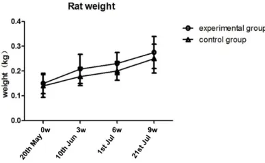
SD rats (14 weeks) was purchased from Sino-British Ltd. Rats were randomly mixed, and divided into two groups, one group of the exper-imental group and another group as the control group. The experimental group of rats fed the way to feed the oil 6 ml per day per rat and mixed with oil as the sole feed. Rat food of con-trol group received no treatment. The two groups of rats were drinking was deionized water. Rats were fed three months. Three-month period, every two weeks for a body weight of rats were weighed. Monthly anatomi-cal one pair of rats gets back dorsal aorta blood vessels and heart. They were divided into three parts, one was for HE staining pathology, one was for qPCR, and one was for western bloting. Rats were weighed periodically data tables and statistics SD rat animal level, the initial weight of rat weight changed growing. And the overall-like recorded rats.
Primary cell culture
Vascular endothelial cell line was obtained from the Shanghai Cinoaisa Institute, and they respectively come from a test group SD rat and a control group SD rat. Vascular en-dothelial
make Cell permeability test by transwell. Add 50 ul Inulin-FITC (Gibco) into every under test hole, finish this process in one minute. After adding Inulin-FITC (gibco), take out every hole down solution 50 ul at 1 min, 5 min, 15 min, 30 min, 45 min, 1 h, 2 h. Separately add them into 96-well-plate, and then dropwise add complete medium 50 ul in the 96-well-plate. At last the number of OD is determined by enzyme-labeled instrument in 500 nm wavelength.
Real time quantitative PCR
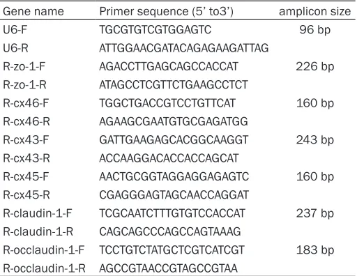
[image:3.612.91.347.84.282.2]Using Trizol regent to isolate the heart and blood vessel’s total RNA from tissue samples of Rattusnorvegicus, the quantity and quality of RNA were confirmed with a NanoDrop 1000 (NanoDrop, Thermo Scientific-Waltham, MA, USA), the primers were designed using primer primer6.0 software and synthesized from Generay Biotech (Table 1). For gene specific reverse transcription, based on the specifica-tion of ReverTta Ace qPCR RT Kit (Toyobo) syn-thesized the first strand. Quantitative real time PCR were conducted on FTC-3000 (Funglyn, Canada) with SYBR Green Fast qPCR kit (KAPA), thermal cycling parameters were as follow. Under the following conditions: 95°C for 3 min (enzyme activation), the next stage were repeat-ed 40 times : 95°C for 5 s (denaturation), and 60°C for 30 s (annealing/extension/data acqui-sition). Data were analyzed by the 2-ΔΔCt algorithm.
Table 1. The list of primers
Gene name Primer sequence (5’ to3’) amplicon size
U6-F TGCGTGTCGTGGAGTC 96 bp
U6-R ATTGGAACGATACAGAGAAGATTAG
R-zo-1-F AGACCTTGAGCAGCCACCAT 226 bp R-zo-1-R ATAGCCTCGTTCTGAAGCCTCT
R-cx46-F TGGCTGACCGTCCTGTTCAT 160 bp R-cx46-R AGAAGCGAATGTGCGAGATGG
R-cx43-F GATTGAAGAGCACGGCAAGGT 243 bp R-cx43-R ACCAAGGACACCACCAGCAT
R-cx45-F AACTGCGGTAGGAGGAGAGTC 160 bp R-cx45-R CGAGGGAGTAGCAACCAGGAT
R-claudin-1-F TCGCAATCTTTGTGTCCACCAT 237 bp R-claudin-1-R CAGCAGCCCAGCCAGTAAAG
R-occlaudin-1-F TCCTGTCTATGCTCGTCATCGT 183 bp R-occlaudin-1-R AGCCGTAACCGTAGCCGTAA
cells were grown in Dul-becco’s Modified Eagle Medium/F12 (DMEM/ F12) (Gibco) with 20% Fetal Bovine Serum (FBS) (Gibco), streptomycin (10 mg/mL) (Sangon)/penicillin (10 KU/mL) (Sangon)/am-photericinB (2- 50 ug/ml) (Sangon) at 37°C with 5% CO2 supplement.
Transwell paracellular permeability assay
Western bolt
Cell lysates were prepared using RIPA buffer (Sigma) containing protease inhibitors (Roche), subsequently agitated on ice for 30 minutes. Pierce™ BCA Protein Assay Kit (Pierce) was used to determine the protein concentration. Protein electrophoresis was performed with Mini-PROTEA III (Bio-Rad). In 10% polyacryam-ide gels (Tris/glycine), proteins were separated and transferred onto polyvinylidene fluoride membrane (Bio-Rad). Primary and secondary antibodies were labeled subsequently. Anti-bodies included Cx43 (rabbit polyclonal anti-Cx43, 1:1000, Sigma-Aldrich), Occludin-1 (rab-bit polyclonal anti-Occludin-1, 1:1000, Abcam), GAPDH (rabbit polyclonal anti-GAPDH; 1:2500, Abcam). Goat anti-rabbit IgG-HRP secondary antibody was purchased from Santa Cruz. Experiments were performed in triplicate.
Immunofluorescence stain
Sections were fixed in 10% formalin for 12 hours, and then embedded in paraffin. 3-μm sections were stained with primary antibody and fluorescent labeled secondary antibody. 100 times magnification field microscopy is applied to capture typical images through Olympus IX71 microscope and Q-iMAGE cam-era. Antibodies included Cx43 (rabbit polyclonal anti-Cx43, 1:1000, Sigma), Zo-1 (rabbit poly-clonal anti-Zo-1, 1:1000, Abcam) and CD14 antibody (rabbit polyclonal anti-CD14, 1:1000, Bioss).
Results
Compared with the control group, the trend of rat weight of which group is treated by high fat diet show higher body weight than that of nor-mal diet. (Figure 1) However no significant
dif-ference was found between the two groups probably due to a small number of samples. In histological section, sub-endothelial fat infiltra-tion and atheromas were shown which indicat-ed success of establishment of model. (Figure 2B) FITC is one of the polymers which can pass through the TJPs formed cell-cell layer. We used Spectraphotometer to determine the concen-tration of Inulin - FITC passing through the cel-lular layer from upper well to lower one. Once the TJPs between endothelial cells are destroyed, Inulin-FITC can easily through the cell layer as (Figure 3). Dyeing cell culture in dif-ferent time point, the value of relative fluores-cence intensity was measured. The RFI value in the control group and treatment group increased slowly with the passage of time in 0 d and 3 d two time points and showed no sig-nificant difference between control group and high fat diet group. However, in 6 d and 9d tests, several time points were revealed to have a higher permeability in high fat diet group. Thus it can draw the conclusion: two groups of cells in 3 d and 6 d were to form the tight junc-tion protein which show higher permeability while cells of high fat diet treatment group in 6 d and 9 d showed dysfunctional paracellular barrier.
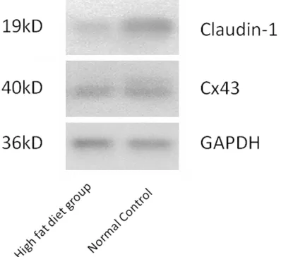
By qRT-PCR assay, the expression of Claudin-1, Cx43, Cx45 and Cx46 amount is lower in the treatment group than the control group in heart coronary artery. Occludin-1 and ZO-1 showed no difference in this tissue between two groups. While it is interesting that only expression quan-tity of Occludin-1 showed significant lower in aorta endothelia in the high fat diet group. These results suggested that coronary artery is more sensitive to endothelial lesion and athero-sclerosis formation than aorta (Figure 4). We select Cx43 and Claudin-1 as TJP and GJP rep-resentatives for western blotting validation on coronary artery tissues (Figure 5).
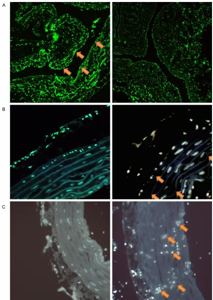
By immunofluorescence staining, while the expression of Zo-1, Cx43 is lower and discon-tinuous in the treatment group than the control group in heart coronary artery, CD14 positive monocyte is shown prone to infiltrate aorta wall in experimental group (Figure 6).
Discussion
[image:4.612.91.285.72.191.2]In this study we found that a high-fat diet lead to quite a lot histological and functional chang-es in both coronary and aorta artery, which
affect pathogenesis of atherosclerosis. We suc-cessfully establish a rat high fat diet model, which helped us conduct further researches on the biological mechanism of atherosclerosis pathogenesis. Adipose infiltration and athero-mas were served as markers of the model.
[image:5.612.92.522.73.232.2]qRT-PCR and WB experiments were performed to evaluated three major proteins of GJP, Cx43, Cx45, Cx46, and three TJPs for ZO-1, Claudin-1 and Occludin-1. Remarkably, we found that all GJPs and Claudin-1 were down-regulated in high-fat diet rat group, compared with control
Figure 2. Sub-endothelial fat infiltration and atheromas were shown which indicated success of establishment of model. High fat diet model was successfully established as shown in (B) compared to normal control shown in (A).
[image:5.612.94.525.287.581.2]group. This was the first time that TJPs family were observed to have a lower expression pat-tern in AS rat model. However interestingly, only Occludin-1 in aorta artery of high fat diet group was observed to have a lower expression sig-nificantly. This may explain the reason why AS lesion mainly occurs in coronary artery. To fur-ther elucidate the function of TJPs, we per-formed FITC-Inulin transwell assay which gave a better understanding of the loss of Tight Junction formed barrier. Results of primary cor-onary arterial or aorta endothelial culture fur-ther demonstrated that gap junction and tight junction loss could further strengthen the abili-ty of paracellular permeabiliabili-ty even in vitro. However, endothelial lesion and dysfunction might be the initial steps of atherosclerosis. Further studies should focus on loss of TJPs and GJPs such as increased infiltration of monocyte or macrophage. Also in vivo GJP and
Figure 4. The expression of Claudin-1, Cx43, Cx45 and Cx46 amount is lower than the control group in heart coro-nary artery in high fat diet group. However, only Occludin-1 showed significantly lower in aorta endothelia in the high fat diet group compared to normal control.
[image:6.612.90.289.468.650.2]TJP functional assay is badly needed in athero-sclerosis pathogenesis.
Acknowledgement
This work is supported by National Nature Sci-ence Foundation of China (NSFC 81300150).
Disclosure of conflict of interest
None.
Address correspondence to: Cuimei Zhao and Feifei Huang, Department of Cardiology, Tongji Hospital, Tongji University, Shanghai 200065, China; E-mail: [email protected] (GMZ); E-mail: af4782@ sina.com (FFH)
References
[1] Scheckenbach KE, Crespin S, Kwak BR, Chan -son M. Connexin channel-dependent signaling pathways in inflammation. J Vasc Res 2011; 48: 91-103.
[2] Berardi DE and Tarbell JM. Stretch and Shear Interactions Affect Intercellular Junction Pro-tein Expression and Turnover in Endothelial Cells. Cell Mol Bioeng 2009; 2: 320-331. [3] Morel S, Burnier L, Kwak BR. Connexins par
-ticipate in the initiation and progression of ath-erosclerosis. Semin Immunopathol 2009; 31: 49-61.
[4] Zhou T, He Q, Tong Y, Zhan R, Xu F, Fan D, Guo X, Han H, Qin S, Chui D. Phospholipid transfer protein (PLTP) deficiency impaired blood-brain barrier integrity by increasing cerebrovascular oxidative stress. Biochem Biophys Res Com-mun 2014; 445: 352-6.
[5] Wong CW, Burger F, Pelli G, Mach F, Kwak BR. Dual Benefit of Reduced Cx43 on Atherosclero -sis in LDL Receptor-Deficient Mice. Cell Com -mun Adhes 2003; 10: 395-400, 2003. [6] Wei JM, Wang X, Gong H, Shi YJ, Zou Y. Experi
-mental research Ginkgo suppresses athero-sclerosis through downregulating the expres-sion of connexin 43 in rabbits. Arc Med Sci 2013; 2: 340-346.
[7] Lehman DM, Leach RJ, Johnson-Pais T, Ham -lington J, Fowler S, Almasy L, Duggirala R, Stern MP, Abboud HE. Evaluation of tight junc-tion protein 1 encoding zona occludens 1 as a candidate gene for albuminuria in a Mexican American population. Exp Clin Endocrinol Dia -betes 2006; 114: 432-7.
[8] Gan H, Wang G, Hao Q, Wang QJ, Tang H. Pro -tein kinase D promotes airway epithelial barri -er dysfunction and p-ermeability through down-regulation of claudin-1. J Biol Chem 2013; 288: 37343-54.
[9] Zhou T, He Q, Tong Y, Zhan R, Xu F, Fan D, Guo X, Han H, Qin S, Chui D. Phospholipid transfer
barrier integrity by increasing cerebrovascular oxidative stress. Biochem Biophys Res Com-mun 2014; 352-6.
[10] Ren Q, Riquelme MA, Xu J, Yan X, Nicholson BJ, Gu S, Jiang JX. Cataract-causing mutation of human connexin 46 impairs gap junction, but increases hemichannel function and cell death. PLoS One 2013; 8: e74732.
[11] Yuan D, Wang Q, Wu D, Yu M, Zhang S, Li L, Tao L, Harris AL. Monocyte-endothelial adhesion is modulated by Cx43-stimulated ATP release from monocytes. Biochem Biophys Res Com-mun 2012; 420: 536-41.
[12] Retamal MA. Connexin and Pannexin hemi-channels are regulated by redox potential. Front Physiol 2014; 5: 80.
[13] Chadjichristos CE. Reduced Connexin43 Ex-pression Limits Neointima Formation After Bal-loon Distension Injury in Hypercholesterolemic Mice. Circulation 2006; 113: 2835-2843. [14] Polacek D, Bech F, McKinsey JF, Davies PF.
Connexin43 Gene Expression in the Rabbit Ar-terial Wall:Effects of Hypercholesterolemia, Balloon Injury and Their Combination. J Vasc Res 1997; 34: 19-30.
[15] Li YY, Qian Y and Zhou CW. Lack of association between the C1019T gene polymorphism and coronary artery disease in a Chinese popula-tion: Meta-analysis of 2,206 subjects. Biomed Rep 2013; 1: 464-468.
[16] Ebihara L, Korzyukov Y, Kothari S, Tong JJ. Cx46 hemichannels contribute to the sodium leak conductance in lens fiber cells. Am J Physiol Cell Physiol 2014; 306: C506-13. [17] Gan H, Wang G, Hao Q, Wang QJ, Tang H. Pro
-tein kinase D promotes airway epithelial barri -er dysfunction and p-ermeability through down-regulation of claudin-1. J Biol Chem 2014; 289: 20489.
[18] Izraely S, Sagi-Assif O, Klein A, Meshel T, Ben-Menachem S, Zaritsky A, Ehrlich M, Prieto VG, Bar-Eli M, Pirker C, Berger W, Nahmias C, Cour -aud PO,Hoon DS, Witz IP. The Metastatic Mi -croenvironment: Claudin-1 Suppresses the Malignant Phenotype of Melanoma Brain Me-tastasis. Int J Cancer 2015; 136: 1296-307. [19] Sappayatosok K and Phattarataratip E. Overex
-pression of Claudin-1 is Associated with Ad-vanced Clinical Stage and Invasive Pathologic Characteristics of Oral Squamous Cell Carci -noma. Head Neck Pathol 2015; 9: 173-80. [20] Yoda S, Soejima K, Hamamoto J, Yasuda H, Na
-kayama S, Satomi R, Terai H, Ikemura S, Sato T, Naoki K, Betsuyaku T. Claudin-1 is a novel target of miR-375 in non-small-cell lung can-cer. Lung Cancer 2014; 85: 366-72.