Original Article
Effect of miR-200b on retinal endothelial cell function
under high glucose environment
Qun Jiang1, Fei Zhao1, Xinmin Liu1, Rongrong Li2, Jianming Liu2
1Department of Ophthalmology,The Second Xiangya Hospital of Central South University, Changsha 410011, China; 2The Third Xiangya Hospital of Central South University, Changsha 410013, China
Received July 28, 2015; Accepted August 28, 2015; Epub September 1, 2015; Published September 15, 2015
Abstract: As one of the important complications of diabetes, diabetic retinopathy (DR) presented high incidence worldwide. Hyperglycemia is an important promoting factor for DR occurrence and development. It can damage retinal endothelial cell, resulting in retinal structure and function disorder. Studies have shown that miR-200b may
involve in regulating DR occurrence and development, but its specific function and mechanism have not been eluci -dated. This study aimed to investigate miR-200b effect and mechanism on human retinal endothelial cells (hRECs) under high glucose environment. hRECs were cultured under high glucose or normal environment. Real time PCR was applied to detect miR-200b expression. MiR-200b was transfected to hRECs and MTT was used to detect its effect on hRECs proliferation under high glucose environment. Real time PCR and Western blot were performed to
determine VEGF and TGFβ1 expression in the retina endothelial cells. MiR-200b expression decreased significantly
under high glucose environment, whereas hRECs proliferated obviously. Compared with normal control, VEGF and
TGFβ1 mRNA and protein expression increased markedly (P < 0.05). After miR-200b transfection, miR-200b
expres-sion increased, while VEGF and TGFβ1 mRNA and protein expresexpres-sion decreased obviously. Compared with high
glucose group, hRECs proliferation was inhibited (P < 0.05). MiR-200b can regulate RECs growth and proliferation
by changing VEGF and TGFβ1 expression to delay DR.
Keywords: Diabetic retinopathy, retinal endothelial cell, VEGF, TGFβ1
Introduction
As one of the most important diabetes compli-cations, diabetic retinopathy (DR) is the leading cause of blindness in adult [1, 2]. According to WHO survey, there were up to 360 million patients suffered from diabetes worldwide. It was expected to reach 1 billion till 2030 [3, 4]. The study found that diabetic retinopathy (DR) was closely related to retinal microvascular sys-tem damage caused by high glucose environ-ment [5]. DR can change retinal structure, lead-ing to its metabolism and function disorder. Retinal microvascular endothelial cells were responsible to supply nutritional requirements for retina nerve. They played a key role in the protection of the vision by maintaining blood-retinal barrier and removing toxins and inflam-matory factors [6, 7].
MicroRNAs, also known as miRNAs or small RNAs, widely existed in animals and plants that
dys-function and DR occurrence. At the same time, vascular endothelial growth factor (VEGF) and transforming growth factor β1 (TGFβ1) were confirmed to be involved in the regulation of DR lesions [13, 14]. This study tended to study miR-200b impact on RECs under high glucose and effect in regulating VEGF and TGFβ1, to analyze its role in DR.
[image:2.612.87.377.82.344.2]cultured under normal condition; High glucose group: high glucose environment was applied to induced culture cells, and the hRECs in loga-rithmic stage was maintained in microvascular endothelia cells medium with 33 mmol/L glu-cose after 72 h; MiR-200b: RGC-5 cells trans-fected with miR-200b were cultured under high glucose.
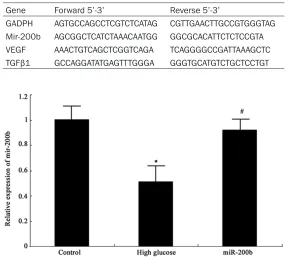
Table 1. Primer sequence
Gene Forward 5’-3’ Reverse 5’-3’
GADPH AGTGCCAGCCTCGTCTCATAG CGTTGAACTTGCCGTGGGTAG Mir-200b AGCGGCTCATCTAAACAATGG GGCGCACATTCTCTCCGTA VEGF AAACTGTCAGCTCGGTCAGA TCAGGGGCCGATTAAAGCTC
TGFβ1 GCCAGGATATGAGTTTGGGA GGGTGCATGTCTGCTCCTGT
Materials and methods
Reagents and instruments
Human retinal endothelial cells were bought from Angio-Proteomie (USA). Human micro-vascular endothelial cell medi-um was got from Cell App- lications. Penicillin-streptomy- cin and EDTA were purchased from Hyclone (USA). DMSO and MTT were bought from Gibco. Enzyme-EDTA was acquired from Sigma. PVDF membrane was got from Pall Life Science. RNA extraction kit, reverse transcription kit, and lipo2000 were bought from Invitrogen. Western blot related reagents were from Beyotime (Shanghai, China). ECL reagent was got from Amersham Biociences. Rabbit anti human VEGF and TGFβ1 antibodies, and HRP tagged IgG secondary antibody was got from Cell signaling (USA). DNA amplifier was bought from PE Gene Amp PCR System 2400 (USA). Other common reagents were from Sangon (Shanghai, China).
hRECs culture and grouping
[image:2.612.90.370.399.569.2]hRECs at 3rd-8th generations were seeded in dish at 1×10⁶ cells/cm2 with neural cell medi-um containing no fetal bovine serum DMEM medium (con-taining 100 U/ml penicillin and 100 μg/ml streptomycin), 5.5 mmol/L glucose. The cells were randomly divided into three groups, including normal control group: the cells were Figure 1. MiR-200b expression in hRECs *P < 0.05, compared with normal
control; #P < 0.05, compared with high glucose group.
Figure 2. MiR-200b impact on hRECs proliferation *P < 0.05, compared
MiR-200b mimics tranfection
MiR-200b mimics was synthesized by Gene- pharma in Shanghai. The sequence of
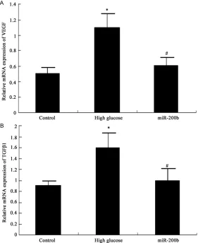
miR-supernatant was moved to new Ep tube and stored at -20°C. The protein was separated by 10% SDS-PAGE electrophoresis and trans-Figure 3. MiR-200b impact on VEGF and TGFβ1 mRNA expression in hRECs
A MiR-200b impact on VEGF mRNA in hRECs B MiR-200b impact on TGFβ1
mRNA in hRECs *P < 0.05, compared with normal control; #P < 0.05,
[image:3.612.92.373.73.417.2]com-pared with high glucose group.
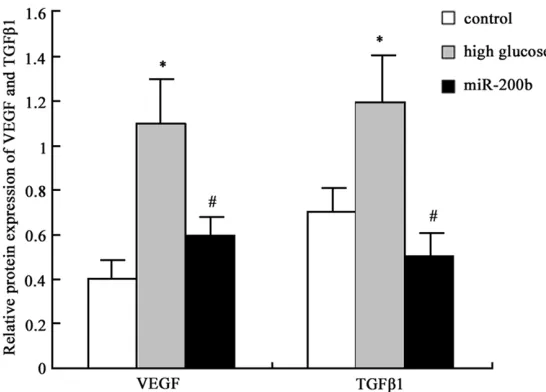
Figure 4. MiR-200b impact on VEGF and TGFβ1 protein expression in hRECs.
1 control; 2, high glucose group; 3, miR-200b group.
200b mimics was 5’-UAAU- ACUGCCUGGUAAUGAUGAC- 3’. Lipo2000 reagent was used to transfect miR-200b to hRECs under high glucose environment. hRECs in loga-rithmic growth phase seeded in 6-well plate at 3×10⁶ cells/ cm2 and maintained in 5% CO
2 incubator at 37°C for 12 h. Then lipo2000 mixed with miR-200b mimics were added to the cells and incubated for 6 h. The cells were further cul-tured after changing the medium.
Real-time PCR
mRNAs were extracted from hRECs by Trizol. Real-time PCR was applied to detect target gene expression using the primers in Table 1. PCR reac-tion contained 52°C for 1 min, followed by 35 cycles includ-ing 90°C for 30 s, 58°C for 50 s and 72°C for 35 s. Gene expression levels were quanti-fied relative to the expression of GAPDH using an optimized comparative Ct (2-ΔCt) value
method.
MTT
The cells were seeded in 96-well plate at 3000/well with five replication. 20 μl MTT solution at 5 g/L was added to each well and the plate was incubated for 4 h. After remov-ing the supernatant, 150 μl DMSO was added for 10 min and the absorbance value at 570 nm was read to calculate cell proliferation rate.
Western blot
[image:3.612.96.374.493.632.2]skim milk for 2 h, the membrane was incubated with VEGF antibody (1:1000) or TGFβ1 antibody (1:2000) at 4°C overnight. After washed by PBST, the membrane was further incubated with goat anti rabbit secondary antibody (1:2000) for 30 min and imaged with chemilu-minescent agent. The band was calculated by Quantity one software with four replication.
Statistical analysis
All statistical analyses were performed using SPSS16.0 software (Chicago, IL). Numerical data were presented as means and standard deviation (_x ± S). Differences between multiple groups were analyzed by one-way ANOVA. P < 0.05 was considered as significant difference.
Results
MiR-200b expression in hRECs
Real-time PCR was applied to detect miR-200b expression in hRECs. As shown in Figure 1, miR-200b expressed highly in normal hRECs, while it decreased obviously under high glu-cose environment (P < 0.05). MiR-200b mimics transfection to hRECs under high glucose envi-ronment can elevate miR-200b level signifi-cantly (P < 0.05).
MiR-200b effect on hRECs proliferation
MTT was performed to test miR-200b effect on hRECs proliferation under high glucose. It was
VEGF and TGFβ1 mRNA expression in hRECs elevated significantly under high glucose (P < 0.05). MiR-200b mimics transfection can sup-press VEGF and TGFβ1 mRNA exsup-pression in hRECs under high glucose obviously (P < 0.05) (Figure 3).
MiR-200b impact on VEGF and TGFβ1 protein expression in hRECs
Western blot was further applied to determine miR-200b mimics transfection effect on VEGF and TGFβ1 protein expression in hRECs and similar results with mRNA expression was observed. VEGF and TGFβ1 protein expression level elevated in hRECs under high glucose (P < 0.05), and their levels were restrained after miR-200b mimics transfection (P < 0.05) (Figures 4 and 5). It suggested that high glu-cose environment can inhibit miR-200b expres-sion in hRECs, and increase VEGF and TGFβ1 mRNA and protein expression, further cause retinal endothelial cell structure and function disorder. Targeting miR-200b to promote its expression can downregulate VEGF and TGFβ1 mRNA and protein expression, thus improve DR.
Discussion
[image:4.612.92.365.70.266.2]DR was a common diabetic microvascular com-plication that can lead to retinal microvascular progressive damage. It seriously affected the Figure 5. MiR-200b impact on VEGF and TGFβ1 protein expression in
hRECs analysis *P < 0.05, compared with normal control; #P < 0.05,
com-pared with high glucose group.
found that high glucose envi-ronment promote hRECs prolif-eration significantly (P < 0.05). After transfecting miR-200b mimics, hRECs proliferation under high glucose was sup-pressed markedly (P < 0.05) (Figure 2). It revealed that miR-200b reduction can promote retinal endothelial cell growth significantly, and targeting miR-200b can inhibit retinal endo-thelial cell proliferation.
MiR-200b impact on VEGF and TGFβ1 mRNA expression in hRECs
patients’ physical and mental health, and brought heavy mental and economic burden to the society [15]. Although medical kept on prog-ress, DR treatment effect was still not satisfied. High glucose environment in diabetic patients can lead to retinal endothelial cells suffered a series of endocrine metabolism changes. High blood glucose is an important factor for diabe-tes complications occurrence and development that can cause organ structure and function abnormity [16, 17]. Retinal endothelial cells structure and function changes were the main pathological mechanism in the chronic process of diabetes. High blood glucose can result in retinal endothelial cell dysfunction including promoting cell proliferation and neovascular-ization through regulating endothelin and VEGF [18, 19].
As voted one of the top ten important discover-ies, miRNAs participated in various processes including cell proliferation, apoptosis, signal transduction, differentiation, hormone secre-tion, lipometabolism and maintaining the potential of embryonic stem cells. It can regu-late the body’s growth and development to make the body adapt to the environment. Recently, multiple studies focused on miRNAs effect in the regulation of tumor occurrence, development, invasion, metastasis, and other biological features [9]. Few researches investi-gated miRNAs in DR. MiR-200b was thought to be involved in regulating diabetes occurrence and development [10], but its role in DR had not been elucidated. Through hRECs culture and high glucose environment treatment, our study confirmed that miR-200b level decreased in hRECs under high glucose state. It was fur-ther revealed that miR-200b can inhibit hRECs abnormal proliferation by transfecting miR-200b mimics.
VEGF had a variety of subtypes that can play its role by binding with the corresponding receptor (VEGFR). Most VEGFR located on endothelial cell surface. Their specific binding can change vascular permeability and promote blood ves-sel formation. Thus, VEGF regulated early stage DR by changing vascular permeability, whereas it participated in the late stage DR through pro-moting neovascularization [20, 21]. TGFβ1 was a kind of isomer of transforming growth factor that belonged to the polypeptide growth factor. It widely distributed in the numerous cells and had many kinds of biological functions. It also played an important role in DR formation by
regulating cell growth, differentiation and migration process, adjusting extracellular matrix synthesis and secretion, and participat-ing in immunity. TGFβ1 is a type of strong regu-latory factor to promote cell proliferation. It also can upregulate integrin expression, promote extracellular matrix secretion, regulate the interactions between cells and matrix, and che-motaxis macrophages to facilitate neovascular-ization and fibroblast growth. It also can play a role of immunosuppression by inhibiting anti-body formation and lymphocyte proliferation to suppress the cytotoxic effect of CTL, NK, and LAK cells [22, 23]. However, there is still lack of investigation about TGFβ1 role in DR. Thus, we intended to study miR-200b effect in retinal endothelial cells. It was confirmed that sup-pressing miR-200b expression under high glu-cose environment may promote VEGF and TGFβ1 mRNA and protein expression to facili-tate endothelial cell proliferation and neovas-cularization, leading to retinopathy. Upregulating miR-200b expression by mimics can reduce VEGF and TGFβ1 mRNA and protein expression, and improve endothelial cell structure and function to postpone DR progress.
To sum up, miR-200b can delay DR progress by changing VEGF and TGFβ1 expression to medi-ate retinal endothelial cell growth and proliferation.
Acknowledgements
Hunan Science and Technology Office of China Scientific Research Project (2014TT2024) and Hunan Science and Technology Office of China Scientific Research Project (2012TT2029).
Disclosure of conflict of interest
None.
Address correspondence to: Dr. Jianming Liu, The Third Xiangya Hospital of Central South University, Changsha 410013, China. Tel: +86-731-88638888; Fax: +86-731-88638888; E-mail: jianming9288@ sina.com
References
[1] Antonetti DA, Klein R, Gardner TW. Diabetic retinopathy. N Engl J Med 2012; 366: 1227-1239.
Shanab AY, Espinosa-Heidmann DG, El-Remessy AB. Imbalance of the nerve growth
factor and its precursor as a potential biomark -er for diabetic retinopathy. Biomed Res Int 2015; 2015: 571456.
[3] Park YH, Shin JA, Han JH, Park YM, Yim HW. The association between chronic kidney dis -ease and diabetic retinopathy: the Korea National Health and Nutrition Examination Survey 2008-2010. PLoS One 2015; 10: e0125338.
[4] Boynton GE, Stem MS, Kwark L, Jackson GR,
Farsiu S, Gardner TW. Multimodal character-ization of proliferative diabetic retinopathy re-veals alterations in outer retinal function and structure. Ophthalmology 2015; 122: 957-967.
[5] Garcia de la Torre N, Fernandez-Durango R, Gomez R, Fuentes M, Roldan-Pallares M,
Donate J, Barabash A, Alonso B, Runkle I,
Duran A, Rubio MA, Calle-Pascual AL. Ex- pression of Angiogenic MicroRNAs in Endo- thelial Progenitor Cells From Type 1 Diabetic Patients With and Without Diabetic Re- tinopathy. Invest Ophthalmol Vis Sci 2015; 56: 4090-4098.
[6] Savage SR, McCollum GW, Yang R, Penn JS.
RNA-seq identifies a role for the PPARbeta/
delta inverse agonist GSK0660 in the
regula-tion of TNFalpha-induced cytokine signaling in
retinal endothelial cells. Mol Vis 2015; 21: 568-576.
[7] Loukovaara S, Gucciardo E, Repo P, Vihinen H, Lohi J, Jokitalo E, Salven P, Lehti K. Indications
of lymphatic endothelial differentiation and en-dothelial progenitor cell activation in the pa-thology of proliferative diabetic retinopathy. Acta Ophthalmol 2015; 93: 512-23.
[8] Orang AV, Barzegari A. MicroRNAs in colorectal cancer: from diagnosis to targeted therapy. Asian Pac J Cancer Prev 2014; 15: 6989-6999.
[9] Gandhi NS, Tekade RK, Chougule MB.
Nanocarrier mediated delivery of siRNA/miR-NA in combination with chemotherapeutic agents for cancer therapy: current progress and advances. J Control Release 2014; 194: 238-256.
[10] Ruiz MA, Feng B, Chakrabarti S. Polycomb re -pressive complex 2 regulates MiR-200b in reti-nal endothelial cells: potential relevance in di-abetic retinopathy. PLoS One 2015; 10: e0123987.
[11] Cao Y, Feng B, Chen S, Chu Y, Chakrabarti S.
Mechanisms of endothelial to mesenchymal transition in the retina in diabetes. Invest Ophthalmol Vis Sci 2014; 55: 7321-7331. [12] Murray AR, Chen Q, Takahashi Y, Zhou KK,
Park K, Ma JX. MicroRNA-200b downregulates
oxidation resistance 1 (Oxr1) expression in the retina of type 1 diabetes model. Invest Ophthalmol Vis Sci 2013; 54: 1689-1697. [13] Shin ES, Sorenson CM, Sheibani N. Diabetes
and retinal vascular dysfunction. J Ophthalmic Vis Res 2014; 9: 362-373.
[14] Rajamani U, Jialal I. Hyperglycemia induces
Toll-like receptor-2 and -4 expression and ac -tivity in human microvascular retinal endothe-lial cells: implications for diabetic retinopathy. J Diabetes Res 2014; 2014: 790902.
[15] Liu L, Wu J, Yue S, Geng J, Lian J, Teng W,
Huang D, Chen L. Incidence Density and Risk
Factors of Diabetic Retinopathy Within Type 2 Diabetes: A Five-Year Cohort Study in China (Report 1). Int J Environ Res Public Health 2015; 12: 7899-7909.
[16] Castilho A, Madsen E, Ambrosio AF, Veruki ML,
Hartveit E. Diabetic hyperglycemia reduces Ca2+ permeability of extrasynaptic AMPA re-ceptors in AII amacrine cells. J Neurophysiol 2015; 114: 1545-1553.
[17] Baptista FI, Castilho AF, Gaspar JM, Liberal JT, Aveleira CA, Ambrosio AF. Long-term exposure to high glucose increases the content of sev-eral exocytotic proteins and of vesicular GABA transporter in cultured retinal neural cells. Neurosci Lett 2015; 602: 56-61.
[18] Monaghan K, McNaughten J, McGahon MK, Kelly C, Kyle D, Yong PH, McGeown JG, Curtis TM. Hyperglycemia and Diabetes Downregulate the Functional Expression of TRPV4 Channels in Retinal Microvascular Endothelium. PLoS One 2015; 10: e0128359.
[19] Behl T, Kaur I, Kotwani A. Implication of oxida-tive stress in progression of diabetic retinopa-thy. Surv Ophthalmol 2015; [Epub ahead of print].
[20] Behl T, Kaur I, Goel H, Kotwani A. Significance
of the antiangiogenic mechanisms of thalido-mide in the therapy of diabetic retinopathy. Vascul Pharmacol 2015; [Epub ahead of print]. [21] Jiang Y, Zhang Q, Steinle JJ. Beta-adrenergic
receptor agonist decreases VEGF levels through altered eNOS and PKC signaling in dia-betic retina. Growth Factors 2015: 1-8. [22] Ko H. Geraniin inhibits TGF-beta1-induced
epi-thelial-mesenchymal transition and suppress-es A549 lung cancer migration, invasion and
anoikis resistance. Bioorg Med Chem Lett
2015; 25: 3529-34.
[23] Kamath VV, Krishnamurthy S, Satelur KP,