Original Article
Decitabine induces G2/M cell cycle arrest
by suppressing p38/NF-κB signaling in
human renal clear cell carcinoma
Donghao Shang1, Tiandong Han1, Xiuhong Xu1, Yuting Liu2
1Department of Urology, Friendship Hospital, Capital Medical University, Beijing 100050, China; 2Department of Pathology, Capital Medical University, Beijing 100069, China
Received June 8, 2015; Accepted July 21, 2015;Epub September 1, 2015; Published September 15, 2015
Abstract: Objective:The anti-neoplastic effects of decitabine, an inhibitor of DNA promoter methylation, are ben-eficial for the treatment of renal cell carcinoma (RCC); however, the mechanism of action of decitabine is unclear. We analyzed gene expression profiling and identified specific pathways altered by decitabine in RCC cells. Methods: Four human RCC cell lines (ACHN, Caki-1, Caki-1, and A498) were used in this study; growth suppression of RCC cells by decitabine was analyzed using the WST-1 assay. Apoptosis and cell cycle arrest were examined using flow cytometric analysis. Gene expression of RCC cells induced by decitabine was evaluated with cDNA microarray, and potential biological pathways were selected using Ingenuity Pathway Analysis. The activity of the p38-NF-κB path-way regulated by decitabine was confirmed by Western blotting. Results: Decitabine suppresses the proliferation of RCC cells in vitro. Although decitabine did not significantly induce apoptosis, decitabine caused cell cycle arrest at G2/M in a dose-dependent manner. Gene expression regulated by decitabine in RCC cells was investigated using microarray analysis. Ubiquitin carboxyl terminal hydrolase 1 (UCHL1), interferon inducible protein 27 (IFI27), and cell division cycle-associated 2 (CDCA2) may be involved in growth suppression of RCC cells by decitabine. The phosphorylation of p38-NF-κB pathway was suppressed by decitabine in RCC cells. Conclusions: We investigated gene expression profiling and pathways modulated by decitabine in RCC cells. Decitabine was shown to suppress the growth of RCC cells via G2/M cell cycle arrest and the p38-NF-κB signaling pathway may play a role in the anti-neoplastic effect of decitabine in RCC cells.
Keywords: Renal cell carcinoma, decitabine, apoptosis, cell cycle arrest, gene expression, p38-NF-κB pathway
Introduction
Renal cell carcinoma (RCC) is the most com-mon tumor involving the adult kidney. RCC is characterized by diverse histologic features and clinical phenotypes, including clear cell RCC (ccRCC), chromophobe RCC, and papillary RCC (PRCC), which originate from the epitheli-um of renal tubules [1, 2]. ccRCC is the major histologic sub-type, accounting for > 75% of all cases [3]. Due to technologic advances, an increased number of patients with RCC are diagnosed at an early stage and < 10% will suc-cumb due to this malignancy [4]. Approximately 30% of patients diagnosed with localized RCC
will undergo relapse after nephrectomy.
More-over, patients with metastatic RCC have a poor 5-year survival rate, ranging from 0-20% [5].
Hypermethylation of cancer-associated genes has been observed in various malignancies, and DNA methylation plays a critical role in human carcinogenesis [6]. Frequently, gene methylation, such as death-associated protein kinase (DAPK) 1, von Hippel-Lindau (VHL) tumor suppressor, tumor suppressor protein RDA32 (RASSFIA), and tissue inhibitor of metallo-
proteinase (TIMP) 3, have been confirmed in
human RCC [7, 8]. The anti-neoplastic effects of 5-aza-2’-deoxycytidine (decitabine), an inhibitor of DNA promoter methylation, appear to be
ben-eficial for treatment of different tumors [9, 10]. Moreover, the effects of concomitant inhibition
with decitabine and chemotherapeutic drugs were also examined in human tumors. Lou et al.
[11] suggested that the gefitinib and
activity and might be considered as a novel therapeutic regimen for treating colon cancer. Another study indicated that the combination of decitabine and panitumumab is well-tolerat-ed and has activity against metastatic colorec-tal cancer [12]. We also investigated the growth suppression of decitabine and chemotherapeu-tic drugs in urinary tract carcinoma, and showed that combination decitabine and paclitaxel (PTX) caused synergistic growth suppression of RCC and prostate carcinoma (PC), suggesting that decitabine and PTX might be a novel strategy to improve the clinical response rate of RCC and PC [13, 14]. In addition, we found that
decitabine could significantly increase the
susceptibility of transitional cell carcinoma to cisplatin, and synergistic growth suppression
by two agents was confirmed in all TCC cell
lines tested [15].
The mechanism of action of decitabine is complicated and controversial. Even though decitabine, a DNA methylation inhibitor, has been reported to suppress tumor cell prolifera-tion by inducing genome-wide demethylaprolifera-tion or
reactivating the expression of specific methyl -ated genes [16, 17], another study indic-ated that decitabine enhances anti-neoplastic activ-ity by causing DNA damage [18]. A recent study
showed that decitabine treatment can signifi -cantly increase CD80 gene expression, and the CTL response elicited by the overexpression of CD80 in a variety of human cancer cells sug-gests that decitabine is also involved in cancer immunotherapy [19]. Another study reported that combined chemotherapy with decitabine and vorinostat could sensitize the tumor cell to Fas-mediated apoptosis and CTL immunother-apy in colon cancer metastasis [20]. Our study indicated that combination treatment with
decitabine and PTX causes significant down-regulation of LEF1/phospho-β-catenin expres
-sion, and the LEF1/β-catenin complex plays a
vital role in the synergy of two agents against RCC [21]. The mechanism of action of deci- tabine in RCC is unclear and should be further investigated.
In this study we analyzed gene-expression
profiling and identified specific pathways
altered by decitabine in RCC cells. Our results suggest that complex networks and numerous pathways linked to decitabine induce growth
inhibition of RCC cells. Moreover, these data confirmed that the p38/NF-κB pathway is involved in G2/M cell cycle arrest of RCC cells
induced by decitabine.
Materials and methods
All procedures followed were in accordance with the Ethics Committee of Friendship
Hospital, Capital Medical University and with
the Helsinki Declaration. Informed consent was obtained from all patients for being included in the study.
Cell culture and agents
Four human ccRCC cell lines (ACHN, Caki-1, Caki-2, and A498) were obtained from the
American Type Culture Collection (Manassas,
VA, USA), and all RCC cell lines were cultured
with complete medium consisting of RPMI-1640 (Gibco, Bio-cult, Glasgow, Scotland), 25 mM HEPES, 1% non-essential amino acids, 100 units/ml of penicillin, 100 μg/ml of str-eptomycin, 2 mM L-glutamine, and 10%
heat-inactivated fetal bovine serum. Cells were maintained as monolayers in 10-cm plastic
dishes and incubated at 37°C in a humidified
incubator containing 5% CO2. RCC cells were detached using 0.25% trypsin/0.01% EDTA. Decitabine was purchased from Sigma (St.
Louis, MO, USA).
Cell proliferation assay
Cell proliferation was analyzed using the WST-1 assay according to the manufacturer’s instruc-tions. RCC cells were planted into 96-well plates and cultured for 24 h, and then the cells were treated with decitabine (1, 2, 4, 8, and 16
μM) for 72 h. After incubation, the cells were treated with 10 μl of WST-1 (Roche, Penzberg,
Germany) and the incubation was continued for 2 h. Finally, the absorbance of each well was measured at 450 nm using a microculture plate reader (Immunoreader, Tokyo, Japan). Three independent experiments were performed and untreated cells were used as blank controls.
Flow cytometric analysis
RCC cells were detached using 0.25% tryp-sin/0.01% EDTA, and 2×10⁵ RCC cells were treated with decitabine for 72 h. Cells were
collected and fixed with 70% ethanol at -20°C
for 8 h, then washed with phosphate-buffered saline and incubated for 30 min with 7-AAD
staining solution (BD Biosciences Pharmingen,
San Diego, CA, USA). The cells in each phase of the cell cycle were counted with FACSCalibur
(Becton Dickinson and analyzed using Cell
cDNA microarray
Total RNA from the four RCC cell lines treated with decitabine was isolated using Trizol accord-ing to the manufacturer’s instructions. After RNA measurement and denaturing gel
electro-phoresis, the RNA samples were amplified and labeled with Cy5 and Cy3 fluorescent dye using
the Agilent Quick Amp labeling kit. Hybridizations were performed with Agilent whole genome oligo microarray, and the slides were scanned by the Agilent DNA microarray scanner. We per-formed sort analysis after pre-treatment to
select good spots for scanning or normalization of the array data, and a number of genes poten-tially relevant to decitabine treatment were
identified from our gene array screens.
Western blotting
Western blot was performed according to the manufacturer’s instructions. Total protein was isolated from RCC cells treated with decitabine for 72 h. The total protein concentration was
measured using a BCA protein assay kit (Pierce,
[image:3.612.95.522.73.532.2]separated by SDS-PAGE and transferred onto
PVDF membranes (Amersham,
Buckingham-shire, UK). Antibodies against p38 and
phos-pho-p38, NF-κB, and phospho-NF-κB were
purchased from Cell Signaling Technology
(Danvers, MA, USA). An anti-β-actin monoclonal
antibody (Abcam, Cambridge, UK) was applied as a loading control. The immune reaction complexes were visualized with an ECL system (Amersham, Aylesbury, UK).
Statistical analysis
Statistical analysis was determined using SPSS (version 19.0; SPSS, Inc., Chicago, IL, USA). All data were calculated in triplicate and the re- sults are represented as the mean ± standard
deviation. Measurement data was investigated
using an independent two-tailed t test and a value of P < 0.05 was considered statistically
significant.
Results
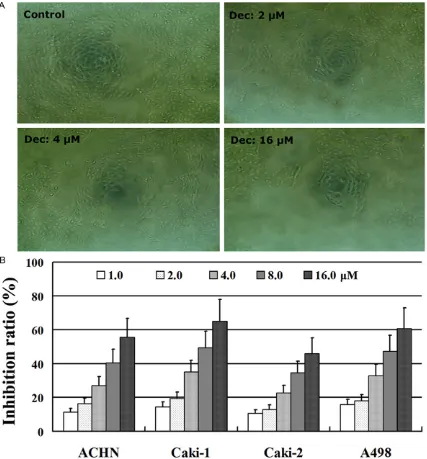
Growth inhibition by decitabine in RCC cells
The growth inhibitory effect of decitabine on RCC cells was analyzed in the four RCC cell lines using the WST-1 assay. RCC cells treated with decitabine had low proliferative ability compared to untreated control cells (Figure 1A). Moreover, decitabine inhibited the growth
[image:4.612.91.521.70.483.2]of RCC cells in a dose-dependent manner (Figure 1B). Thus, targeting DNA methylation with decitabine may be a novel therapy by which to improve prognosis of patients with advanced RCC.
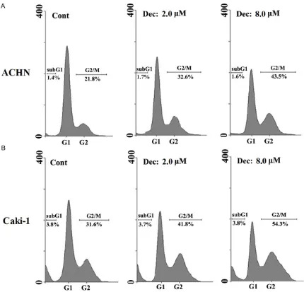
Apoptotic properties of RCC cells treated by decitabine
We analyzed the induction of apoptosis and cell cycle arrest by decitabine in four RCC cell lines. Flow cytometric analysis indicated that
decitabine did not significantly induce apopto
-sis, even at 8 μM, in all RCC cell lines; however, decitabine caused cell cycle arrest at G2/M in
a dose-dependent manner (Figure 2 [the ACHN and Caki-1 data are shown]).
cDNA microarray analysis
Gene expression regulated by decitabine in RCC cells was investigated using microarray analysis. Top 10 up- or down-regulated genes by decitabine are shown in Table 1. The expres-sion of each gene was determined by calculat-ing the average of four RCC cell lines and repre-sented as fold changes compared to untreated control cells. We also selected the possible biological pathways modulated by decitabine
in RCC cells using Ingenuity Pathway Analysis (IPA, version 3.0; Table 2). The probable path-ways induced by decitabine in RCC cells were ranked following the P value; a low P value rep-resents that the pathway is closely correlated with the effect of decitabine against RCC cells.
Our results indicated that the NF-κB pathway,
hypoxia, and p53 in the cardiovascular system may be involved in anti-proliferation of decita- bine in RCC cells.
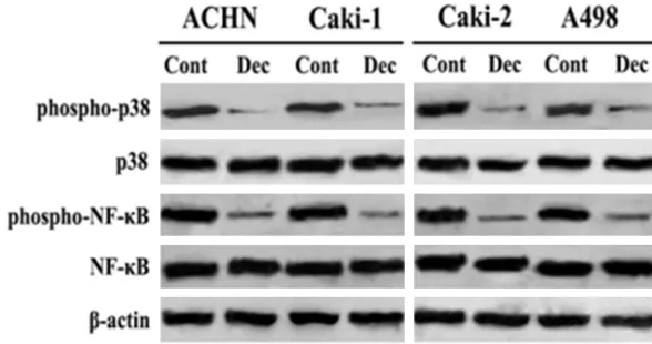
Suppression of p38-NF-κB activity by
decitabine in RCC cells
To demonstrate that the NF-κB signaling path -way is associated with the effect of decitabine
on RCC cells, the activity of the p38-NF-κB
signaling pathway was investigated after sti- mulation with decitabine. In four RCC cell lines, although decitabine did not regulate the total
[image:5.612.91.522.96.435.2]expression of p38 or NF-κB, it suppressed the phosphorylation of p38 and NF-κB (Figure 3). Table 1. Gene expression regulated by decitabine in RCC cells was investigated using microarray analysis
TOP 10 up-regulated genes by decitabine
Probe location Gene bank No. Gene location Gene name [Decitabine vs. Control]Log2 ratio A_23_P132956 NM_004181 chr4: 40965097..40965156 UCHL1 5.64
A_23_P48513 NM_005532 chr14: 93652682..93652741 IFI27 5.33 A_23_P148541 NM_139250 chrX: 153468202..153468261 CTAG1A 3.91 A_23_P73429 NM_005335 chr3: 122833026..122832969 HCLS1 3.49 A_23_P16523 NM_004864 chr19: 18360890..18360949 GDF15 3.18 A_23_P133408 NM_000758 chr5: 131439351..131439410 CSF2 3.17 A_23_P15542 NM_000413 chr17: 37960565..37960624 HSD17B1 3.16 A_23_P216501 NM_213674 chr9: 35672147..35672088 TPM2 3.14 A_23_P146946 NM_001323 chr11: 65537414..65537473 CST6 2.98 A_23_P76078 NM_016584 chr12: 55020350..55020409 IL23A 2.74
TOP 10 down-regulated genes by decitabine
Probe location Gene bank No. Gene location Gene name [Decitabine vs. Control]Log2 ratio A_24_P916585 AF058296 chr20: 25155371..25155312 ENTPD6 -4.45
These results suggest that the p38-NF-κB sig -naling pathway was suppressed by decitabine
and p38-NF-κB may play a vital role in the
action of decitabine against RCC cells. Discussion
The prognosis of RCC is poor because most patients are at an advanced stage at the time of diagnosis and current treatments are not
effective [22]. Thus, finding new anti-neoplastic
drugs for RCC therapy has the potential to improve the clinical strategy and outcomes in patients with RCC. Decitabine, a DNA methyl-transferase inhibitor, has been shown to have anti-tumor activities in human carcinoma [23]. Clinical trials with decitabine have also been conducted in patients with RCC [24, 25]; how-ever, the mechanism of action of decitabine against RCC is not entirely clear.
In this study we investigated the effect of decitabine on the growth of RCC cells and
iden-genes were identified and will be investigated
in the future. Our results indicated that ubiqui-tin carboxyl terminal hydrolase (UCHL) 1 and
interferon inducible protein (IFI) 27 are signifi -cantly up-regulated by decitabine in RCC cells. Although UCHL1 may contribute to colorectal
cancer progression by activating the β-catenin/
TCF pathway [26], UCHL1 has been reported as a tumor suppressor and promoter methylation has been detected in ovarian and prostate can-cers [27, 28]. Aberrant CpG methylation of the
[image:6.612.91.522.86.241.2]UCHL1 promoter is also significantly associat -ed with transcriptional silencing in breast can-cer tissues [29]. Interferons (IFNs) manifest their functions by regulating the expression of target genes, knownas IFN-stimulated genes (ISGs). IFI27 (ISG12) is commonly induced by IFNs and sensitizes cells to apoptotic stimuli by mitochondrial membrane destabilization [30]. IFI27 has been reported to have low expression in leiomyomas and plays a role in the IFN path-way during leiomyoma development [31]. Table 2. Pathways modulated by decitabine in RCC cells were selected by Ingenuity pathway analysis
TOP 10 pathways modulated by decitabine Genes in pathwaythe Genes in chip the Matched with microarray P value
NF-κB pathway 214 191 19 0.008
Hypoxia and p53 in the cardiovascular system 27 16 4 0.009
amb2 integrin signaling 56 20 5 0.012 TRAIL signaling pathway 633 246 20 0.013 Class I PI3K signaling events 449 181 15 0.02
PDGF signaling 45 35 5 0.025
IL2-mediated signaling events 179 64 9 0.026 eNOS activity regulation 27 7 2 0.035 mTORC1-mediated signaling 12 7 2 0.035 IL23-mediated signaling events 99 40 5 0.036
Figure 3. Decitabine suppressed the phosphorylation of p38 and NF-κB.
tified specific pathways
al-tered by decitabine in RCC cells. Our results indicated that decitabine inhibits the proliferation of RCC cells. Even though decitabine
cannot significantly induce
apoptosis in RCC cells, decitabine causes cell
cycle arrest at G2/M in a
[image:6.612.90.384.264.419.2]Another study indicated that IFI27 is involved in the proliferation of skin keratinocytes and that IFI27 might be a new target for development of a novel anti-psoriasis therapy [32]. Our results also indicated that the expression of cell
divi-sion cycle-associated (CDCA) 2 is significantly
down-regulated by decitabine in RCC cells. CDCA2 plays a critical role in the DNA damage response and regulates the activation of p53. A recent study indicated that the expression of CDCA2 is over-expressed in oral squamous cell carcinoma and is associated with tumor pro-gression [33]. Low expression of CDCA2 can suppress proliferation in the G1 phase [33]; however, the expression of UCHL1, IFI27, and CDCA2 in RCC is unclear, thus, the role of these genes in RCC and growth inhibition induced by decitabine should be further investigated. In this study we further investigated the biologi-cal pathways regulated by decitabine in RCC cells using IPA analysis. Our results indicated
that the NF-κB pathway is suppressed signifi -cantly and is involved in the anti-proliferative effect of decitabine on RCC cells. To evaluate
the role of the NF-κB pathway in RCC cells treat -ed with decitabine, the activity of the
p38-NF-κB signaling pathway was analyzed after stimu -lation with decitabine. Although decitabine
can-not modulate the expression of p38 or NF-κB, it
decreased the phosphorylation of p38 and
NF-κB. Our results suggest that the p38-NF-κB
pathway is suppressed by decitabine and
p38-NF-κB may play a critical role in the anti-prolifer -ative effect of decitabine against RCC cells. In conclusion, our study demonstrated that decitabine suppresses the proliferation of RCC
cells via G2/M cell cycle arrest by suppressing p38-NF-κB activity. Moreover, targeting DNA
methylation with decitabine may be a useful approach for improving prognosis in patients with advanced RCC.
Acknowledgements
Our research was supported by a grant from National Natural Science Foundation of China
(81572502) and Beijing Municipal Administ-ration of Hospitals Clinical Medicine
Deve-lopment of Special Funding Support, code: ZYLX201604.
Disclosure of conflict of interest
None.
Address correspondence to: Dr. Yuting Liu, De- partment of Pathology, Capital Medical University, Beijing 100069, China. Tel: 86-10-6313-8553; Fax: 86-10-6313-8553; E-mail: yutingliu889@163.com
References
[1] Arai E, Kanai Y. Genetic and epigenetic altera-tions during renal carcinogenesis. Int J Clin Exp Pathol 2010; 4: 58-73.
[2] Gupta K, Miller JD, Li JZ, Russell MW, Charbonneau C. Epidemio-logic and socioeco-nomic burden of metastatic renal cell carcino-ma (mRCC): a literature review. Cancer Treat Rev 2008; 34: 193-205.
[3] Rini BI, Campbell SC, Escudier B. Renal cell carcinoma. Lancet 2009; 373: 1119-1132. [4] Joseph RW, Kapur P, Serie DJ, Eckel-Passow
JE, Parasramka M, Ho T, Cheville JC, Frenkel E, Rakheja D, Brugarolas J, Parker A. Loss of BAP1 protein expression is an independent marker of poor prognosis in patients with low-risk clear cell renal cell carcinoma. Cancer 2013; 120: 1059-1067.
[5] Stewart GD, O’Mahony FC, Powles T, Riddick AC, Harrison DJ, Faratian D. What can molecu-lar pathology contribute to the management of renal cell carcinoma? Nat Rev Urol 2011; 8: 255-265.
[6] Chalitchagorn K, Shuangshoti S, Hourpai N, Kongruttanachok N, Tangkijvanich P, Thong-ngam D, Voravud N, Sriuranpong V, Mutirangura A. Distinctive pattern of LINE-1 methylation level in normal tissues and the association with carcinogenesis. Oncogene 2004; 23: 8841-8846.
[7] Banks RE, Tirukonda P, Taylor C, Hornigold N, Astuti D, Cohen D, Maher ER, Stanley AJ, Harnden P, Joyce A, Knowles M, Selby PJ. Genetic and epigenetic analysis of von Hippel-Lindau (VHL) gene alterations and relationship with clinical variables in sporadic renal cancer. Cancer Res 2006; 66: 2000-2011.
[8] Morris MR, Hesson LB, Wagner KJ, Morgan NV, Astuti D, Lees RD, Cooper WN, Lee J, Gentle D, Macdonald F, Kishida T, Grundy R, Yao M, Latif F, Maher ER. Multigene methylation analysis of Wilms’ tumour and adult renal cell carcinoma. Oncogene 2003; 22: 6794-6801.
[9] Suh I, Weng J, Fernandez-Ranvier G, Shen WT, Duh QY, Clark OH, Kebebew E. Antineoplastic effects of decitabine, an inhibitor of DNA promoter methylation, in adrenocortical carci-noma cells. Arch Surg 2010; 145: 226-232. [10] Viet CT, Dang D, Achdjian S, Ye Y, Katz SG,
Schmidt BL. Decitabine rescues cisplatin resistance in head and neck squamous cell carcinoma. PLoS One 2014 12; 9: e112880. [11] Lou YF, Zou ZZ, Chen PJ, Huang GB, Li B, Zheng
DNA methylation inhibitor decitabine exerts synergistic anti-cancer activity in colon cancer cells. PLoS One 2014; 9: e97719.
[12] Garrido-Laguna I, McGregor KA, Wade M, Weis J, Gilcrease W, Burr L, Soldi R, Jakubowski L, Davidson C, Morrell G, Olpin JD, Boucher K, Jones D, Sharma S. A phase I/II study of decitabine in combination with panitumumab in patients with wild-type (wt) KRAS metastatic colorectal cancer. Invest New Drugs 2013; 31: 1257-1264.
[13] Shang D, Ito N, Kamoto T, Ogawa O. Demethylating agent 5-aza-2’-deoxycytidine enhances susceptibility of renal cell carcinoma to paclitaxel. Urology 2007; 69: 1007-1012. [14] Shang D, Liu Y, Liu Q, Zhang F, Feng L, Lv W,
Tian Y. Synergy of 5-aza-2’-deoxycytidine (DAC) and paclitaxel in both androgen-dependent and -independent prostate cancer cell lines. Cancer Letters 2009; 278: 82-87.
[15] Shang D, Liu Y, Matsui Y, Ito N, Ogawa O. Demethylaing agent 5-aza-2’-deoxycytidine en-hances susceptibility of bladder transitional cell carcinoma to cisplatin. Urology 2008; 71: 1220-1225.
[16] Shi H, Wei SH, Leu YW, Rahmatpanah F, Liu JC, Yan PS, Nephew KP, Huang TH. Triple analysis of the cancer epigenome: an integrated micro-array system for assessing gene expression, DNA methylation, and histone acetylation. Cancer Res 2003; 63: 2164-2171.
[17] Alleman WG, Tabios RL, Chandramouli GV, Aprelikova ON, Torres-Cabala C, Mendoza A, Rogers C, Sopko NA, Linehan WM, Vasselli JR. The in vitro and in vivo effects of re-expressing methylated von Hippel-Lindau tumor suppres-sor gene in clear cell renal carcinoma with 5-aza-2’-deoxycytidine. Clin Cancer Res 2004; 10: 7011-7021.
[18] Zhu WG, Hileman T, Ke Y, Wang P, Lu S, Duan W, Dai Z, Tong T, Villalona-Calero MA, Plass C, Otterson GA. 5-aza-2’-deoxycytidine activates the p53/p21Waf1/Cip1 pathway to inhibit cell proliferation. J Biol Chem 2004; 279: 15161-15166.
[19] Wang LX, Mei ZY, Zhou JH, Yao YS, Li YH, Xu YH, Li JX, Gao XN, Zhou MH, Jiang MM, Gao L, Ding Y, Lu XC, Shi JL, Luo XF, Wang J, Wang LL, Qu C, Bai XF, Yu L. Low dose decitabine treatment induces CD80 expression in cancer cells and stimulates tumor specific cytotoxic T lympho-cyte responses. PLoS One 2013; 8: e62924. [20] Yang D, Torres CM, Bardhan K, Zimmerman M,
McGaha TL, Liu K. Decitabine and vorinostat cooperate to sensitize colon carcinoma cells to Fas ligand-induced apoptosis in vitro and tumor suppression in vivo. J Immunol 2012; 188: 4441-4449.
[21] Shang D, Liu Y, Xu X, Han T, Tian Y. 5-aza-2’-deoxycytidine enhances susceptibility of renal cell carcinoma to paclitaxel by decreasing
LEF1/phospho-β-catenin expression. Cancer Letters 2011; 311: 230-236.
[22] Escudier B, Eisen T, Porta C, Patard JJ, Khoo V, Algaba F, Mulders P, Kataja V; ESMO Guidelines Working Group. Renal cell carcinoma: ESMO Clinical Practice Guidelines for diagnosis, treatment and follow-up. Ann Oncol 2012; 23 Suppl 7: vii65-71.
[23] Shin DY, Kim GY, Kim CG, Kim WJ, Kang HS, Choi YH. Anti-invasive effects of decitabine, a DNA methyltransferase inhibitor, through tight-ening of tight junctions and inhibition of matrix metalloproteinase activities in AGS human gastric carcinoma cells. Oncol Rep 2012; 28: 1043-1050.
[24] Gollob JA, Sciambi CJ, Peterson BL, Richmond T, Thoreson M, Moran K, Dressman HK, Jelinek J, Issa JP. Phase I trial of sequential low-dose 5-aza-2’-deoxycytidine plus high-dose intrave-nous bolus interleukin-2 in patients with mela-noma or renal cell carcimela-noma. Clin Cancer Res 2006; 12: 4619-4627.
[25] Abele R, Clavel M, Dodion P, Bruntsch U, Gundersen S, Smyth J, Renard J, van Glabbeke M, Pinedo HM. The EORTC Early Clinical Trials Cooperative Group experience with 5-aza-2’-deoxycytidine (NSC 127716) in patients with colo-rectal, head and neck, renal carcinomas and malignant melanomas. Eur J Cancer Clin Oncol 1987; 23: 1921-1924.
[26] Zhong J, Zhao M, Ma Y, Luo Q, Liu J, Wang J, Yuan X, Sang J, Huang C. UCHL1 acts as a colorectal cancer oncogene via activation of the β-catenin/TCF pathway through its deubiq-uitinating activity. Int J Mol Med 2012; 30: 430-436.
[27] Jin C, Yu W, Lou X, Zhou F, Han X, Zhao N, Lin B. UCHL1 Is a Putative Tumor Suppressor in Ovarian Cancer Cells and Contributes to Cisplatin Resistance. J Cancer 2013; 4: 662-670.
[28] Ummanni R, Jost E, Braig M, Lohmann F, Mundt F, Barett C, Schlomm T, Sauter G, Senff T, Bokemeyer C, Sültmann H, Meyer-Schwesinger C, Brümmendorf TH, Balabanov S. Ubiquitin carboxyl-terminal hydrolase 1 (UCHL1) is a potential tumour suppressor in prostate cancer and is frequently silenced by promoter methylation. Mol Cancer 2011; 10: 129.
[29] Trifa F, Karray-Chouayekh S, Jmaa ZB, Jmal E, Khabir A, Sellami-Boudawara T, Frikha M, Daoud J, Mokdad-Gargouri R. Frequent CpG methylation of ubiquitin carboxyl-terminal hy-drolase 1 (UCHL1) in sporadic and hereditary Tunisian breast cancer patients: clinical signifi-cance. Med Oncol 2013; 30: 418.
6-16 and ISG12/IFI27) in innate immunity and cancer. J Interferon Cytokine Res 2011; 31: 173-181.
[31] Mihalich A, Viganò P, Gentilini D, Borghi MO, Vignali M, Busacca M, Di Blasio A. Interferon-inducible genes, TNF-related apoptosis-induc-ing ligand (TRAIL) and interferon inducible protein 27 (IFI27) are negatively regulated in leiomyomas: implications for a role of the interferon pathway in leiomyoma development. Gynecol Endocrinol 2012; 28: 216-219. [32] Hsieh WL, Huang YH, Wang TM, Ming YC, Tsai
CN, Pang JH. IFI27, a novel epidermal growth factor-stabilized protein, is functionally involv- ed in proliferation and cell cycling of human epidermal keratinocytes. Cell Prolif 2015; 48: 187-197.