Original Article
Promoting peripheral nerve regeneration with
biodegradable poly (DL-lactic acid) f lms
Ruijun Li1, Lei Chen1, Jinling Fu2, Zhigang Liu1, Shuang Wang1, Yuehai Pan1
1Department of Hand Surgery, The First Hospital of Jilin University, Changchun, China; 2Department of
Ophthalmology, The Second Hospital of Jilin University, Changchun, China
Received May 12, 2015; Accepted June 25, 2015; Epub July 1, 2015; Published July 15, 2015
Abstract: Regeneration and repair of peripheral nerve injury has always been a major problem in the clinic. The conventional technique based on suturing the nerve ends to each other coupled with the implantation of nerve conduits outside is associated with postoperative adhesions and scar problems. Recently, a novel biodegradable poly (DL-lactic acid) (PDLLA) film has been introduced. This novel anti-adhesion film has a porous structure with better mechanical properties, better flexibility, and more controllable degradation as compared to traditional non-porous nerve conduits. However, little is known about the effects of such PDLLA films on regeneration and repair of peripheral nerve injury in vivo. In this study, we evaluated the effects of PDLLA films implantation after sciatic nerve transection and anastomosis on subsequent sciatic nerve regeneration in vivo, using a rat sciatic nerve injury model. Sciatic nerve transection surgery coupled with direct suturing only, suturing and wrapping with traditional nerve conduits, or suturing and wrapping with PDLLA films was performed on adult Wistar rats. The additional wrapping with PDLLA films inhibited the nerve adhesion after 12 weeks recovery from surgery. It also increased the compound muscle action potentials and tibialis and gastrocnemius muscle wet weight ratio following 8 weeks recovery from surgery. Regenerated nerve fibers were relatively straight and the aligned structure was complete in rats with implantations of PDLLA films. The results suggested that PDLLA films can improve the nutritional status in the muscles innervated by the damaged nerves and promote nerve regeneration in vivo.
Keywords: Biodegradable materials, poly (DL-lactic acid) film, peripheral nerve, regeneration
Introduction
Regeneration and repair of peripheral nerve injury has always been a major problem in the clinic. Whether peripheral nerve can achieve successful regeneration after peripheral nerve injury depends on the availability of suitable microenvironment for their re-growth [1]. More recently, due to rapid advances in microsurgi-cal techniques, nerve anastomosis quality has been greatly improved [1-3]. However, the post-operative adhesions and scar problems have not been well solved. Therefore, it is highly important to prevent the nerve adhesion and provide good anastomotic microenvironment to promote nerve regeneration and repair.
One of the exciting and promising therapeutic strategies is to wrap the anastomosis with nerve conduits [1, 2, 4]. The ideal conduit not only gives physical support to injured nerves,
but also provides anastomosis with a relatively concealed microenvironment. The conduits guide the axial growth of neuronal axons. The conduit structure with the appropriate porosity and the size of pores allows free exchange of
tissue fluids and nutrients, as well as accumula -tion of neurotrophic factors that are required for nerve regeneration [3, 5]. Over the years, the nerve conduits have evolved from silicone
based materials, non-absorbable artificial
materials, or biological based material (intrave-nous, amniotic membrane, etc.) to the modern biodegradable polymers [2, 6].
duits separate the microenvironment of axon regeneration from the surrounding environ-ment, thus avoiding the incorrect growth and invasive growth of non-neural tissues. Third, the biodegradable polymeric materials are often non-toxic, non-irritating, and considered to be the ideal biomaterials for nerve repairing [6, 7].
The synthetic PLA has been approved by US Food and Drug Administration (FDA). It has good biocompatibility, biodegradability and bio-absorbability [8, 9]. Lactic acid, the degrada-tion producdegrada-tion of PLA, is an intermediate metabolite in citric acid cycles in vivo. The mechanism of lactic acid absorption and metabolism has been clearly demonstrated. Due to its reliability and biosafety, PLA polyes-ters are the most extensively studied and the most widely used biodegradable materials to
date. Their application in the field of biomedical
engineering became particularly popular in the recent years. For instance, resorbable PLA
bar-rier film can act as an adhesion barbar-rier to pre
-vent posterior spinal scar formation [7]. It has
been found that PLA film used as an absorb -able adhesion barrier effectively reduced post-surgery adhesion and minimized safety issues [7, 10].
Recently, a novel biodegradable poly (DL-lactic
acid) (PDLLA) film has been prepared by using a
phase transformation method with biodegrad-able polylactic acid polymer as starting
materi-al [11]. This novel anti-adhesion film has a
porous structure, which provides better
mechanical properties, better flexibility, and
more controllable degradation as compared to
traditional non-porous films. However, little is known about the effects of such PDLLA films on
regeneration and repair of peripheral nerve injury in vivo. Therefore, in the present study,
we evaluated the effects of PDLLA films implan
-001). Animals were housed in a temperature- and humidity-controlledvivarium under a 12 h light/dark cycle. All animals had free access to food daily with water ad libitum. All animal experiments were approved by the local Institutional Animal Care and Use Committee. The housing and treatment of the animals fol-lowed the Guidance Suggestions for the Care and Use of Laboratory Animals, formulated by the Ministry of Science and Technology of China.
Sciatic nerve transection and anastomosis
Adult Wistar rats (n = 24) were randomly divid-ed into three groups (n = 8/group). Rats were anesthetized by 10% chloral hydrate (0.3 ml/ kg; i.p.). After shaving the hair from the mid back to hind limb area, rats were then placed in the prone position on the surgery table with a heating pad underneath for maintaining the body temperature at approximately 37°C. To allow the exposure of the sciatic nerve at the dorsocaudal region, an approximately 3 cm-long incision was made starting at 0.5 cm later-ally from the rat midline toward the tibiofemoral articulation, followed by separations of the femoral biceps and gluteus muscles. A micro-surgical scissor was used to make unilateral left side sciatic nerve transection. The nerve was then repaired by epineural microsutures using 9-0 nylon sutures (Johnson & Johnson medical equipment, Shanghai, China) under
the magnification of a binocular loupe. Group 1
received sciatic nerve transection and
anasto-mosis with no additional treatment. Group 2
received sciatic nerve transection and
anasto-mosis with 6 mm artificial nerve conduits (Tian Xin Fu Medical Appliance, Beijing, China). Group
3 received nerve transection and anastomosis
with 6 mm biodegradable PDLLA films wrap
non-transected rats were kept as a control group.
Gross observation
Following the surgery, the rats were observed daily. We also recorded the appearance of the left lower limb, any signs of tissue swelling and infection, as well as the healing of the wound. In some rats, the ulcers on the side toe of foot that received the sciatic surgery appeared dur-ing the early recovery. We recorded the time when the ulcers appeared and healed. Finally, after 8 or 12 weeks of recovery, the conditions
of the implanted PDLLA films and the sciatic
nerve adhesions were examined. Neuroelectrophysiological examination
Eight weeks after the surgery, the transected
rounding connective tissues and exposed in each group of rats (n = 6/group). The normal sciatic nerves on the left side of non-transect-ed control rats (sham group; n = 6) were also
exposed. The stimulation electrodes (fine
non-insulated platinum needles) were placed along
the proximal sciatic nerve. The final position of
electrodes was chosen in a manner that allowed obtaining an electrical response in the tested muscle on the weakest stimulus, usually less than 0.5 mA. Stimulating rectangular puls-es of 0.05 ms in duration were delivered
through a Viking IV electromyelography (EMG)
machine (Keypoint, Frederiksberg, Denmark). The intensity of the stimulus used throughout the investigations was 5 mA. A platinum needle ground electrode was placed subcutaneously in the distal part of the paw. The compound muscle action potentials (CMAPs) recorded
[image:3.612.91.524.70.442.2]from the flexor (gastrocnemius and soleus) and
us) were measured using two subcutaneous platinum needles, one positioned over the bulk of the muscle, and the other placed distally at the level of the ankle. The latency period and the peak amplitude were recorded, and the motor nerve conduction velocity (MNCV) was calculated on the normal and regenerated nerves.
Tibialis and gastrocnemius muscle wet weight ratio
Recovery assessment was also indexed using the weight ratio of the tibialis and gastrocne-mius muscles 8 weeks after surgery.
Immediately after animals were sacrificed, the
tibialis and gastrocnemius muscles were dis-sected and carefully harvested from both intact and injured rats, and weighed while still wet using an electronic balance. All measurements were made by two blinded observers. Values were expressed as a ratio of the wet weight of the tibialis and gastrocnemius muscles to the body weight of the rat.
Histological evaluation
Following 12 weeks of recovery from surgery, the rats were euthanized and a 1 cm-long seg-ment of the sciatic nerve containing the
anasto-mosis site was removed. The conditions of nerve regeneration at the anastomosis site and scar tissue formations were examined.
Data analysis
The experimental data were expressed as
means ± SD. The statistical significance of dif
-ferences between groups was determined by a one-way analysis of variance (ANOVA) followed by Duncan’s test for multiple comparisons. A value of P < 0.05 was considered statistically
significant. Statistical analyses were performed
using SPSS version 19.0 (SPSS, Inc., Chicago, IL, USA).
Results
Gross observation
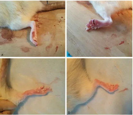
No postoperative mortality occurred. None of the rats had infections on the surgical incisions, and the incision sutures were off on their own. However, 14 rats in all three groups displayed toe or foot ulcers at about 10 days after surgery (Figure 1A and 1B). The ulcer healing time was 10.2 weeks on average in animals that received suture only. However, the ulcer healing time was 8.7 or 8.5 weeks in rats that received
[image:4.612.90.524.72.301.2]implantation of nerve conduits or PDLLA films,
respectively (Figure 1C and 1D). In general, ulcer healing time was much shorted in animals that received either nerve conduits or PDLLA
films implantation following the nerve anasto
-mosis, as compared to those with the sutures only.
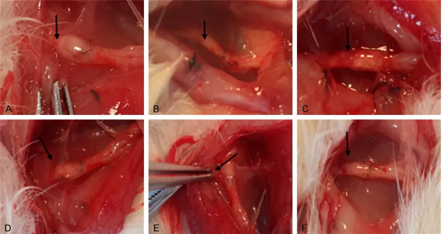
After 8 weeks of recovery, the implanted nerve
conduits or PDLLA films did not shed, and both
types of implantations covered the nerve anastomosis nicely. In contrast, group 1 of rats
with suture only showed significant
posto-perative perineural adhesions, proximal nerve enlargement, and the formation of traumatic neuromas around the site of nerve anastomo-sis (Figure 2A). However, the animals from groups 2 and 3 with the implantations of either
nerve conduits or PDLLA films had no signifi -cant adhesion appearance between the periph-eral nerve tissues and the biomaterials.
Specifically, PDLLA film was clearly softened
and closely attached to the nerves (Figure 2B and 2C).
Similarly, after 12 weeks of recovery, the
implanted nerve conduits or PDLLA films did
not shed, and both types of implantations covered the nerve anastomosis nicely. In contrast, group 1 of rats with suture only
showed significant postoperative perineural
adhesions, proximal nerve enlargement, and the formation of traumatic neuromas around the site of nerve anastomosis (Figure 2D). However, group 2 or group 3 of rats with the implantations of either nerve conduits or PDLLA
films had no significant adhesion appearance
between peripheral nerve tissues and the
bio-materials (Figure 2E and 2F). While the bioma-terials were almost degraded, a thin layer of loose connective tissues formed surrounding the sciatic nerve anastomosis. There was no compression on the nerve itself.
Neuroelectrophysiological examination
Eight weeks after surgery, we measured the compound muscle action potentials (CMAPs), recorded the latency period, and calculated the motor nerve conduction velocity (MNCV). The sciatic transection surgery reduced the amplitude of action potentials in all rats, as compared with sham rats (F(3, 20) = 9.13; P < 0.0001; Figure 3A). In the sham group, the mean amplitude of action potentials was 25.4 ± 8.9 mV. However, in the group of rats receiv-ing suture only after sciatic nerve transection
(i.e., Group 1), the mean amplitude of action
potentials was 5.2 ± 1.45 mV. Furthermore,
implantations of nerve conduits (i.e., Group 2;
6.4 ± 2.8 mV) failed to enhance the amplitude
of action potentials, as compared with Group 1
(Figure 3A). However, implantations of PDLLA
films (i.e., Group 3; 9.5 ± 4.1 mV) increased the
amplitude of action potentials, as compared
with Group 1 (Figure 3A).
Furthermore, the sciatic transection surgery reduced the MNCV in all rats, as compared with sham rats (F(3, 20) = 6.41; P < 0.001; Figure 3B). In the sham group, the mean nerve conduction velocity was 24.8 ± 5.12 m/s. However, in the group of rats receiving suture only after sciatic
nerve transection (i.e., Group 1) the mean
nerve conduction velocity was 14.5 ± 2.89
m/s. Furthermore, implantations of nerve
con-duits (i.e., Group 2; 17.84 ± 4.05 m/s) failed to
enhance the nerve conduction velocity, as
com-pared with Group 1 (Figure 3B). Similarly,
implantations of PDLLA films (i.e., Group 3;
16.9 ± 4.28 m/s) failed to increase the nerve
conduction velocity, as compared with Group 1
(Figure 3B).
Tibialis and gastrocnemius muscle wet weight ratio
Eight weeks after surgery, we also measured the wet weight ratio of the tibialis and gastroc-nemius muscles (Figure 3C). The sciatic tran-section surgery reduced the wet weight ratio in all rats, as compared with sham rats (F(3, 20) = 7.28; P < 0.01; Figure 3C). In the sham group, the mean wet weight ratio was 0.81 ± 0.05%. However, in the group of rats receiving suture
only after sciatic nerve transection (i.e., Group
1), the mean wet weight ratio was 0.32 ± 0.04%. Furthermore, implantations of nerve
conduits (i.e., Group 2; 0.38 ± 0.03%) failed to
enhance the wet weight ratio, as compared
with Group 1 (Figure 3B). However,
implanta-tions of PDLLA films (i.e., Group 3; 0.48 ±
0.05%) increased the wet weight ratio, as
com-pared with Group 1 (Figure 3B).
fibers were relatively straight, and the aligned
structure was complete (Figure 4B). In addition,
little inflammation was observed.
Discussion
In the present study, we demonstrated that
implantation of a novel PDLLA film following sci -atic nerve transection surgery promoted the regeneration of injured nerve tissues in rats.
Specifically, rats with the implantations of either traditional nerve conduits or PDLLA films had no significant adhesion appearance
between peripheral nerve tissues and the
bio-materials. Furthermore, PDLLA film was clearly
softened and closely attached with nerves after 12 weeks recovery from surgery. These
results suggest that PDLLA films have better
biocompatibility and biodegradability. Addi-
tionally, while implantations of PDLLA films
were not able to increase the nerve conduction velocity, this therapeutic method increased the amplitude of action potentials following 8 weeks recovery from sciatic nerve transection
surgery. These results indicate that PDLLA films
may promote axon regeneration in vivo.
Moreover, implantations of PDLLA films
increased the tibialis and gastrocnemius mus-cle wet weight ratio, suggesting that PDLLA
films can improve the nutritional status of the
muscles innervated by the damaged nerves. Last but not the least, in rats with
implanta-tions of PDLLA films, the regenerated nerve fibers were relatively straight and the aligned
structure was complete. In addition, little
inflammation was observed. Taken together, our results suggest that PDLLA films promote
the nerve regeneration in vivo.
The formation of adhesions with surrounding
tissues and nerve scars are difficult
complications after surgery [1, 2]. The prolifer- ating loose connective tissue between the nerve ends continuously grow into the
[image:6.612.89.290.70.316.2]rium, forming scar tissue [12]. The formation of scar tissue reduces the cross-sectional area and represents a major obstacle for axon regeneration. In order to prevent the adhesions between peripheral nerve and surrounding tis-sues and inhibit the formation of scar tistis-sues, various surgical techniques, such as
intrave-nous wrapping, muscle flaps, and free fat trans
-plantation, as well as all kinds of biomaterials including a variety of biodegradable polymers, have been used in nerve regeneration [13-17].
Specifically, the collagen nerve conduits have
been shown to have protective effect on the epineurium suture, improve the proliferation of connective tissue, and serve a similar protec-tive role as sheath by itself [18]. Application of
PLA film to the sciatic nerve in rats has been
demonstrated to reduce the scar tissue
forma-tion [19]. In the present study, PDLLA films
implantation resulted in the minimal formation
of scar tissues, suggesting that PDLLA films
can promote nerve regeneration by inhibiting the scar tissue formation process.
In recent years, PLA has become a hot topic in
the field biomedical materials due to its
non-toxicity, good biodegradability, and proven safety. The polymer has been widely used in abdominal surgery [20]. In addition, it has been
shown that PLA film effectively reduces the pro
-liferation and inflammation of loose connective
tissue, and prevents peripheral nerve adhesion [21]. However, the degradation of PLA is rela-tively slow, and the material remains in the body for a long time. This is the main disadvan-tage that limits its application [21-24]. Meanwhile, non-permeable medical materials could hinder the nutrient and oxygen exchange between peripheral nerves and surrounding tis-sues, thus subsequently impairing the axon regeneration [25-27].
To overcome these disadvantages of PLA, a
novel polylactic-DL-acid absorbable film (PDLLA film) was developed by Changchun Sino
Biomaterials by using a phase inversion
meth-od. The novel anti-adhesion medical film has a
porous structure. Our experimental results demonstrated that anastomosis coupled with a
porous PDLLA film wrapping after sciatic nerve
injury in rats resulted in diminished connective tissue adhesions, as compared with no wrap-ping or nerve conduit wrapwrap-ping groups.
Histological examination of nerve fibers also
confirmed that better nerve fiber regeneration occurred in the PDLLA film group, as compared
with the other two groups. Furthermore, func-tional examination of the sciatic nerve showed that sciatic nerve MNCV and CMAP recovered
better in the PDLLA film implanted group. Finally, PDLLA film implanted group had higher
muscle relative wet weight, which is an indirect index of axons and target organ function,
sug-gesting that PDLLA film implantation can serve
as a better therapeutic method for nerve regeneration.
The therapeutic effects of PDLLA films may
also be ascribed to the good biodegradability. After 12 weeks recovery from surgery, implant-
ed PDLLA films were substantially degraded,
indicating good biodegradability of this type of biomaterials. PDLLA belongs to polylactic acid family of polymers [28]. Due to the irregular arrangement of the asymmetric carbon atoms in the polymer chain, PDLLA is a type of amor-phous polymer with a Tm of 65°C [8, 29, 30]. The degradation and absorption rate of PDLLA is fast, generally around 3-6 months, which is superior to other PLA materials with slow deg-radation rates [8, 29, 30]. In addition, com-pared to the non-porous products, the porous
PDLLA films maintain good mechanical proper
-ties and have a good balance between flexibili
-ty and controllabili-ty of degradation [8].
Furthermore, similar to other PLA films, PDLLA film has good barrier effect. It wraps on nerve
anastomosis ends to form a concealed
micro-environment. Therefore, PDLLA film can form a
physically more resilient regeneration pipeline than epineurium. It prevents the proliferation of connective tissue ingrowth, inhibits the
over-flow of nerve growth factors, and creates a bet
-ter micro-environment for nerve regeneration. Additionally, compared with other non-perme-able materials, the presence of porous
struc-ture of PDLLA films allows smooth liquid flow across the film, which is beneficial to nerve
regeneration. Thus, in the present study, the
regenerated nerve fibers were relatively straight
and the aligned structure was complete after
the implantation of PDLLA films, as compared
with direct suturing.
In summary, the novel absorbable PDLLA film
None.
Address correspondence to: Dr. Yuehai Pan, Depart- ment of Hand Surgery, The First Hospital of Jilin University, No. 71 Xinmin Street, Changchun130021, China. E-mail: [email protected]
References
[1] Grinsell D, Keating CP. Peripheral nerve recon-struction after injury: a review of clinical and experimental therapies. Biomed Res Int 2014; 2014: 698256.
[2] Pfister BJ, Gordon T, Loverde JR, Kochar AS, Mackinnon SE, Cullen DK. Biomedical engi-neering strategies for peripheral nerve repair: surgical applications, state of the art, and fu-ture challenges. Crit Rev Biomed Eng 2011; 39: 81-124.
[3] Vleggeert-Lankamp CL, de Ruiter GC, Wolfs JF, Pego AP, van den Berg RJ, Feirabend HK, Malessy MJ, Lakke EA. Pores in synthetic nerve conduits are beneficial to regeneration. J Biomed Mater Res A 2007; 80: 965-982. [4] Aldini N, Fini M, Rocca M, Giavaresi G, Giardino
R. Guided regeneration with resorbable con-duits in experimental peripheral nerve injuries. Int Orthop 2000; 24: 121-125.
[5] Rodriguez FJ, Gomez N, Perego G, Navarro X. Highly permeable polylactide-caprolactone nerve guides enhance peripheral nerve regen-eration through long gaps. Biomaterials 1999; 20: 1489-1500.
[6] Stang F, Keilhoff G, Fansa H. Biocompatibility of Different Nerve Tubes. Materials 2009; 2: 1480-1507.
[7] Welch WC, Thomas KA, Cornwall GB, Gerszten PC, Toth JM, Nemoto EM, Turner AS. Use of polylactide resorbable film as an adhesion bar-rier. J Neurosurg 2002; 97: 413-422.
[8] Ohya Y. Synthesis of nobel poly (lactic acid)-based biodegradable polymers and their appli-cation as biomaterials. Kobunshi Ronbunshu 2002; 59: 484-498.
[9] Omidi Y, Davaran S. Impacts of Biodegradable Polymers: Towards Biomedical Applications.
[13] Ohsumi H, Hirata H, Nagakura T, Tsujii M, Sugimoto T, Miyamoto K, Horiuchi T, Nagao M, Nakashima T, Uchida A. Enhancement of peri-neurial repair and inhibition of nerve adhesion by viscous injectable pure alginate sol. Plast Reconstr Surg 2005; 116: 823-830.
[14] Dam-Hieu P, Lacroix C, Said G, Devanz P, Liu S, Tadie M. Reduction of postoperative perineural adhesions by Hyaloglide gel: an experimental study in the rat sciatic nerve. Neurosurg 2005; 56: 425-433.
[15] Ikeda K, Yamauchi D, Osamura N, Hagiwara N, Tomita K. Hyaluronic acid prevents peripheral nerve adhesion. Br J Plast Surg 2003; 56: 342-347.
[16] Merle M, Lallemand B, Lim A, Gantois G. Experimental and clinical evaluation of an ab-sorbable biomaterial inducing an anti-adhe-sive barrier (Divide (R)). Eur J Orthop Surg Tr 2008; 18: 255-263.
[17] Xu J, Varitimidis SE, Fisher KJ, Tomaino MM, Sotereanos DG. The effect of wrapping scarred nerves with autogenous vein graft to treat re-current chronic nerve compression. J Hand Surg Am 2000; 25: 93-103.
[18] Kim PD, Hayes A, Amin F, Akelina Y, Hays AP, Rosenwasser MP. Collagen nerve protector in rat sciatic nerve repair: A morphometric and histological analysis. Microsurg 2010; 30: 392-396.
[19] Okui N, Yamamoto M, Fukuhira Y, Kaneko H, Hirata H. A new nerve coaptation technique us-ing a biodegradable honeycomb-patterned film. Microsurg 2012; 32: 466-474.
[20] Bostman OM. Absorbable implants for the fixa-tion of fractures. J Bone Joint Surg Am 1991; 73: 148-153.
[21] Scherman P, Kanje M, Dahlin LB. Local effects on triiodothyronine-treated polyglactin sutures on regeneration across peripheral nerve de-fects. Tissue Eng 2004; 10: 455-464.
[22] Scherman P, Kanje M, Dahlin LB. Bridging short nerve defects by direct repair under ten-sion, nerve grafts or longitudinal sutures. Restor Neurol Neurosci 2004; 22: 65-72. [23] Fujiwara T, Matsuda K, Kubo T, Tomita K,
Axonal supercharging technique using reverse end-to-side neurorrhaphy in peripheral nerve repair: an experimental study in the rat model. J Neurosurg 2007; 107: 821-829.
[24] Turgut M, Uysal A, Pehlivan M, Oktem G, Yurtseven ME. Assessment of effects of pine-alectomy and exogenous melatonin adminis-tration on rat sciatic nerve suture repair: an electrophysiological, electron microscopic, and immunohistochemical study. Acta Neurochir (Wien) 2005; 147: 67-77.
[25] Cemil B, Ture D, Cevirgen B, Kaymaz F, Kaymaz M. Comparison of collagen biomatrix and omentum effectiveness on peripheral nerve regeneration. Neurosurg Rev 2009; 32: 355-362.
[26] Lundborg G. Ischemic nerve injury. Experi- mental studies on intraneural microvascular pathophysiology and nerve function in a limb subjected to temporary circulatory arrest. Scand J Plast Reconstr Surg Suppl 1970; 6: 3-113.
[27] Lundborg G. Structure and function of the in-traneural microvessels as related to trauma, edema formation, and nerve function. J Bone Joint Surg Am 1975: 57: 938-948.
[28] Sodergard A, Stolt M. Properties of lactic acid based polymers and their correlation with composition. Prog Polym Sci 2002; 27: 1123-1163.
[29] Zhao J, Liao W, Wang Y, Pan J, Liu F. Preparation and degradation characteristic study of bone repair composite of DL-polylactic acid/hydroxy-apatite/decalcifying bone matrix. Chin J Traumatol 2002; 5: 369-373.