Original Article
PAK4 enhances TGF-β1-induced
epithelial-mesenchymal transition
through activating β-catenin signaling
pathway in renal tubular epithelial cells
Yan Fan1, Xv Wang2, Yang Li3, Xing Zhao1, Jieqing Zhou1, Xiaoxue Ma1, Dong An1, Hong Jiang1
1Department of Pediatrics, The First Hospital of China Medical University, Shenyang 110001, Liaoning, China; 2Department of Breast Surgery and Surgical Oncology, Research Unit of General Surgery, The First Hospital of
China Medical University, Shenyang, China; 3Department of Cell Biology, Key Laboratory of Cell Biology, Ministry of
Public Health, Key Laboratory of Medical Cell Biology, Ministry of Education, China Medical University, Shenyang, Liaoning, China
Received February 21, 2018; Accepted March 25, 2018; Epub June 1, 2018; Published June 15, 2018
Abstract: Epithelial-mesenchymal transition (EMT) of renal tubular epithelial cells contributes to development and
progression of renal interstitial fibrosis in CKD. p21-activated kinase 4 (PAK4) is a member of serine/threonine protein kinases but the role of PAK4 in renal fibrosis remains unknown. In this study, we investigated the effects of PAK4 on transforming growth factor-β1 (TGF-β1)-treated human renal tubular epithelial cells (HK-2 cells) and aimed to elucidate probable mechanisms for its fibrogenic effects. Our results revealed that PAK4 was highly expressed in TGF-β1-treated HK-2 cells. Overexpressing PAK4 could further decrease TGF-β1-induced E-cadherin expression and increase TGF-β1-induced fibronectin and vimentin expression in HK-2 cells. In addition, overexpressing PAK4 could promote the translocation of β-catenin from cell membranes into the nucleus in TGF-β1-treated HK-2 cells. These results indicate that PAK4 could enhance TGF-β1-induced EMT in renal tubular epithelial cells. Our findings indicate that PAK4 may promote renal interstitial fibrosis by activating β-catenin signaling pathway. Thus, we suggest that PAK4 might be a potential therapeutic target for ameliorating renal interstitial fibrosis.
Keywords: p21-activated kinase 4 (PAK4), transforming growth factor beta 1 (TGF-β1), epithelial-mesenchymal transition (EMT), beta-catenin, renal interstitial fibrosis (RIF)
Introduction
Chronic kidney disease (CKD) is a progressive irreversible process whose final outcome is end-stage renal disease (ESRD) [1, 2]. The fatality rate of CKD in children with renal dis-ease is 9% and that of ESRD is extremely high [3]. There are various causes of CKD, with renal interstitial fibrosis as the most important path-ological feature [4-7]. It is believed that the extent of renal interstitial fibrosis is closely related to prognosis of CKD. The pathogenesis of renal interstitial fibrosis is very complex and remains unclear. Therefore, we need to deeply study the mechanism of renal interstitial fibro-sis and provide a theoretical bafibro-sis for amelio-rating progression of CKD. Recently, great efforts have been made to explore the mecha-nism of renal interstitial fibrosis.
EMT occurs in different contexts of embryonic development, tissue fibrosis, and tumorigene-sis [8] and contributes to renal interstitial fibro-sis [9-11]. EMT can be caused by massive me- diators such as various cytokines and growth factors, among which TGF-β is considered the key fibrogenic growth factor [9]. TGF-β expres-sion has been detected in damaged kidneys, both in patients suffering from kidney diseases and in animal models with renal injuries [12, 13]. Clinical studies have also found elevated TGF-β expression in urine of patients with kid-ney diseases and level of TGF-β has been posi-tively related to severity of renal interstitial fibrosis [12, 13].
Pase signaling. PAKs can phosphorylate mas-sive substrates and affect a wide range of bio-logical processes including cytoskeletal con-struction, cell mitosis, proliferation, motility, transformation, stress, inflammation, and gene expression [14]. PAKs consist of 6 members that are divided into two categories: type I PA- Ks (PAK1, PAK2 and PAK3) and type II PAKs (PAK4, PAK5, and PAK6), according to their structural characteristics and similarity [15]. PAK4 is the most representative of the type II PAKs. One study has shown that reducing PAK4 expression inhibited occurrence of EMT in pros-tate cancer cells and colon cancer cells [16]. PAK4 has been suggested to promote the EMT process of tumor cells. However, the effect of PAK4 on EMT of renal tubular epithelial cells, a potential mechanism of renal interstitial fibro-sis, remains unknown.
In this study, we investigated the effect of PAK4 on EMT of renal tubular epithelial cells and illu-minated the molecular mechanism. We found that PAK4 could induce EMT and enhance TGF-β1-induced EMT by activating β-catenin signal-ing pathway in renal tubular epithelial cells. These data indicate that PAK4 might play a crucial role in renal interstitial fibrosis through enhancing the EMT process, providing a thera-peutic target for CKD.
Materials and methods
Cell culture
Normal human renal tubular epithelial cell line HK-2 was cultured in Dulbecco’s Modified
Eag-le Medium: Nutrient Mixture F-12 (Gibco, Los Angeles, CA, USA) containing 10% fetal bovine serum (FBS), incubated at 37°C in a humidified incubator with 5% CO2.
Real-time PCR
Total RNA was extracted using TRIzol Reagent (Invitrogen, Carlsbad, USA) from HK-2 cells. Total RNA (1 μg) was used for synthesis of cD- NA using PrimeScriptTM RT Reagent Kit (TaKa- Ra, China). Sequences (5’-3’) of the primers for Fibronectin were ACAACACCGAGGTGACTGAGAC (F) and GGACACAACGATGCTTCCTGAG (R), Vi- mentin were AGGCAAAGCAGGAGTCCACTGA (F) and ATCTGGGCGTTCCAGGGACTCAT (R), E-ca- dherin were GCCTCCTGAAAAGAGAGTGGAAG (F) and TGGCAGTGTCTCTCCAAATCCG (R), PAK4 were GATGATTCGGGACAACCTGCCA (F) and AGGAATGGGTGCTTCAGCAGCT (R), GAPDH we-
re GTCTCCTCTGACTTCAACAGCG (F) and AC- CACCCTGTTGCTGTAGCCAA (R). Real-Time PCR was performed using SYBR green mix (TaKaRa, China). Reactions were performed with a 7500 Fast Real-Time PCR System (Applied Biosy- stems, La Jolla, CA).
Western blot assay
HK-2 cells were lysed in RIPA lysis buffer sup-plemented with protease inhibitors (Roche, USA). Nuclear protein was extracted through a Nuclear and Cytoplasmic Protein Extraction Kit (Beyotime, China). Total protein extracts were separated by 8% SDS-PAGE and then trans-ferred to a PVDF membrane (Millipore, USA). The membranes were incubated with anti-E-cadherin (DB, 1:5000), anti-Fibronectin (Sigma, 1:5000), Vimentin (Abcam, 1:1000), anti-PAK4 (Cell Signal, 1:1000), anti-Flag (Shang Hai Ruixing, 1:2000), anti-β-catenin (Abcam, 1:1000), anti-GAPDH (KangChen, China, as a loading control, 1:15000), and anti-Lamin B (Abcam, as a loading control, 1:1000) anti- bodies.
Transwell migration assay
Lentivirus-infected HK-2 cells (5 × 10⁴ shPAK4 and NC) were respectively placed into the upper chambers of Transwell chambers (8 μm BioCoat Control Inserts, Corning Costar, USA). The lower chambers were filled with 600 μl DMEM/F12 medium added with 10% FBS. After incubation for 24 hours at 37°C, the cells were fixed by 4% paraformaldehyde and stained with 0.4% try-pan blue. Cells in the upper chambers were removed with cotton swabs, gently, and count-ed (five random fields per well at 100X magnifi-cation) under a light microscope.
Lentiviral infection
Immunofluorescence
HK-2 cells were fixed with me- thanol at room temperature for 20 minutes and blocked with normal goat serum for 30 minutes. The cells were incu-bated with primary antibody at 4°C overnight and with the secondary antibody conjugat-ed with Alexa Fluor 488 (Gr- een) or 594 (red) dye from Molecular Probes, after wash-ing three times in PBT (PBS with 1‰ triton x-100). DNA dye DAPI (Molecular Probes) was used (blue). Confocal scanning analysis system was performed using a Leica laser confocal scanning microsco- pe, in accordance with estab-lished methods, utilizing se- quential laser excitation to minimize the possibility of flu-orescent emission bleed-thr- ough.
Statistical analysis
Differences between the two groups were evaluated with one-way ANOVA and t-test. All data were analyzed using Statistical SPSS Version 17.0. P value <0.05 was considered statistically significant. P value <0.01 was considered remark-ably statistically significant.
Results
TGF-β1 induced morphologi-cal changes and EMT in HK-2 cells and PAK4 was involved in TGF-β1-induced EMT in HK-2 cells
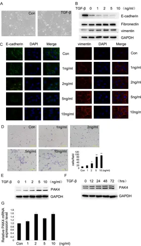
Since TGF-β is recognized as the major factor causing EMT, regular cultivating HK-2 cells Figure 1. TGF-β1 induced morphological changes and EMT in HK-2 Cells
and expression of PAK4 in TGF-β1-treated HK-2 C cells. (A) HK-2 cells were treated with 5 ng/mL TGF-β1 for 72 h. The morphological change was ob
-served with inverted microscope (10 ×). (B) HK-2 cells were treated with 0, 1, 2, 5, 10 ng/ml TGF-β1 for 48 h. Western Blot assay was carried out using anti-E-cadherin, fibronectin, vimentin, GAPDH antibodies. (C) HK-2 cells were treated as (B) and analyzed by immunofluorescence using anti-E-cadherin,
vimentin antibodies followed by Alexa Flour 488 (green) or 594 (red) anti-body and nucleus was stained by DAPI (blue) (60 ×). (D) HK-2 cells were
treated as (B). Effects of different concentration of TGF-β1 on the migration
of cultured HK-2 cells were examined by Transwell migration assays. Results
are representative of three independent experiments. Migrated cells were
plotted as the average number per field of view. *P<0.05, **P<0.01. (E, F)
HK-2 cells were treated with 0, 1, 2, 5, 10 ng/ml TGF-β1 for 48 h (E) and with 5 ng/mL TGF-β1 for 0, 12 h, 24 h, 48 h and 72 h (F). Protein expression
was detected by Western Blot
as-say with anti-PAK4, GAPDH anti
-bodies. (G) The same stimulation
as (B). Real-Time PCR was per-formed to measure the mRNA of
[image:3.612.91.371.71.560.2]Figure 2. PAK4 induced morphological changes and EMT in HK-2 Cells. (A) Empty vector, Flag-PAK4, NC, and shPAK4 were stably expressed in HK-2 cells through lentivirus. (B) Empty vector and Flag-PAK4 stably expressed HK-2 cells
were treated with 5 ng/mL TGF-β1 for 72 hours. Morphological changes were observed by ph- ase contrast microscope. Compared with nor-mal cultured HK-2 cells, TGF-β1-treated HK-2 cells were remodeled, the morphology was found to be spindle-shaped with increased in- tercellular space instead of a polygonal shape (Figure 1A). HK-2 cells were incubated with TGF-β1 (0, 1, 2, 5 and 10 ng/mL) for 48 hours. The results of Western Blot assay showed that TGF-β1 downregulated protein expression of E-cadherin and upregulated protein expression of fibronectin and vimentin in a dose depen-dent manner (Figure 1B). Furthermore, protein expression was detected by immunofluores-cence. Results showed that E-cadherin pre-sented a continuously linear distribution on cell membranes while Vimentin was few in normal HK-2 cells. With increasing concentrations of TGF-β1, protein expression of E-cadherin de- creased while that of Vimentin increased in a dose dependent manner (Figure 1C). Transwell migration assay was performed and our resul- ts revealed that HK-2 cell migration ability was obviously enhanced along with increasing con-centrations of TGF-β1 (Figure 1D). All of these results suggest that TGF-β1 could, indeed, induce EMT in HK-2 cells. Further Western Blot assay showed that with the increase of TGF-β1 concentration and incubating time, protein ex- pression levels of PAK4 gradually increased (Figure 1E, 1F). PAK4 mRNA expression ch- anged without statistical significance, however, after treatment with TGF-β1 (Figure 1G).
PAK4 induced morphological changes and EMT in HK-2 cells
In order to detect the effects of PAK4 on EMT in renal tubular epithelial cells, we successfully constructed stable infected HK-2 cells with overexpressing PAK4 and silencing PAK4 using lentivirus. Western Blot assay detected that Flag-PAK4 protein expression was efficiently upregulated compared to that in Flag vacant vector group. Protein expression of PAK4 in shPAK4 group was significantly lower than in
NC group (Figure 2A). Compared with Flag va- cant vector group, HK-2 cells with overexpress-ing PAK4 appeared as a spindle shape with increased intercellular space (Figure 2B). The results of Western blot and real-time PCR re- vealed that overexpressing PAK4 downregulat-ed protein and mRNA expression of E-cadherin and upregulated protein and mRNA expression of fibronectin and vimentin. However, silencing PAK4 had the exact opposite results (Figure 2C, 2D). Moreover, immunofluorescence assay and Transwell assay were used to further verify effects. Immunofluorescence results revealed that overexpressing PAK4 reduced E-cadherin expression while silencing PAK4 increased E-cadherin expression, remarkably (Figure 2E). Under an inverted microscope, much more cells migrated to the lower layer of microporo- us membrane, showing that overexpressing PAK4 increased cell migration ability and silen- cing PAK4 reduced cell migration ability (Figure 2F).
PAK4 enhanced TGF-β1-induced EMT process in HK-2 cells
In previous results, we found that PAK4 was involved in TGF-β1- induced EMT in HK-2 cells. In the following study, we further explored the role of PAK4 in this process. Stably infected HK-2 cells that overexpress and silence PAK4 were still used. Western blot assay showed that overexpression of PAK4 further reduced pro-tein expression of E-cadherin and increased that of fibronectin and vimentin induced by TGF-β1. Protein expression of E-cadherin in- duced by TGF-β1 was not obviously changed but protein expression of fibronectin and vimen-tin decreased in silencing PAK4 group (Figure
3A). Real-time PCR results revealed that over-expression of PAK4 further reduced mRNA expression of E-cadherin and increased mRNA expression of fibronectin and vimentin induced by TGF-β1. Decreased mRNA expression of E-cadherin and increased expression of fibro-nectin and vimentin induced by TGF-β1 were significantly reversed by silencing PAK4 (Figure
to examine the EMT markers in the Empty vector and Flag-PAK4, or NC and shPAK4 stably expressed HK-2 cells.
*P<0.05, **P<0.01. (E) The Empty vector, Flag-PAK4, NC, and shPAK4 stably expressed HK-2 cells were used to
detected E-cadherin with immunofluorescence by Alexa Flour 488 (green) antibody and nucleus was stained by DAPI (blue) (60 ×). (F) The same cells as (E) were used to observe the migration activity by Transwell migration assays. Results are representative of three independent experiments. Migrated cells were plotted as the average number of
3B). In addition, immunofluorescence assay results presented that silencing PAK4 attenu-ated protein expression of vimentin induced by TGF-β1 (Figure 3C). Alterations of cell migration ability were also tested by Transwell migration assay. Overexpression of PAK4 could increase the number of cells migrating to the lower layer of microporous membranes, illustrating that overexpressing PAK4 could efficiently increase
the migration ability of HK-2 cells (Figure 3D). All of the above data suggests that PAK4 en- hances TGF-β1-induced EMT in HK-2 cells.
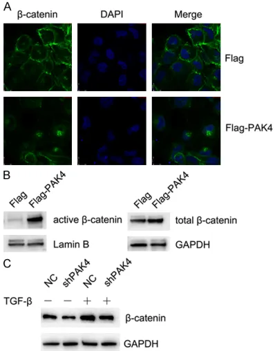
PAK4 enhanced TGF-β1-induced EMT process in HK-2 cells through activating β-catenin signaling pathway
Research has pointed out that activation of Wnt/β-catenin signaling pathways contributes to renal interstitial fibrosis [17]. Therefore, we further investigated whether β-catenin partici-pates in the process of promoting TGF-β1-induced EMT by PAK4. Immunofluorescence assay was used to observe the changes of β- catenin. The images of confocal laser micros-copy showed that overexpressing PAK4 made β-catenin proteins translocate from cell mem-branes into the nucleus in TGF-β1-treated HK-2 cells (Figure 4A). Total protein and nuclear pro-tein of β-catenin in HK-cells were extracted, respectively, using a protein extraction kit. We found that overexpressing PAK4 could remark-ably increase active β-catenin protein expres-sion and total β-catenin protein expresexpres-sion increased a little bit more (Figure 4B). Western Blot results demonstrated that silencing PAK4 caused the reduction of β-catenin in HK-2 cells induced by TGF-β1 (Figure 4C). Thus, we sug-gest that PAK4 enhances TGF-β1-induced EMT process in HK-2 cells through activating β-ca- tenin signaling pathway.
Discussion
[image:7.612.89.288.191.445.2]Renal interstitial fibrosis is a biological proce- ss managed by interaction between different cellular constituents and intricate networks of signaling pathways [18]. The pathogenesis of renal interstitial fibrosis is very complex and remains unclear. EMT in renal tubular epithelial cells plays a crucial role during the process of renal interstitial fibrosis [9-11]. EMT has been identified in tubular epithelial cells of human kidney biopsies with different kidney diseases Figure 3. PAK4 promotes TGF-β1-induced EMT process in HK-2 cells through activating β-catenin signaling pathway. (A) Western Blot assay was carried out to examine the EMT markers in a group of Mock/-, Mock/+, Empty vector/+, Flag-PAK4/+ HK-2 cells and another group of Mock/-, Mock/+, NC/+, shPAK4/+ HK-2 cells. (B) A group of Flag/-, Flag-PAK4/-, Flag/+, Flag-PAK4/+ HK-2 cells and another group of NC/-, shPAK4/-, NC/+, shPAK4/+ HK-2 cells were collected for measuring the EMT markers by Real-time PCR. *P<0.05, **P<0.01, #P<0.05, ##P<0.01. (C)
NC/-, shPAK4/-, NC/+, shPAK4/+ HK-2 cells were used to detected vimentin with immunofluorescence by Alexa Flour 594 (red) antibody and nucleus was stained by DAPI (blue) (60 ×). (D) The same cells as (B) were cultured
to observe the migration activity by Transwell migration assays. Results are representative of three independent
experiments. Migrated cells were plotted as the average number of cells per field of view. *P<0.05, **P<0.01,
#P<0.05, ##P<0.01.
Figure 4. PAK4 promotes the TGF-β1-induced EMT process in HK-2 cells through activating β-catenin signaling pathway. (A) Empty vector and Flag-PAK4 stably expressed HK-2 cells were used to observe the translocation of β-catenin with immunofluores -cence by Alexa Flour 488 (green) antibody and nu-cleus was stained by DAPI (blue) (60 ×). (B) Western
Blot assay was employed to measure the active and
total β-catenin expression separately in Empty vector and Flag-PAK4 stably expressedHK-2 cells. (C) NC/-, shPAK4/-, NC/+, shPAK4/+ HK-2 cells were collect
and has been correlated with the degree of dis-eases [19, 20]. TGF-β is regarded as the princi-ple fibrogenic growth factor involved in the EMT process in a variety of diseases [21-24]. In our study, we confirmed that TGF-β1 could cause morphological changes and enhance migratory capacity of HK-2 cells. TGF-β1 could also induce EMT of HK-2 cells through downregulating E- cadherin expression while upregulating fibro-nectin and vimentin expression in a dose-de- pendent manner. The results of our study are similar to the results of Brockhausen et al. [21].
During the process of TGF-β1-induced EMT, we stumbled upon a situation in which PAK4 over-expressed in TGF-β1-treated HK-2 cells. Cur- iously, PAK4 mRNA expression changed, with-out statistical significance, after treatment with TGF-β1. This implies that regulation of PAK4 expression in TGF-β1-treated renal tubular epi-thelial cells might occur after translation and posttranslational modification. As one of the type II PAKs, PAK4 participates in massive bio-logical processes. PAK4 plays a vital role in tumors, not only in tumorigenesis and progres-sion, but also in invasion and metastasis [25-28]. So far, the effects of PAK4 on renal intersti-tial fibrosis have remained undefined. We detected that PAK4 could bring changes of EMT markers in HK-2 cells. Researches have dem-onstrated that PAK4 could take part in TGF-β causing biological effects in gastric cancer cells [28]. Thus, we speculated that PAK4 might be involved in TGF-β1-induced EMT in renal tubular epithelial cells. Our results revealed that overexpression of PAK4 further decreased E-cadherin protein expression and increased fibronectin and vimentin protein expression induced by TGF-β1 in HK-2 cells. These data suggest that PAK4 enhances the process of TGF-β1-induced EMT in renal tubular epithelial cells. Nonetheless, unnoticeable variance of E-cadherin protein expression induced by TGF-β1 was detected, after silencing PAK4. E-ca- dherin is a transmembrane glycoprotein, con-necting epithelial cells together at adherent junctions, and loss of E-cadherin represents occurrence of EMT. Since TGF-β is regarded as the primary fibrogenic growth factor [29], we suspected that TGF-β was so powerful in reduc-ing E-cadherin that silencreduc-ing PAK4 could not reverse this effect. Therefore, further evidence from future studies is needed to investigate this phenomenon.
TGF-β-induced fibrosis is generally mediated by Smad-dependent and Smad-independent signaling pathways [30-32]. Wnt/β-catenin be- longs to one of the Smad-independent signal-ing pathways. As a principal mediator of can- onical Wnt signaling pathways, β-catenin plays a crucial role in governing organ development, tissue homoeostasis, and the pathologic pro-cess of diverse human disorders [33]. Activa- tion of Wnt/β-catenin signaling pathways con-tributes to renal interstitial fibrosis [34]. We de- tected, in the immunofluorescence assay, that overexpressing PAK4 made the β-catenin pro-tein translocate from cell membranes into the nucleus in TGF-β-treated HK-2 cells. Further- more, results of Western Blot assay revealed that overexpressing PAK4 could remarkably increase active β-catenin protein expression. Once stimulated by upstream mediators, the β-catenin protein, which is free in the cyto-plasm, was stabilized. Stable accumulation of β-catenin translocates into the nucleus and binds to T-cell factor (TCF)/lymphoid enhancer-binding factor (LEF) transcription factor family, then triggers the transcription of downstream target genes (such as c-myc, cyclin D1, etc.) [35]. Previous studies have presented similar results indicating that activation of β-catenin signaling induces tubular EMT and enhances the magnitude of EMT induced by TGF-β1 [36, 37]. Consistently, inhibiting Wnt/β-catenin sig-naling ameliorates kidney injuries and miti-gates renal fibrotic lesions in chronic kidney disease [38]. β-catenin signaling could be a converging effector of several basic fibrotic sig-naling pathways. Therefore, inhibiting β-catenin transcription activity may be a promising meth-od to attenuate renal interstitial fibrosis [17, 37].
In summary, our present study provides evi-dence of a novel role for PAK4 in TGF-β1-induced-EMT and suggests that it is a potential therapeutic target for ameliorating renal inter-stitial fibrosis of CKD.
Acknowledgements
This work was supported by a grant from the National Natural Foundation of China [Grant number: 81601292].
Disclosure of conflict of interest
Address correspondence to: Dr. Hong Jiang, De- partment of Pediatrics, The First Hospital of China Medical University, Shenyang 110001, Liaoning,
China. Tel: +86 13940195519; Fax: +0086 24
83282527; E-mail: jianghong724@163.com
References
[1] Gerson AC, Butler R, Moxey-Mims M, Wentz A,
Shinnar S, Lande MB, Mendley SR, Warady BA, Furth SL and Hooper SR. Neurocognitive
out-comes in children with chronic kidney disease: current findings and contemporary endeavors.
Ment Retard Dev Disabil Res Rev 2006; 12: 208-215.
[2] Kari J. Epidemiology of chronic kidney disease
in children. J Nephropathol 2012; 1: 162-163. [3] Yadav SP, Shah GS, Mishra OP and Baral N.
Pattern of renal diseases in children: a
devel-oping country experience. Saudi J Kidney Dis
Transpl 2016; 27: 371-376.
[4] Ardissino G, Dacco V, Testa S, Bonaudo R, Claris-Appiani A, Taioli E, Marra G, Edefonti A, Sereni F and ItalKid P. Epidemiology of chronic renal failure in children: data from the ItalKid
project. Pediatrics 2003; 111: e382-387. [5] Bielesz B, Sirin Y, Si H, Niranjan T, Gruenwald
A, Ahn S, Kato H, Pullman J, Gessler M, Haase VH and Susztak K. Epithelial notch signaling regulates interstitial fibrosis development in the kidneys of mice and humans. J Clin Invest 2010; 120: 4040-4054.
[6] Liu Y. Cellular and molecular mechanisms of
renal fibrosis. Nat Rev Nephrol 2011; 7:
684-696.
[7] Mong Hiep TT, Ismaili K, Collart F, Van Damme-Lombaerts R, Godefroid N, Ghuysen MS, Van Hoeck K, Raes A, Janssen F and Robert A.
Clinical characteristics and outcomes of
chil-dren with stage 3-5 chronic kidney disease.
Pediatr Nephrol 2010; 25: 935-940.
[8] Kalluri R and Weinberg RA. The basics of epi -thelial-mesenchymal transition. J Clin Invest 2009; 119: 1420-1428.
[9] Bani-Hani AH, Campbell MT, Meldrum DR and
Meldrum KK. Cytokines in epithelial-mesen -chymal transition: a new insight into obstruc-tive nephropathy. J Urol 2008; 180: 461-468. [10] Massague J and Wotton D. Transcriptional
con-trol by the TGF-beta/Smad signaling system. EMBO J 2000; 19: 1745-1754.
[11] Zeisberg M and Kalluri R. The role of epithelial-to-mesenchymal transition in renal fibrosis. J
Mol Med (Berl) 2004; 82: 175-181.
[12] Bottinger EP and Bitzer M. TGF-beta signaling
in renal disease. J Am Soc Nephrol 2002; 13: 2600-2610.
[13] Yamamoto T, Noble NA, Cohen AH, Nast CC,
Hishida A, Gold LI and Border WA. Expression
of transforming growth factor-beta isoforms in
human glomerular diseases. Kidney Int 1996;
49: 461-469.
[14] Kumar R, Sanawar R, Li X and Li F. Structure, biochemistry, and biology of PAK kinases. Gene 2017; 605: 20-31.
[15] Manser E, Leung T, Salihuddin H, Zhao ZS and
Lim L. A brain serine/threonine protein kinase
activated by Cdc42 and Rac1. Nature 1994; 367: 40-46.
[16] Jin R, Liu W, Menezes S, Yue F, Zheng M, Kova -cevic Z and Richardson DR. The metastasis
suppressor NDRG1 modulates the phosphory -lation and nuclear translocation of beta-catenin through mechanisms involving FRAT1
and PAK4. J Cell Sci 2014; 127: 3116-3130.
[17] He W, Dai C, Li Y, Zeng G, Monga SP and Liu Y.
Wnt/beta-catenin signaling promotes renal
in-terstitial fibrosis. J Am Soc Nephrol 2009; 20:
765-776.
[18] Lovisa S, Zeisberg M and Kalluri R. Partial epi -thelial-to-mesenchymal transition and other
new mechanisms of kidney fibrosis. Trends En -docrinol Metab 2016; 27: 681-695.
[19] Murakami K, Takemura T, Hino S and Yoshioka K. Urinary transforming growth factor-beta in
patients with glomerular diseases. Pediatr Nephrol 1997; 11: 334-336.
[20] Sanderson N, Factor V, Nagy P, Kopp J, Kon
-daiah P, Wakefield L, Roberts AB, Sporn MB
and Thorgeirsson SS. Hepatic expression of mature transforming growth factor beta 1 in transgenic mice results in multiple tissue le-sions. Proc Natl Acad Sci U S A 1995; 92: 2572-2576.
[21] Brockhausen J, Tay SS, Grzelak CA, Bertolino P, Bowen DG, d’Avigdor WM, Teoh N, Pok S, Shackel N, Gamble JR, Vadas M and Mc
-Caughan GW. miR-181a mediates
TGF-beta-induced hepatocyte EMT and is dysregulated in cirrhosis and hepatocellular cancer. Liver Int 2015; 35: 240-253.
[22] Katsuno Y, Lamouille S and Derynck R.
TGF-beta signaling and epithelial-mesenchymal
transition in cancer progression. Curr Opin On -col 2013; 25: 76-84.
[23] Li C, Wan L, Liu Z, Xu G, Wang S, Su Z, Zhang Y,
Zhang C, Liu X, Lei Z and Zhang HT. Long
non-coding RNA XIST promotes TGF-beta-induced
epithelial-mesenchymal transition by regulat-ing miR-367/141-ZEB2 axis in non-small-cell lung cancer. Cancer Lett 2018; 418: 185-195. [24] Mamuya FA and Duncan MK. aV integrins and
TGF-beta-induced EMT: a circle of regulation. J
Cell Mol Med 2012; 16: 445-455.
kinase-mediated SCG10 phosphorylation involved in gastric cancer metastasis. Oncogene 2014;
33: 3277-3287.
[26] Li X, Ke Q, Li Y, Liu F, Zhu G and Li F. DGCR6L, a novel PAK4 interaction protein, regulates PAK4-mediated migration of human gastric cancer cell via LIMK1. Int J Biochem Cell Biol
2010; 42: 70-79.
[27] Murray BW, Guo C, Piraino J, Westwick JK, Zhang C, Lamerdin J, Dagostino E, Knighton D, Loi CM, Zager M, Kraynov E, Popoff I, Chris
-tensen JG, Martinez R, Kephart SE, Marakovits J, Karlicek S, Bergqvist S and Smeal T. Small-molecule p21-activated kinase inhibitor
PF-3758309 is a potent inhibitor of oncogenic signaling and tumor growth. Proc Natl Acad Sci U S A 2010; 107: 9446-9451.
[28] Wang C, Li Y, Zhang H, Liu F, Cheng Z, Wang D,
Wang G, Xu H, Zhao Y, Cao L and Li F. Onco
-genic PAK4 regulates Smad2/3 axis involving gastric tumorigenesis. Oncogene 2014; 33:
3473-3484.
[29] Biernacka A, Dobaczewski M and Frangogi
-annis NG. TGF-beta signaling in fibrosis. Growth
Factors 2011; 29: 196-202.
[30] Cai H, Su S, Li Y, Zeng H, Zhu Z, Guo J, Zhu Y, Guo S, Yu L, Qian D, Tang Y and Duan J. Protec -tive effects of salvia miltiorrhiza on adenine-induced chronic renal failure by regulating the
metabolic profiling and modulating the NADPH oxidase/ROS/ERK and TGF-beta/Smad signal -ing pathways. J Ethnopharmacol 2018; 212: 153-165.
[31] Cai T, Sun D, Duan Y, Qiu Y, Dai C, Yang J and
He W. FHL2 promotes tubular epithelial-to-mesenchymal transition through modulating beta-catenin signalling. J Cell Mol Med 2018; 22: 1684-1695.
[32] Zhou XL, Xu P, Chen HH, Zhao Y, Shen J, Jiang C, Jiang S, Ni SZ, Xu B and Li L. Thalidomide
inhibits TGF-beta1-induced epithelial to mes -enchymal transition in alveolar epithelial cells via smad-dependent and smad-independent signaling pathways. Sci Rep 2017; 7: 14727. [33] Clevers H and Nusse R. Wnt/beta-catenin
sig-naling and disease. Cell 2012; 149: 1192-1205.
[34] Tan RJ, Zhou D, Zhou L and Liu Y.
Wnt/beta-catenin signaling and kidney fibrosis. Kidney
Int Suppl (2011) 2014; 4: 84-90.
[35] Rao TP and Kuhl M. An updated overview on
Wnt signaling pathways: a prelude for more. Circ Res 2010; 106: 1798-1806.
[36] Garcia de Herreros A and Baulida J. Coopera
-tion, amplifica-tion, and feed-back in
epithelial-mesenchymal transition. Biochim Biophys Acta 2012; 1825: 223-228.
[37] Hao S, He W, Li Y, Ding H, Hou Y, Nie J, Hou FF,
Kahn M and Liu Y. Targeted inhibition of
beta-catenin/CBP signaling ameliorates renal
inter-stitial fibrosis. J Am Soc Nephrol 2011; 22:
1642-1653.
[38] Xu H, Li Q, Liu J, Zhu J, Li L, Wang Z, Zhang Y, Sun Y, Sun J, Wang R and Yi F. β-Arrestin-1 de
-ficiency ameliorates renal interstitial fibrosis by blocking Wnt1/β-catenin signaling in mice. J