Original Article
Suppression of miR-301a alleviates LPS-induced
inflammatory injury in ATDC5 chondrogenic
cells by targeting Sirt1
Hongwei Chen1*, Jie Qi2*, Qing Bi3, Shimin Zhang4
1Department of Orthopedic Surgery, Yiwu Central Hospital, Affiliated Hospital of Wenzhou Medical University,
Yiwu 322000, Zhejiang, China; 2Department of Orthopedics, Shaanxi Provincial People’s Hospital, Xi’an 710068,
China; 3Department of Orthopedics and Joint Surgery, Zhejiang Provincial People’s Hospital, Hangzhou 310014,
China; 4Department of Orthopedic Surgery, Yangpu Hospital, School of Medicine, Tongji University, Shanghai
200090, China. *Co-first authors.
Received May 9, 2017; Accepted June 20, 2017; Epub August 1, 2017; Published August 15, 2017
Abstract: Osteoarthritis is one of the most common joint diseases and is characterized by joint inflammation. Mi -croRNAs (miRNA) play an important role in osteoarthritis. In this study, we examined the role of miR-301a in lipo-polysaccharide (LPS)-treated murine chondrogenic ATDC5 cells. LPS at 10 µg/mL concentration was used to induce
inflammatory injury in chondrogenic cells. Cell Counting Kit-8 assay was used to measure cell viability and flow cytometry was used to measure cell apoptosis. Effect of miR-301a on concentrations of inflammatory cytokines was measured using ELISA, and on mRNA expressions was measured using qRT-PCR. The miR-301 target was identified
by luciferase reporter assay. Western blot analysis was used to measure the expressions of apoptotic proteins, Sirt1,
and PI3K/AKT and NF-κB pathways proteins. Treatment with LPS decreased cell viability, and increased apoptosis, inflammatory cytokines (IL-1β, IL-6, IL-8, and TNF-α) level, and miR-301a expression. Overexpression of miR-301a aggravated the effects of LPS on the chondrogenic cells and the inflammatory cytokines by negatively regulating Sirt1 expression. Sirt1 was identified as a target of miR-301. Suppression of miR-301a showed the opposite effects. Western blot showed that suppression of miR-301a increased the expression of PI3K/AKT and NF-κB pathways
proteins. Suppression of miR-301a expression alleviated LPS-induced chondrogenic cell injury by upregulating Sirt1
and activating the PI3K/AKT and NF-κB signal pathways.
Keywords: MicroRNA-301a, osteoarthritis, lipopolysaccharide, inflammation, chondrogenic cell injury, Sirt1
Introduction
Osteoarthritis is the most common adult joint disease, involving degeneration of articular car-tilage, bone remodeling and joint inflammation [1, 2]. Degeneration of articular cartilages of joints such as the hip and knee is hallmark of osteoarthritis. These degenerations lead to complete loss of the cartilage surface and exposure of the subchondral bones [3]. Mechanical and enzymatic factors are believed to impair chondrocyte function and damage the matrix [2]. A variety of enzymatic and mechani-cal factors stimulate cartilage cells leading to the secretion of cytokines, cartilage degenera-tion protease, and inflammatory mediators, which in turn cause cartilage degeneration [4]. The clinical manifestations of osteoarthritis include joint pain, tenderness, stiffness, joint
swelling, limitation of motion, and joint defor-mity [2, 5]. Treatments used for osteoarthritis ranges from drugs, orthopedic aids, physiother-apy to surgery [2]. Osteoarthritis is not yet a cur-able disease, and its pathogenesis is yet to be understood fully. Therefore, it is very important to study the underlying molecular mechanism of inflammatory injury of the cartilage cells for better management of osteoarthritis.
and chondrogenic differentiation and prolifera-tion, thereby influencing the catabolism and anabolism of bone and cartilage [9-11]. It has been suggested that miRNAs have important diagnostic and therapeutic potential, and can act as a novel therapeutic option for treating osteoarthritis [12].
MiR-301a, a newly discovered miRNA, is known to be a positive regulator of the immune re- sponse of leukocytes and is therefore associ-ated with inflammation [13]. MiR-301a is also involved in many cellular processes, such as tumor cell proliferation [14]. However, the role of miR-301a in osteoarthritis has not yet been studied. In the present study, we investigated the role of miR-301a in LPS-treated murine chondrogenic ATDC5 cells.
Materials and methods
Cell culture and treatment
The murine chondrogenic ATDC5 cell line was purchased from American Type Culture Co- llection (ATCC, Manassas, VA, USA), and was cultured in DMEM (Nutrient Mixture F-12 [DMEM/F-12], Thermo Scientific, Rockford, IL, USA), supplemented with 2 mM glutamine (Sigma-Aldrich, St. Louis, MO, USA) and 10% (for cell growth) or 2% (for cell maintaining) fetal bovine serum (FBS; HyClone, Logan, UT, USA) at 37°C in a humidified 5% CO2 incubator. Cells with more than 75% confluence were split in 1:2 using 0.25% trypsin (Ameresco, Fram- ingham, MA, USA). Cells were treated with lipo-polysaccharide (LPS) in a series of concentra-tion (1, 5, and 10 µg/mL) for 5 hrs.
Cell Counting Kit-8 (CCK-8) assay
Cell proliferation was assessed by a Cell Co- unting Kit-8 (CCK-8, Dojindo Molecular Tec- hnologies, Gaithersburg, MD). Cells were seed-ed in a 96-well plate, with 5000 cells/well. After stimulation, the CCK-8 solution was ad- ded to the culture medium, and the cultures were incubated for 1 hr at 37°C in humidified 95% air and 5% CO2. Absorbance was mea-sured at 450 nm using a Microplate Reader (Bio-Rad, Hercules, CA).
Apoptosis assay
Flow cytometry analysis was done to identify and quantify the apoptotic cells using Annexin
V-FITC/PI apoptosis detection kit (Beijing Bi- osea Biotechnology, Beijing, China). The cells (1,00,000 cells/well) were seeded in a 6 well-plate. Treated cells were washed twice with cold phosphate buffered saline and re-sus-pended in buffer. The adherent and floating cells were combined and treated according to the manufacturer’s instruction and measured with flow cytometer (Beckman Coulter, USA) to differentiate apoptotic cells (Annexin-V positive and PI-negative) from necrotic cells (Annexin-V and PI-positive).
Enzyme-linked immunosorbent assay (ELISA)
Culture supernatant was collected from 24-well plates and concentrations of inflammatory cytokines were measured by ELISA (R&D Systems, Abingdon, UK) as per the manufac-turer’s protocols and normalized to cell protein concentrations.
Cell transfection
MiR-301a mimic, miR-301a inhibitor (si-miR-301a), si-Sirt1, and negative control (NC) were synthesized by GenePharma Co. (Shanghai, China). Cells were transfected using Lipo- fectamine 3000 reagent (Invitrogen) following the manufacturer’s protocol.
Quantitative reverse transcription polymerase chain reaction (qRT-PCR)
Total RNA was extracted from cells using Trizol reagent (Life Technologies Corporation, Car- lsbad, CA, USA) according to the manufactur-er’s instructions. For the measurement of mRNA expression level of miR-301a, TaqMan MicroRNA Reverse Transcription Kit and Ta- qMan Universal Master Mix II were used; U6 (Applied Biosystems, Foster City, CA, USA) was used as internal control.
Dual luciferase activity assay
Western blot analysis
The proteins used for western blotting were extracted using RIPA lysis buffer (Beyotime Biotechnology, Shanghai, China) supplemented with protease inhibitors (Roche, Guangzhou, China). The proteins were quantified using BCA™ Protein Assay Kit (Pierce, Appleton, WI, USA). Western blot system was established
Results
LPS induces cell injury and increases expres-sion of inflammatory cytokines in chondro-genic cells
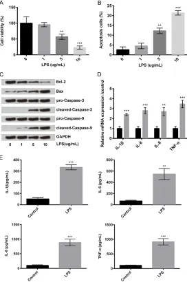
[image:3.612.95.367.74.484.2]To measure the effects of LPS on cell viability and apoptosis in ATDC5 chondrogenic cells, the cells were treated with LPS at different con- Figure 1. LPS induces cell injury and increases expression of inflammatory
cytokines in chondrogenic cells. ATDC5 chondrogenic cells were treated
with LPS (1, 5, or 10 µg/mL). (A) Cell viability. (B) Cell apoptosis. (C) West-ern blot was used to measure the protein expression of Bcl-2, Bax, cleaved caspase-3, and cleaved caspase-9. (D) RT-PCR was used to measure the mRNA expression and (E) ELISA was used to measure the concentration
of IL-1β, IL-6, IL-8, and TNF-α in LPS-treated and control cells. **P<0.01, ***P<0.001. ELISA: enzyme-linked immunosorbent assay; GAPDH: glycer
-aldehyde-3-phosphate dehydrogenase; IL: interleukin; LPS: lipopolysaccha
-ride; RT-PCR: reverse transcription polymerase chain reaction; TNF-α: tumor necrosis factor-α.
using a Bio-Rad Bis-Tris Gel system according to the man-ufacturer’s instructions. GA- PDH antibody was purchased from Sigma. Primary antibod-ies were prepared in 5% blo- cking buffer at adilution of 1:1000. Primary antibody was incubated with the membrane at 4°C overnight, followed by wash and incubation with sec-ondary antibody marked by horseradish peroxidase for 1 hour at room temperature. After rinsing, the polyvinyli-dene difluoride membrane-carried blots and antibodies were transferred into Bio-Rad ChemiDoc™ XRS system, and then 200 μl Immobilon We- stern Chemiluminescent HRP Substrate (Millipore, MA, USA) was added to cover the mem-brane surface. The signals were captured and the inten-sity of the bands was quanti-fied using Image Lab™ So- ftware (Bio-Rad, Shanghai, China).
Statistical analysis
All experiments were repea- ted three times. The results of multiple experiments are presented as mean ± stan-dard error. Statistical analys- es were performed using SP- SS 19.0 statistical software.
centrations (1, 5, and 10 µg/mL) for 5 hrs. Compared to the control group of cells, LPS sig-nificantly decreased cell viability at 5 µg/mL (P<0.01) and 10 µg/mL (P<0.01; Figure 1A); and increased apoptosis at 5 µg/mL (P<0.01) and 10 µg/mL (P<0.001; Figure 1B). Western blot analysis for apoptosis showed that LPS decreased the protein expression of Bcl-2 (anti-apoptotic protein) and increased the expres-sion of Bax, cleaved caspase-3, and cleaved caspase-9 (pro-apoptotic proteins) in chondro-genic cells at 5 µg/mL and 10 µg/mL compared to the control group of cells (Figure 1C). In the further experiments, LPS was used at 5 µg/mL concentration.
As osteoarthritis is associated with synovial inflammation, we measured the effects of LPS on inflammatory cytokines, interleukin (IL)-1β, IL-6, IL-8, and tumor necrosis factor-alpha (TNF-α). The relative mRNA expressions of these pro-inflammatory cytokines were measured by RT-PCR and the concentration levels of these cytokines were measured by ELISA. Compared to the control group of cells, LPS increased the mRNA expressions (Figure 1D) and concen-trations (Figure 1E) of these inflammatory cytokines.
These results indicated that LPS induced cell injury and increased expression of inflammato-ry cytokines in chondrogenic cells.
LPS increases the expression of miR-301a
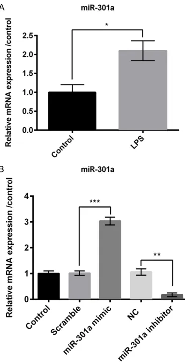
We then measured effects of LPS on expres-sion of miR-301a in chondrogenic cells using RT-PCR. The results showed that LPS signifi-cantly increased the relative mRNA expression of miR-301a in chondrogenic cells compared to the control group (P<0.05; Figure 2A).
We transfected the chondrogenic cells with miR-301a mimic to increase the expression of miR-301a and si-miR-301a to decrease its expression, and measured their efficiency using RT-PCR. As shown in Figure 2B, trans- fection with miR-301a mimic significantly in- creased the mRNA expression of miR-301a (P<0.001) as compared to the scramble; and si-miR-301a significantly decreased the mRNA expression of miR-301a (P<0.01) as compared to the negative control.
Overexpression of miR-301a aggravates LPS-induced cell injury and increases expression of inflammatory cytokines
As LPS increased the expression of miR-301a, we measured the effects of altered expression of miR-301a on cell viability, apoptosis, and inflammatory cytokines. For these tests, chon-drogenic cells were transfected with LPS, LPS+ scramble, LPS+miR-301a mimic, LPS+NC, or LPS+si-miR-301a; untransfected cells served as control. The results obtained from miR-301a mimic group were compared with those of LPS+ scramble, whereas those obtained from si-miR-301a group were compared with those of LPS+NC.
[image:4.612.95.281.71.434.2]Overexpression of miR-301a significantly de- creased cell viability (Figure 3A) and increased Figure 2. LPS increases the expression of miR-301a.
RT-PCR was used to measure the relative mRNA ex-pression of miR-301a in (A) LPS-treated or control cells; and in (B) cells transfected with scramble, miR-301a mimic, NC, or miR-301a inhibitor and
con-trol cells. *P<0.05, **P<0.01, ***P<0.001. LPS:
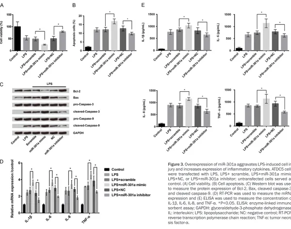
Figure 3. Overexpression of miR-301a aggravates LPS-induced cell
in-jury and increases expression of inflammatory cytokines. ATDC5 cells
were transfected with LPS, LPS+ scramble, LPS+miR-301a mimic, LPS+NC, or LPS+miR-301a inhibitor; untransfected cells served as control. (A) Cell viability. (B) Cell apoptosis. (C) Western blot was used to measure the protein expression of Bcl-2, Bax, cleaved caspase-3, and cleaved caspase-9. (D) RT-PCR was used to measure the mRNA expression and (E) ELISA was used to measure the concentration of
IL-1β, IL-6, IL-8, and TNF-α. *P<0.05. ELISA: enzyme-linked immuno -sorbent assay; GAPDH: glyceraldehyde-3-phosphate dehydrogenase;
IL: interleukin; LPS: lipopolysaccharide; NC: negative control; RT-PCR: reverse transcription polymerase chain reaction; TNF-α: tumor necro
apoptosis (Figure 3B), while suppression of miR-301a showed opposite results (P<0.05 for
all). Western blot analysis for apoptosis showed that overexpression of miR-301a decreased the protein expression of Bcl-2 and increased the expression of Bax, cleaved caspase-3, and cleaved caspase-9 in chondrogenic cells, wh- ereas suppression of miR-301a showed oppo-site results (Figure 3C). Moreover, overexpres-sion of miR-301a significantly increased the mRNA expression (Figure 3D) and concentra-tion (Figure 3E) of the inflammatory cytokines (IL-1β, IL-6, IL-8, and TNF-α), while suppression of miR-301a showed opposite results (P<0.05 for all).
These results indicated that overexpression of miR-301a aggravated LPS-induced cell injury and increases expression of inflammatory cyto-kines, while suppression of miR-301a reversed these effects.
miR-301a negatively regulates Sirt1 expres-sion
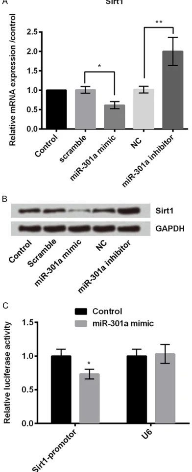
We then measured the effects of miR-301a on Sirt1 expression, which is shown to be involved in cartilage biology [15]. RT-PCR was used to measure the mRNA expression of Sirt1 and western blot was used to measure the protein expression of Sirt1 in chondrogenic cells. For these assays, chondrogenic cells were trans-fected with scramble, miR-301a mimic, NC, or si-miR-301a; untransfected cells served as control. As shown in Figure 4A and 4B, overex-pression of miR-301a significantly decreased the mRNA and protein expression of Sirt1 com-pared to the scramble group (P<0.05), while suppression of miR-301a significantly incre- ased the mRNA and protein expression of Sirt1 as compared to the NC group (P<0.01). We also measured the relative luciferase activity of Sirt1 in chondrogenic cells, and found that miR-301a mimic significantly decreased the lucifer-ase activity of Sirt1 promotor in chondrogenic cells compared to the control (P<0.05; Figure 4C).
These results indicated that miR-301a negat- ively regulated Sirt1, which was a target of miR- 301a.
Suppression of miR-301a reduces cell injury by upregulating Sirt1
As Sirt1 was identified as a target of miR-301a in chondrogenic cells, we measured cell viabili-ty, cell apoptosis, and expressions of inflamma-Figure 4. miR-301a negatively regulates Sirt1. (A)
Quantitative RT-PCR was used to measure relative mRNA expression and (B) western blot was used to measure the protein expression of Sirt1 in ATDC5 cells transfected with scramble, miR-301a mimic, NC, or miR-301a inhibitor; untransfected cells served as control. (C) Relative luciferase activity of Sirt1 was measured in ATDC5 cells transfected with control or
miR-301a mimic. *P<0.05 **P<0.01. NC: negative
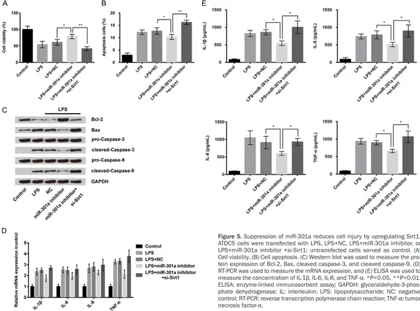
[image:6.612.91.287.78.559.2]Figure 5. Suppression of miR-301a reduces cell injury by upregulating Sirt1. ATDC5 cells were transfected with LPS, LPS+NC, LPS+miR-301a inhibitor, or LPS+miR-301a inhibitor +si-Sirt1; untransfected cells served as control. (A) Cell viability. (B) Cell apoptosis. (C) Western blot was used to measure the pro-tein expression of Bcl-2, Bax, cleaved caspase-3, and cleaved caspase-9. (D) RT-PCR was used to measure the mRNA expression, and (E) ELISA was used to
measure the concentration of IL-1β, IL-6, IL-8, and TNF-α. *P<0.05, **P<0.01. ELISA: enzyme-linked immunosorbent assay; GAPDH: glyceraldehyde-3-phos
tory cytokines in chondrogenic cells with knock-down of miR-301a and Sirt1 expressions. For these tests, chondrogenic cells were transfect-ed with LPS, LPS+NC, LPS+si-miR-301a, or LPS+si-miR-301a+si-Sirt1; untransfected cells served as control.
As discussed earlier, suppression of miR-301a expression increased cell viability, decreased apoptosis, and also decreased the concentra-tions of the inflammatory cytokines (IL-1β, IL-6, IL-8, and TNF-α) in chondrogenic cells
com-pared to the negative control group (P<0.05 for all; Figure 5A-E). As compared to these effect of miR-301a suppression, down-regulation of both miR-301a and Sirt1 significantly decr- eased cell viability (P<0.01; Figure 5A), in- creased apoptosis (P<0.01; Figure 5B), and increased mRNA expression (Figure 5D), and concentration level (P<0.05 for all; Figure 5E) of the inflammatory cytokines in chondrogenic cells. Additionally, western blot analysis for apoptosis showed that down-regulation of both miR-301a and Sirt1 decreased the protein expression of Bcl-2 and increased the expres-sion of Bax, cleaved caspase-3, and cleaved caspase-9 (Figure 5C).
These findings indicated that suppression of miR-301a alleviated cell injury and decreased inflammatory cytokines by increasing the ex- pression of Sirt1.
Suppression of miR-301a alleviated LPS-in-duced cell injury by activation of PI3K/AKT and NF-κB pathways
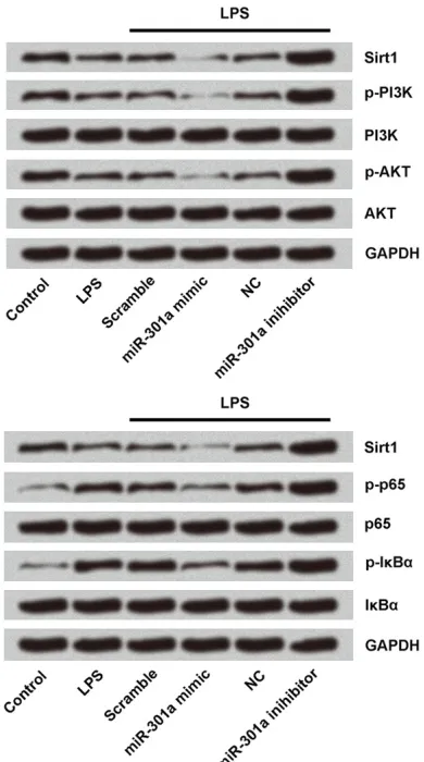
Finally, we explored the mechanism underlying the effects of miR-301a in chondrogenic cells by measuring the expressions of the proteins associated with PI3K/AKT and NF-κB pathways using western blot. For this analysis, chondro-genic cells were transfected with LPS, LPS+ scramble, LPS+miR-301a mimic, LPS+NC, or LPS+si-miR-301a; untransfected cells served as control. As shown in Figure 6, oversion of miR-301a mimic decreased the expres-sions of Sirt1, p-PI3K, p-AKT, p-65, and p-lκBα in chondrogenic cells, while suppression of miR-301a showed opposite results.
These results indicated that suppression of miR-301a alleviates LPS-induced cell injury and decreased inflammation by activation of PI3K/AKT and NF-κB pathways.
Discussion
[image:8.612.91.286.72.422.2]In this study, we investigated the effects of miR-301a in the LPS-induced ATDC5 cell injury and the possible underlying mechanism. First, we induced inflammatory injury to the chondro-genic cells by treating them with LPS, and then measured viability and apoptosis of the injured cells, and expressions of inflammatory cyto-kines (IL-1β, IL-6, IL-8, and TNF-α) and miR-301a in the injured cells. Next, we examined Figure 6. Suppression of miR-301a activates PI3K/
AKT and NF-κB pathways. Western blot analysis was used to measure the protein expressions of Sirt1,
PI3K, AKT, p65, IκBα, and GAPDH in the cells trans -fected with LPS, LPS+ scramble, LPS+miR-301a mimic, LPS+NC, or LPS+miR-301a inhibitor;
untrans-fected cells served as control. AKT: serine-threonine kinase; GAPDH: glyceraldehyde-3-phosphate dehy
-drogenase; IκBα: inhibitory κB proteins alpha; LPS: lipopolysaccharide; NC: negative control; NF-κB: nu-
the effect of miR-301a and Sirt1 on the LPS-injured cells and inflammatory cytokines. La- stly, we studied the mechanism underlying these effects by measuring the expressions of the proteins associated with PI3K/AKT and NF-κB pathways in the injured cells.
Clinical osteoarthritis is characterized by in- flammation of the joints. Chronic inflammation is a key driver of progressive cartilage degen-eration in the joints. Lots of biomechanical fac-tors are involved in chronic inflammation [16]. Pro-inflammatory cytokines-IL-1β, IL-6, IL-8, and TNF-α are biomechanical factors which are involved in the development of chronic inflam-mation. Studies showed that IL-6 and IL-8 lev-els are increased in osteoarthritic serum and synovial fluid [17, 18]. In our study, LPS increased the concentration of these inflamma-tory cytokines in the chondrogenic cells.
In our study, overexpression of miR-301a ag- gravated the LPS-induced cell injury and in- creased the expression of inflammatory cyto-kines (IL-1β, IL-6, IL-8, and TNF-α), while sup-pression of miR-301a reversed these effects. Effects of miR-301a on LPS-injured chondro-genic cells have not been studied yet; however, its effect on inflammatory cytokines has been studied. Overexpression of miR-301a activates NF-κB, which produces IL-8, interferon-β, nitric oxide synthase 2A and cytochrome oxidase subunit 2 [13]. He et al showed that miR-301a promotes intestinal mucosal inflammation th- rough induction of IL-17A and TNF-α in inflam-matory bowel disease [19].
In further experiments, we showed that sup-pression of miR-301a exsup-pression reduces cell injury and decreases the expressions of inflam-matory cytokines by increasing the expression of Sirt1. Sirt1 is a member of the sirtuin family, which plays a wide variety of biological func-tions, including cell development and survival, and inflammation [20]. In human chondrocytes, Sirt1 promotes cell survival, even under pro-inflammatory stress [15]. Therefore, Sirt1 could serve as a novel drug target in treating osteoar-thritis. No study has examined the association between miR-301a and Sirt1, except the pres-ent study. Our findings indicate that miR-301a exerts its effect on the chondrogenic cells by targeting Sirt1.
Lastly, we investigated the molecular mecha-nism underlying the effects of miR-301a by
measuring the expression of PI3K/AKT and NF-κB signal pathway associated proteins. The PI3K/AKT signal pathway is an important path-way for cell survival, proliferation, and migra-tion [21]. The PI3K/AKT pathway is involved in the degradation of extracellular matrix and the death of chondrocytes in osteoarthritis [22]. The nuclear factor-kappaB (NF-κB) proteins are a family of transcription factors that play an important role in most immune and inflamma-tory processes. NF-κB proteins have prominent role in cartilage degradation, cell proliferation, angiogenesis, and pannus formation. Inhibitory κB proteins alpha (IκBα) and p65 belong to the NF-κB family. NF-κB signaling pathways medi-ate important events of inflammatory response by chondrocytes, which led to progressive ex- tracellular matrix damage, and cartilage de- struction [23]. MiR-301a down-regulates the expression of NF-κB pathway proteins in im- mune cells, thereby increasing the production of IL-1β and TNF-α [13]. In our study, suppres-sion of miR-301a increased the expressuppres-sion of PI3K/AKT and NF-κB signal pathway proteins.
In conclusion, suppression of miR-301a expres-sion alleviates LPS-induced chondrogenic cell injury by upregulating Sirt1 and activating the PI3K/AKT and NF-κB signal pathways. Results of the current study indicated that miR-301a could serve as a potential novel therapeutic tar-get for osteoarthritis.
Acknowledgements
This study was supported by Key Science and Technology Program of Yiwu City (No.2009- G3-02).
Disclosure of conflict of interest
None.
Address correspondence to: Hongwei Chen, Depart- ment of Orthopedic Surgery, Yiwu Central Hospital,
Affiliated Hospital of Wenzhou Medical University,
No. 699, Jiangdong Road, Yiwu 322000, Zhejiang, China. E-mail: chenhongwei151@126.com
References
[1] Pelletier JP, Martelpelletier J and Abramson
SB. Osteoarthritis, an inflammatory disease:
[2] Michael JW, Schlüterbrust KU and Eysel P. The
epidemiology, etiology, diagnosis, and
treat-ment of osteoarthritis of the knee. Dtsch Arz -tebl Int 2010; 107: 152-162.
[3] The Deterioration of Articular Cartilage in Os-teoarthritis by Corticosteroid Injections. Jour-nal of Prolotherapy. March 2012. http://jou- rnalofprolotherapy.com/the-deterioration-of- articular-cartilage-in-osteoarthritis-by-cortico-steroid-injections/. Accessed April 15, 2017. [4] Sanchezadams J, Leddy HA, Mcnulty AL,
O’Conor CJ and Guilak F. The mechanobiology
of articular cartilage: bearing the burden of osteoarthritis. Curr Rheumatol Rep 2014; 16: 451.
[5] Sharma L. Osteoarthritis year in review 2015: clinical. Osteoarthritis Cartilage 2016; 24: 36-48.
[6] Slezakprochazka I, Durmus S, Kroesen BJ and
van den Berg A. MicroRNAs, macrocontrol: regulation of miRNA processing. RNA 2010; 16: 1087-1095.
[7] Bartel DP. MicroRNAs: genomics, biogenesis, mechanism, and function. Cell 2004; 116: 281-297.
[8] Bhalala OG, Srikanth M and Kessler JA. The
emerging roles of microRNAs in CNS injuries. Nat Rev Neurol 2013; 9: 328-339.
[9] Xu B, Li YY, Ma J and Pei FX. Roles of microRNA
and signaling pathway in osteoarthritis patho-genesis. J Zhejiang Univ Sci B 2016; 17: 200-208.
[10] Sondag GR and Haqqi TM. The role of MicroR-NAs and their targets in osteoarthritis. Curr Rheumatol Rep 2016; 18: 56.
[11] Yu XM, Meng HY, Yuan XL, Wang Y, Guo QY, Peng J, Wang AY and Lu SB. MicroRNAs’ in-volvement in osteoarthritis and the prospects for treatments. Evid Based Complement Alter-nat Med 2015; 2015: 236179.
[12] Yu C, Chen WP and Wang XH. MicroRNA in os-teoarthritis. J Int Med Res 2011; 39: 1-9. [13] Huang L, Liu Y, Wang L, Chen R, Ge W, Lin Z,
Zhang Y, Liu S, Shan Y and Lin Q.
Down-regula-tion of miR-301a suppresses pro-inflammatory cytokines in toll-like receptor-triggered macro -phages. Immunology 2013; 140: 314-322.
[14] Ni Z, Shang XF, Wang YF, Sun YJ and Fu DJ. Up -regulated microRNA-301a in osteosarcoma promotes tumor progression by targeting CD-C14A. Genet Mol Res 2016; 15.
[15] Dvir-Ginzberg M and Steinmeyer J. Towards elucidating the role of SirT1 in osteoarthritis.
Front Biosci (Landmark Ed) 2013; 18:
343-355.
[16] Sokolove J and Lepus CM. Role of inflamma -tion in the pathogenesis of osteoarthritis:
lat-est findings and interpretations. Ther Adv Mus
-culoskelet Dis 2013; 5: 77-94.
[17] Kaneko S, Satoh T, Chiba J, Ju C, Inoue K and Kagawa J. Interleukin-6 and interleukin-8 lev
-els in serum and synovial fluid of patients with osteoarthritis. Cytokines Cell Mol Ther 2009;
6: 71-79.
[18] Attur MG, Patel IR, Patel RN, Abramson SB and Amin AR. Autocrine production of IL-1 beta by human osteoarthritis-affected cartilage and differential regulation of endogenous nitric ox-ide, IL-6, prostaglandin E2, and IL-8. Proc As-soc Am Physicians 1998; 110: 65-72.
[19] He C, Shi Y, Wu R, Sun M, Fang L, Wu W, Liu C,
Tang M, Li Z and Wang P. miR-301a promotes
intestinal mucosal inflammation through in
-duction of IL-17A and TNF-α in IBD. Gut 2016;
65: 1938-1950.
[20] Rahman S and Islam R. Mammalian Sirt1: in-sights on its biological functions. Cell Commun Signal 2011; 9: 11.
[21] Chen CH, Sung CS, Huang SY, Feng CW, Hung HC, Yang SN, Chen NF, Tai MH, Wen ZH and Chen WF. The role of the PI3K/Akt/mTOR path -way in glial scar formation following spinal cord injury. Exp Neurol 2016; 278: 27-41.
[22] Chen J, Crawford R and Xiao Y. Vertical
inhibi-tion of the PI3K/Akt/mTOR pathway for the
treatment of osteoarthritis. J Cell Biochem 2013; 114: 245-249.
[23] Roman-Blas JA, Jimenez SA and Jimenez SA.