Original Article
Emodin inhibits the proliferation and invasion
of bladder cancer cells via down-regulating Notch1
Libin Ma1*, Kean Chen3*, Kang Jiang2*, Gang Deng2, Peiwu Jiang2, Jia Shao2, Zhijian Yu2
1Department of Nephrology, Sir Run Run Shaw Hospital, Zhejiang University School of Medicine, Hangzhou, PR
China; 2Department of Urology, Hangzhou First People’s Hospital, Nanjing Medical University, Hangzhou, Zhejiang,
PR China; 3Hangzhou First People’s Hospital, Zhejiang Chinese Medical University, Hangzhou, Zhejiang, PR China. *Equal contributors.
Received June 6, 2017; Accepted July 20, 2017; Epub September 1, 2017; Published September 15, 2017
Abstract: Emodin exhibits anti-proliferative effects in numerous cancer cell lines via different mechanisms. The present study aimed to study the effects and underlying molecular mechanisms of emodin on bladder cancer cells. We treated two bladder cancer cell lines, T24 and 5637, with different concentrations of emodin (20, 40 and 80 µmol/L) or DMSO (Control). We analyzed the biological effects of emodin on cell growth, invasion, as well as the mRNA and protein expression of Notch1, Jagged1, VEGF, VEGFR2 and MMP2 using Cell Counting Kit-8, transwell, reverse transcription-quantitative PCR and western blot assays. Emodin repressed cell growth, invasion and Notch1 expression in a concentration-dependent fashion. And Notch1 over-expression assay showed that the anti-prolifera-tive and anti-invasive roles of emodin, along with the down-regulated effects on the expression of Notch1, Jagged1, VEGF, VEGFR2 and MMP2 were partially rescued by Notch1 over-expression. In conclusion, Emodin might suppress the progression of bladder cancer via inhibiting the expression of Notch1.
Keywords: Emodin, Notch1, bladder cancer, proliferation, invasion
Introduction
Bladder cancer is the second most common malignancy of the urogenital system. Transi- tional cell carcinoma (TCC), also known as uro-thelial carcinoma, begins from abnormalities of the urothelial cells lining of the bladder. TCC accounts for the vast majority of bladder can-cer [1, 2]. The majority of bladder cancan-cer pa- tients present with superficial tumors, while 20% to 40% of patients either present with or develop invasive disease. Transurethral resec-tion of bladder tumor and immunotherapy are often applied to treat and prevent superficial tumor recurrence. And for muscle invasive dis-ease, the radical ablative surgery and a combi-nation of radiation and chemotherapy have been identified as effective clinical treatmen- ts [3]. The recovery rate of bladder cancer depends on the depth of the tumor invading into the bladder wall. Whereas, in order to improve the overall survival of invasive bladder cancer, new treatment options are urgently needed.
mitochondri-Effects of emodin on bladder cancer cells
al signaling pathway [14]. Although emodin shows its inhibitory action on many cancer cells through different mechanisms, its effects on bladder cancer are still not clear.
In this study, we analyzed the biological effects of emodin on the proliferation and invasion of two bladder cancer cell lines, T24 and 5637. We also examined the relative protein expres-sion which might be regulated by emodin in both bladder cancer cell lines. Taken together, the present study proved that emodin repressed the proliferation and invasion of bladder cancer cells through down-regulating the expression of Notch1.
Materials and methods
Cell culture
The bladder cancer cells, T24 and 5637, were purchased from American Type Culture Coll- ection (ATCC). Cells were cultured in Dulbecco’s Modified Eagle’s Medium (DMEM) supplement-ed with 10% fetal bovine serum (FBS, Gibico) plus 1% antibiotics (100 U/ml penicillin and 100 µg/ml streptomycin), and maintained at 37°C in a 5% CO2 incubator.
Concentration screening
T24 and 5637 cells were cultured in DMEM medium added with 0 µmol/L (DMSO), 20 µmol/L, 40 µmol/L and 80 µmol/L emodin, re- spectively. Cell Counting Kit-8 (CCK-8), tran-swell, reverse transcription-quantitative PCR (qRT-PCR) and western blot assays were per-formed to select the appropriate concentration of emodin.
Notch1 over-expression
Full-length human Notch1 cDNA was synthe-sized and cloned into pCDNA3.1(+) (Addgene). pCDNA3.1(+) was served as negative control. The constructs were transfected into bladder cancer cells using Lipofectamine 2000 (In- vitrogen) per the manufacture’s introductions.
Over-expression assay
The bladder cancer cells were divided into five groups. And the experimental design was as following: 1. Control group: T24 and 5637 cells without any treatments; 2. Negative control (NC) group: T24 and 5637 cells were
transfect-ed with control vector and culturtransfect-ed in DMEM medium added with DMSO; 3. Notch1 group: T24 and 5637 cells were transfected with full-length cDNA of Notch1 and cultured in DMEM medium added with DMSO; 4. NC + Emodin group: T24 and 5637 cells were transfected with control vector and cultured in DMEM medi-um added with 80 µmol/L emodin; 5. Notch1 + Emodin group: T24 and 5637 cells were trans-fected with full-length cDNA of Notch1 and cul-tured in DMEM medium added with 80 µmol/L emodin.
CCK-8 assay
CCK-8 assay was used to analyze the prolifera-tion of bladder cancer cells. T24 and 5637 cells were seeded in 96-well plates and cell growth was measured with commercial CCK-8 Assay Kit (Sigma) at 0 h, 24 h, 48 h and 72 h. Ab- sorbance excited at 450 nm of reacted cells was examined to valuate cell proliferation.
Transwell assay
Transwell assay was performed to evaluate the invasiveness of bladder cancer cells. T24 and 5637 cells were serum starved for 24 h after medication or transfection. Then, the cells were seeded into upper chamber of a 24-well tran-swell chamber (Trueline) which was coated with 50 µl Matrigel (1:2 dilution, BD Biosciences) containing a polycarbonate filter. The lower chamber was filled with 0.75 ml DMEM culture medium. After 24 h of incubation, the non-migrated cells were scraped from the upper chamber, and the adherent cells were stained with crystal violet and photographed.
Reverse transcription-quantitative PCR (qRT-PCR)
sequences were as follow: Notch1 (NM_017- 617.3): Primer F 5’ GACGCACAAGGTGTCTTC 3’, Primer R 5’ TTGCCCAGGTCATCTACG 3’; VEGF (NM_001025366.2), Primer F 5’ ATTTCTGG- GATTCCTGTAG 3’, Primer R 5’ CAGTGAAGACA- CCAATAAC 3’; VEGFR2 (NM_002253.2), Primer F 5’ CTCAGCAGGATGGCAAAG 3’, Primer R 5’ ACTGTCCGTCTGGTTGTC 3’; Jagged1 (NM_00- 0214.2), Primer F 5’ CTTCACGGGAACATACTG 3’, Primer R 5’ GCACTTGTAGGAGTTGAC 3’; MMP-2 (NM_004530.4), Primer F 5’ TTGA- CGGTAAGGACGGACTC 3’, Primer R 5’ GGCGTT- CCCATACTTCACAC 3’; GAPDH (NM_001256- 799.1), Primer F 5’ CACCCACTCCTCCACCTTTG 3’, Primer R 5’ CCACCACCCTGTTGCTGTAG 3’.
Western blot assay
At 48 h post treatments, bladder cancer cells were harvested and subjected to SDS-PAGE. After transferred to a nitrocellulose membrane, the membranes were blocked with 5% skim milk and then incubated with primary antibod-ies. After incubation with secondary antibod- ies, the blots were visualized by the enhanced chemiluminescence system. The antibody list was as follows: Notch1 (1:1000, #3608, Cell Signaling Technology), VEGF (1:1000, Ab46154,
Abcam), VEGFR2 (1:1000, AF6281, Affinity), Jagged1 (1:1000, Ab109536, Abcam), MMP2 (1:1000, Ab92536, Abcam), GAPDH (1:2000, #5174, Cell Signaling Technology).
Statistical analysis
Data analysis was performed by GraphPad Prism software (San Diego, CA). Values were presented as means ± SD. Statistical signifi-cance was determined by two-tailed Student’s t-test. P<0.05 was considered to be statistically significant.
Results
Emodin inhibited proliferation and invasion of bladder cancer cells concentration depend-ently
[image:3.612.93.517.72.325.2]To evaluate the effects of emodin on cell prolif-eration and invasion of bladder cancer cells, CCK-8 and transwell assays were conducted in T24 and 5637 cells respectively. As shown in Figure 1A and 1B, optical density (OD) value was measured at 0 h, 24 h, 48 h and 72 h after the addition of control medium and different concentrations of emodin. Emodin showed a
Effects of emodin on bladder cancer cells
remarkable concentration-dependent inhibito-ry effects on the proliferation of both bladder cancer cells. Then we also estimated the inva-sion cell number of two bladder cancer cells at 48 h post medication. As expected, cell inva-sive capacity was also inhibited by emodin in a concentration-dependent fashion (Figure 1C and 1D). These data suggested that emodin treatment (20-80 µmol/L) significantly sup-pressed the proliferation and invasion of blad-der cancer cells in a concentration-dependent manner.
Emodin inhibited the expression of Notch1 in bladder cancer cells
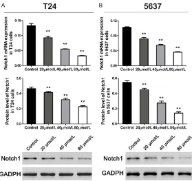
We examined the mRNA expression and protein levels of Notch1 in T24 and 5637 cells using qRT-PCR and western blot at 48 h after differ-ent concdiffer-entrations of emodin medication. As shown in Figure 2, the expression of Notch1 was significantly depressed by emodin addition in both T24 and 5637 cells. And the inhibitory effect of emodin on Notch1 expression was concentration dependent. Therefore, we select-ed the dose of 80 µmol/L to perform further assays.
ing the expression of Notch1 in bladder cancer cells.
Emodin reduced the expression of related pro-teins via inhibiting Notch1 in bladder cancer cells
To further confirm the biological effects of emo-din medication, we then examined the endoge-nous expression of four proteins, which were involved in proliferation and invasion process-es, using qRT-PCR and western blot at 48 h post cDNA transfected and emodin addition. As shown in Figure 4, the mRNA and protein levels of Jagged1, VEGF, VEGFR2 and MMP2 were obviously inhibited after treated with emodin, and such effects were partially rescued in Notch1 over-expression groups. Taken togeth-er, these results indicated that emodin might inhibit cell growth and invasion relating pro-teins via suppressing Notch1 in bladder cancer cells.
Discussion
[image:4.612.92.371.70.333.2]Except of the anti-proliferative effect of emo-din, many studies have focused on the invasion
Figure 2. Emodin inhibited Notch1 expression concentration-dependently in bladder cancer cells. A and B: mRNA and protein expression of Notch1 in T24 and 5637 cell lines (n=3). Data were shown as mean ± S.D., *P<0.05, **P<0.01, ***P<0.001.
Emodin affected cell prolifera-tion and invasion via regulat-ing Notch1 in bladder cancer cells
Effects of emodin on bladder cancer cells
inhibitory effect of emodin on human cancer cells, and different mechanisms have been pro-posed. In squamous cell carcinoma and breast cancer cells, emodin is proved inhibiting cell invasion via suppressing AP-1 and nuclear factor-κB (NF-κB) signaling pathways [15]. Emodin also inhibits CXCL12-induced migration and invasion of prostate and lung cancer cells by blocking the expression of CXC chemokine receptor-4 [16]. Aloe-emodin, a structurally similar compound of emodin, has been report-ed to inhibit invasion in nasopharyngeal carci-noma cell by the down-regulation of MMP-2 through the p38-NF-κB-dependent pathway [17]. In bladder cancer cells, aloe-emodin has also been suggested inhibiting proliferation via activating the p53 dependent apoptotic path-way [18].
In the current study, we found that the prolifera-tion and invasion of bladder cancer cells was dose-dependently inhibited by emodin expo-sure. Our previous study has showed that
emo-din inhibits the growth of prostate cancer cells via the Notch signaling pathway [12]. Notch sig-naling pathway, a highly conserved cell signal-ing system, is actively involved in embryonic development, cell differentiation, cell-cell com-munication, cell proliferation and apoptosis [19]. Aberrantly activated Notch signaling con-tributes to the tumorigenesis of a variety of human cancers [20]. Notch inhibitors, especial-ly γ-secretase inhibitors, have been regarded as targeted therapeutic agents [21, 22]. Here, we found the inhibitory effect of emodin on Notch1 expression. And over-expression assay showed that Notch1 overexpression partially rescued the inhibitory effects of emodin on bladder cancer cell proliferation and invasion. Therefore, the related data suggested that emodin might inhibit the growth and invasive capacity of bladder cancer cell through down-regulating Notch1.
[image:6.612.97.520.127.375.2]We then examined the relative protein expres-sion in bladder cancer cells, containing VEGF, Cell proliferation was inhibited in emodin medication groups (n=3). E: Cell invasion was significantly depressed in emodin medication groups (n=3). Data were shown as mean ± S.D., *P<0.05, **P<0.01 (compared with negative controls); #P<0.05, ##P<0.01 (compared with Notch1 groups).
VEGFR2, Jagged1 and MMP-2. VEGF and VE- GFR2 are identified as metastasis-related ge- nes. A previous study suggested that VEGF expression was an independent prognostic fac-tor of recurrence and metastasis of bladder cancer [23]. Down-regulation of Jagged1, a ligand of Notch1, obviously inhibits cell inva-sion and migration in prostate cancer cells [24, 25]. MMP-2 is also involved in the metastasis of human cancers, including breast, gastric and bladder cancer [26-28]. In this study, the data showed that over-expressed Notch1 signifi-cantly up-regulated the expression of these four proteins. And the down-regulation of No- tch1 induced by emodin medication caused the inhibition of VEGF, VEGFR2, Jagged1 and MMP-2 expression in bladder cancer cells. These results indicated that emodin inhibited cell invasion by related proteins through down-regulating Notch1.
Summarily, our study revealed that emodin inhibited cell proliferation and invasion of blad-der cancer cells in a concentration-dependent way via the down-regulation of Notch1 expres-sion. And the proteins related to cell invasion were also inhibited by emodin through regulat-ing the expression of Notch1.
Acknowledgements
This study was supported by Natural Science Foundation of Zhejiang province (Y2111329 and LY17H050002), Science and Technology Plan Projects of Zhejiang Province (2014C37- 016), Chinese Medicine Science and Technolo- gy Plan Projects of Zhejiang Province (2016- ZB099, 2013ZA107 and 2011ZB099), Medi- cine and health science and technology plan projects of Zhejiang Province (2011KYB066 and 2015KYB295) and Science and Technology Plan Projects of Hangzhou Zhejiang Province (2017A05, 20110833B05 and 20110733Q12). Disclosure of conflict of interest
None.
Address correspondence to: Gang Deng, Depart- ment of Urology, Hangzhou First People’s Hospital, Nanjing Medical University, 261 Huansha Rd., Hangzhou 310006, Zhejiang, PR China. Tel: +86-571-56006932; Fax: +86-571-87914773; E-mail: denggang32@yeah.net
References
[1] Heney NM, Ahmed S, Flanagan MJ, Frable W, Corder MP, Hafermann MD and Hawkins IR. Superficial bladder cancer: progression and recurrence. J Urol 1983; 130: 1083-1086. [2] Vale CL. Adjuvant chemotherapy in invasive
bladder cancer: a systematic review and meta-analysis of individual patient data Advanced Bladder Cancer (ABC) meta-analysis collabora-tion. Eur Urol 2005; 48: 189.
[3] Stein JP, Lieskovsky G, Cote R, Groshen S, Feng AC, Boyd S, Skinner E, Bochner B, Thangathu-rai D and Mikhail M. Radical cystectomy in the treatment of invasive bladder cancer: long-term results in 1,054 patients. J Clin Oncol 2001; 19: 666-675.
[4] Tsai TH. Analytical approaches for traditional chinese medicines exhibiting antineoplastic activity. J Chromatogr B Biomed Sci Appl 2001; 764: 27.
[5] Efferth T, Li PC, Konkimalla VS, Kaina B. From traditional Chinese medicine to rational cancer therapy. Trends Mol Med 2007; 13: 353-361. [6] Wang H, Dong Y and Xiu ZL.
Microwave-assist-ed aqueous two-phase extraction of piceid, resveratrol and emodin from Polygonum cuspi-datum by ethanol/ammonium sulphate sys-tems. Biotechnol Lett 2008; 30: 2079-84. [7] Chang CH, Lin CC, Yang JJ, Namba T and
Hat-tori M. Anti-inflammatory effects of emodin from ventilago leiocarpa. Am J Chin Med 2012; 24: 139-142.
[8] Jayasuriya H, Koonchanok NM, Geahlen RL, Mclaughlin JL and Chang CJ. Emodin, a protein tyrosine kinase inhibitor from Polygonum cus-pidatum. J Nat Prod 1992; 55: 696-8. [9] Zhang L and Hung MC. Sensitization of HER-2/
neu-overexpressing non-small cell lung cancer cells to chemotherapeutic drugs by tyrosine kinase inhibitor emodin. Oncogene 1996; 12: 571-576.
[10] Zhang L, Lau YK, Xia W, Hortobagyi GN and Hung MC. Tyrosine kinase inhibitor emodin suppresses growth of HER-2/neu-overexpress-ing breast cancer cells in athymic mice and sensitizes these cells to the inhibitory effect of paclitaxel. Clin Cancer Res 1999; 5: 343-353. [11] Cha TL, Qiu L, Chen CT, Wen Y and Hung MC.
Emodin down-regulates androgen receptor and inhibits prostate cancer cell growth. Can-cer Res 2005; 65: 2287-2295.
[12] Deng G, Ju X, Meng Q, Yu ZJ and Ma LB. Emo-din inhibits the proliferation of PC3 prostate cancer cells in vitro via the Notch signaling pathway. Mol Med Rep 2015; 12: 4427-4433. [13] Srinivas G, Anto RJ, Srinivas P, Vidhyalakshmi
Effects of emodin on bladder cancer cells
lls through poly(ADP-ribose) polymerase cle- avage and activation of caspase-9. Eur J Phar-macol 2003; 473: 117-125.
[14] Su YT, Chang HL, Shyue SK and Hsu SL. Emo-din induces apoptosis in human lung adeno-carcinoma cells through a reactive oxygen species-dependent mitochondrial signaling pathway. Biochem Pharmacol 2005; 70: 229-241.
[15] Huang Q, Shen HM and Ong CN. Inhibitory ef-fect of emodin on tumor invasion through sup-pression of activator protein-1 and nuclear factor-kappaB. Biochem Pharmacol 2004; 68: 361.
[16] Ok S, Kim SM, Kim C, Nam D, Shim BS, Kim SH, Ahn KS, Choi SH and Ahn KS. Emodin in-hibits invasion and migration of prostate and lung cancer cells by downregulating the ex-pression of chemokine receptor CXCR4. Immu-nopharmacol Immunotoxicol 2012; 34: 768-778.
[17] Lin ML, Lu YC, Chung JG, Wang SG, Lin HT, Kang SE, Tang CH, Ko JL and Chen SS. Down-regulation of MMP-2 through the p38 MAPK-NF-κB-dependent pathway by aloe-emodin le- ads to inhibition of nasopharyngeal carcino- ma cell invasion. Mol Carcinog 2010; 49: 783-797.
[18] Lin JG, Chen GW, Li TM, Chouh ST, Tan TW and Chung JG. Aloe-emodin induces apoptosis in T24 human bladder cancer cells through the p53 dependent apoptotic pathway. J Urol 2006; 175: 343-347.
[19] Zhou ZD, Kumari U, Xiao ZC, Tan EK. Notch as a molecular switch in neural stem cells. IUBMB Life 2010; 62: 618-623.
[20] Bolós V, Grego-Bessa J and de la Pompa JL. Notch signaling in development and cancer. Endocr Rev 2007; 28: 339-363.
[21] Purow B. Notch inhibition as a promising new approach to cancer therapy. In: editors. Notch signaling in embryology and cancer. Springer; 2012. pp. 305-319.
[22] Shih IM and Wang TL. Notch signaling, γ-sec- retase inhibitors, and cancer therapy. Cancer Res 2007; 67: 1879-1882.
[23] Inoue K, Slaton JW, Karashima T, Yoshikawa C, Shuin T, Sweeney P, Millikan R and Dinney CP. The prognostic value of angiogenesis factor ex-pression for predicting recurrence and metas-tasis of bladder cancer after neoadjuvant che-motherapy and radical cystectomy. Clin Cancer Res 2000; 6: 4866-4873.
[24] Zhang Y, Wang Z, Ahmed F, Banerjee S, Li Y, Sarkar FH. Down-regulation of Jagged-1 induc-es cell growth inhibition and S phase arrinduc-est in prostate cancer cells. Int J Cancer 2006; 119: 2071-2077.
[25] Wang Z, Li Y, Banerjee S, Kong D, Ahmad A, Nogueira V, Hay N and Sarkar FH. Down-regu-lation of Notch-1 and Jagged-1 inhibits pros-tate cancer cell growth, migration and inva-sion, and induces apoptosis via inactivation of Akt, mTOR, and NF-κB signaling pathways. J Cell Biochem 2010; 109: 726-736.
[26] Jinga D, Blidaru A, Condrea I, Ardeleanu C, Dragomir C, Szegli G, Stefanescu M and Ma-tache C. MMP-9 and MMP-2 gelatinases and TIMP-1 and TIMP-2 inhibitors in breast cancer: correlations with prognostic factors. J Cell Mol Med 2006; 10: 499-510.
[27] Hwang TL, Lee LY, Wang CC, Liang Y, Huang SF and Wu CM. Claudin-4 expression is associat-ed with tumor invasion, MMP-2 and MMP-9 expression in gastric cancer. Exp Ther Med 2010; 1: 789-97.