Original Article
Transcriptomic analysis of stromal cells
from patients with endometrial carcinoma
Min Li1*, Xiao-Yan Xin2*, Zhi-Shan Jin2, Teng Hua2, Hong-Bin Wang3, Hong-Bo Wang2
1Department of Obstetrics and Gynecology, Xuhui Hospital, Zhongshan Hospital, Fudan University, Shanghai,
China; 2Department of Obstetrics and Gynecology, Union Hospital, Tongji Medical College, Huazhong University
of Science and Technology, Wuhan, China; 3South Branch of The Six People’s Hospital Affiliated to Shanghai Jiao
Tong University, Shanghai, China. *Co-first authors.
Received November 8, 2016; Accepted August 2, 2017; Epub September 1, 2017; Published September 15, 2017
Abstract: Tumor microenvironment plays a critical role in cancer pathogenesis. In this study, we performed tran-scriptomic analysis of stromal cells from patients diagnosed with endometrial carcinoma. Endometrial stromal cells from patients and healthy donors were cultured and total RNA was extracted for RNA integrity examination and gene profiling analysis. Gene ontology (GO) and KEGG analysis were also performed. In this study, we found that, in endo-metrial stromal cells from endoendo-metrial cancer patients, a total of 605 genes were changed (fold change ≥2, p-value <0.05). From these, 275 were up-regulated and 330 were down-regulated genes. In addition, GO analysis showed that the differentially expressed genes (DEGs) were involved in various biological processes including cell adhesion, biological adhesion, bone development and extracellular matrix organization. Furthermore, KEGG analysis of the DEGs identified four pathways including Wnt signaling pathway, cadherin signaling pathway, ECM-receptor interac-tion, and focal adhesion. Our study identified 605 DEGs in stromal cells from endometrial carcinoma which mapped to a variety of biological processes. These results may contribute to understanding the molecular mechanisms o of endometrial carcinoma pathogenesis.
Keywords: Endometrial carcinoma, gene expression profiling, stromal cells, microenvironment
Introduction
Endometrial carcinoma is one of the worldwide leading gynecological malignancies in women. Typical clinical symptoms observed include dys-pareunia, dysmenorrhea, and infertility [1, 2]. In 2013, 49500 new cases and 8200 deaths caused by endometrial cancer were reported in the United States [3]. To date, surgical resec-tion and hormonal suppressant drugs are the primary therapeutic options for endometrial cancer [4]. To date, little is known about
endo-metrial cancer pathogenesis, therefore identifi -cation of altered genes may contribute to understanding the molecular events in endo-metrial cancer.
It is well established that cancer is a systemic and multiple-step disease, encompassing inter-action between tumor cells and host stromal cells [5, 6]. Increasing evidence demonstrates
that tumors are composed of tumor parenchy-ma and stroparenchy-ma, two discrete but closely inter-active parts, and that their crosstalk promotes tumor growth [7, 8]. Recently, many studies support the notion that tumor stromal cells play critical roles in malignant behaviors, such as cell growth, invasion, differentiation, and metastasis [9, 10]. In the current study, we
examined the gene expression profile of stro -mal cells isolated from endometrial carcinoma patients.
Materials and methods
Endometrial tissue isolation
RNA extraction and purification
Total RNA was extracted using TRIZOL Reagent
(Life technologies, Carlsbad, CA, USA) following the manufacturer’s instructions. RNA quality was determined via Agilent Bioanalyzer 2100 (Agilent technology, Santa Clara, CA, USA). Total
RNA was further purified by RNeasy microkit (QIAGEN, GmBH, Germany) and RNase Free DNase Set (QIAGEN, GmBH, Germany).
RNA amplification and labeling
Total RNA was amplified, labeled and purified via GeneChip 3’IVT Express Kit (Affymetrix,
Santa Clara, CA, USA) following the manufac-turer’s instructions to obtain biotin labeled cRNA.
CA, US) and Command Console Software 3.1 (Affymetrix, Santa Clara, CA, US) with default
settings. The enrichment analysis of GO and KEGG pathway annotations were conducted
based on a hypergeometric test.
Results
Transcriptional profiles in stromal cells from endometrial carcinoma patients
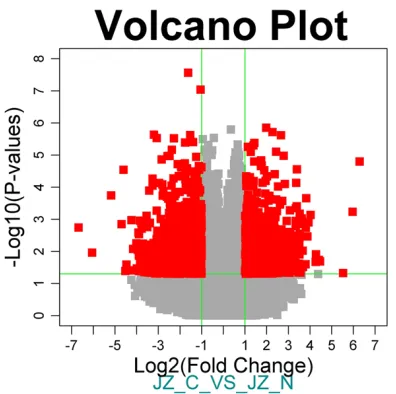
[image:2.612.90.334.86.189.2] [image:2.612.92.290.241.438.2]As previously described, stromal cells were characterized with cytokeratin (epithelial mark-er) and vimentin (stromal markmark-er) [11]. Total RNA was extracted f from healthy donors stro-ma cells and endometrial carcinostro-ma patients, and further subjected to microarray analysis using Affymetrix U133 plus 2.0 array. The con-centrations and A260/280 values of RNA pre-pared for microarray analysis are shown in
Table 1. We found that stromal cells from
endo-metrial cancer patients exhibited significant
variety in gene expression levels when com-pared to healthy donors. As shown in Figures 1
and 2, a total of 605 genes were changed (fold
change ≥2, p-value <0.05). Changes included 275 up-regulated genes and 330 down-regulat-ed genes.
Functional categories of differentially ex-pressed genes
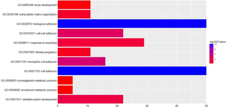
The differentially expressed genes (DEGs) were further analyzed with GO ontology analysis to
determine a correlation between genes and
physiology. We found that DEGs were involved
in various molecular functions and biological processes (Figure 3) including cell adhesion, biological adhesion, bone development and extracellular matrix organization.
Table 1. Preparation of RNAs for microarray analysis Sample Name Concentration (μg/μL) Volume (μL) Total (μg) A260/280
JZ-N-1 0.202 100 20.19 1.79
JZ-N-2 0.213 100 21.31 1.76
JZ-N-3 0.186 100 18.57 1.80
JZ-C-1 0.140 100 14.01 1.71
JZ-C-2 0.136 100 13.57 1.75
JZ-C-3 0.121 100 12.06 1.82
JZ-N represents control and JZ-C means cancer. A total of six samples were subjected to microarray analysis with three samples in each group.
Figure 1. Volcano Plot of differentially expressed genes. X axis represents log2 (fold change) and Y axis is marked with -log10. Differentially expressed genes with fold change ≥2 and P<0.05 are marked in red.
Array hybridization
Array hybridization and wash was
per-formed using GeneChip® Hybridization,
wash and stain Kit (Affymetrix, Santa Clara, CA) in Hybridization Oven 645
(Affymetrix, Santa Clara, CA) and Flui- dics Station 450 (Affymetrix, Santa Cl- ara, CA) following the manufacturer’s instructions.
Data analysis
Slides were scanned by GeneChip®
Differential gene expression profile with KEGG analysis
To further characterize the potential functions
of the stromal genes, the DEGs were subjected to KEGG pathway analysis. As a result, 4 path -ways were assigned including Wnt signaling pathway, cadherin signaling pathway, ECM-receptor interaction, and focal adhesion. An- alysis of the pathways relative to Wnt signaling, cadherin signaling, ECM-receptor interaction
and focal adhesion identified 28, 18, 10, and
11 differentially expressed genes, respectively (Table 2).
transforming growth factor-β (TGF-β) synthe -sized by stromal cells participates in bone resorption induced by oral squamous cell carci-noma [15]. Important biomolecules in endome-trial carcinoma stromal cells include genes that are intricately related to biological functions such as cell growth, apoptosis, and metastasis.
Our previous study demonstrated that stromal
cells of endometrial carcinoma promoted
epi-thelial cell growth via regulating HGF/c-Met/Akt
signaling pathway [11]. However, the
transcrip-tional profile of stromal cells derived from
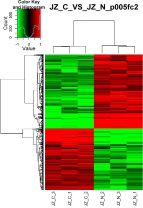
[image:3.612.93.379.75.492.2]endometrial cancer has not been previously reported.
Figure 2. Cluster analysis of transcriptional profiles in stromal cells. Transcrip-tional profiles were measured in stromal cells from endometrial carcinoma (JZ-C-1,2,3) and normal controls (JZ-N-1,2,3). Fold change ≥2, P<0.05.
Discussion
Endometrial carcinoma is a multiple-step process invo- lved in initiation, promotion and progression [5]. Accu- mulating evidence supports the notion that cancer devel-opment is intimately related to the complex tumor micro-environment [7, 12]. The im- portance of the local tumor microenvironment in tumor progression has been recog-nized for many years. In the current study, we examined gene transcription expres-sion levels in stromal cells derived from endometrial carcinoma patients.
The tumor microenvironm- ent, is primarily composed
of immune cells, stromal
fi-broblasts, extracellular mat- rix proteins, and endothelial cells derived from various cell types, which can directly regulate the cell phenotypes, representing a unique app- roach for cancer diagnosis and treatment [13]. The pri-mary cell types in the tumor microenvironment that are required for cancer progres-sion consist of
In the current study, we compared the gene expression levels in endometrial carcinoma-derived stromal cells to those in the healthy
controls. Our results indicated that a total of 605 genes were changed (fold change ≥2,
p-value <0.05). Among these, 275 were up-reg-ulated and 330 were down-regup-reg-ulated genes. In
addition, GO ontology analysis showed that the DEGs were involved in various molecular func -tions and biological processes.
Furthermore, KEGG analysis revealed that DEGs were involved in Wnt signaling pathway,
cadherin signaling pathway, ECM-receptor
interaction, and focal adhesion. More specifi -cally, 28 genes belonged to Wnt signaling path-way. The Wnt signaling is a signal transduction pathway made of proteins that passes signals into a cell through cell surface receptors. In mammals, Wnt signaling is important in body axis patterning, cell growth, and cell migration [16]. Constitutive activation of this pathway is
closely associated with human cancers [17].
Our results indicate that, many genes mapped
to the protocadherins (PCDHs) family including PCDHA1-13. PCDHs are a large family of cad-herin-like cell adhesion proteins that are involved in the establishment and maintenance of cellular connections. Adhesive properties of these proteins are critical for cell rearrange-ment, migration, and cell sorting [18, 19]. In addition, a recent study showed that PCDHs mediate cell growth, cell cycle arrest, apopto-sis, and migration of endometrial cancer, underlining their critical roles in endometrial carcinogenesis [20].
[image:4.612.91.524.74.283.2]In conclusion, our study identified 605 differen -tially expressed genes, mapping to a variety of biological processes including Wnt signaling pathway, cadherin signaling, ECM-receptor in- teraction and focal adhesion. These results may contribute to understanding the molecular mechanisms of endometrial carcinoma.
Figure 3. GO analysis of differentially expressed genes. The Y-axis shows significantly enriched gene ontology (GO) terms relative to the genome, and the X-axis shows the enrichment scores of these terms.
Table 2. KEGG analysis of DEGs
Term Count P Value Genes
Wnt signaling pathway 28 <0.05 PCDHA6, PCDHA7, PCDHA8, PCDHA9, PCDHA2, CORIN, PCDHA3, PCDHA4, PCDHA5, PCDHA1, PCDHAC2, PCDHAC1, HOXC6, PLCB4, HOXC4, PCDHA10, PCDHA11, PCDHA12, PCDHA13, AXIN2, FZD8, TCF7, MYH3, PCDH10, PCDHB2, PCDH9, PCDH7, TLE2, EP300, HOXB7, SFRP1, KREMEN1, CDH10
Cadherin signaling pathway 18 <0.05 PCDHA6, FZD8, PCDHA7, PCDHA8, TCF7, PCDHA9, CORIN, PCDHA2, PCDHA3, PCDHA4,
PCDH10, PCDHA5, PCDHB2, PCDH9, PCDHA1, PCDH7, PCDHAC2, PCDHAC1, PCDHA10, PCDHA11, PCDHA12, PCDHA13, CDH10
ECM-receptor interaction 10 <0.05 COL4A4, LAMA1, TNXB, TNXA, ITGA1, COL6A1, LAMC2, COL1A1, COL11A1, HMMR, SPP1
[image:4.612.90.525.347.452.2]Acknowledgements
The project of Fenxian district Science and Technology Committe [no 20151217].
Disclosure of conflict of interest
None.
Address correspondence to: Hong-Bo Wang, De- partment of Obstetrics and Gynecology, Union Hospital, Tongji Medical College, Huazhong Uni- versity of Science and Technology, Wuhan 430022, China. Tel: +86-21-57424168; E-mail: whongbow@ 163.com; Hong-Bin Wang, South Branch of The Six People’s Hospital Affiliated to Shanghai Jiao Tong University, Shanghai 201400, China. Tel: +86-21-57422115; E-mail: whb417@163.com
References
[1] Morice P, Leary A, Creutzberg C, Abu-Rustum N, Darai E. Endometrial cancer. Lancet 2016; 387: 1094-108.
[2] Stefansson IM, Salvesen HB, Akslen LA. Prog-nostic impact of alterations in P-cadherin ex-pression and related cell adhesion markers in endometrial cancer. J Clin Oncol 2004; 22: 1242-52.
[3] Peng Q, Mo C, Qin A, Lao X, Chen Z, Sui J, Wu J, Zhai L, Yang S, Qin X, Li S. MDM2 SNP309 polymorphism contributes to endometrial can-cer susceptibility: evidence from a meta-analy-sis. J Exp Clin Cancer Res 2013; 32: 85. [4] Carlson MJ, Thiel KW, Leslie KK. Past, present,
and future of hormonal therapy in recurrent endometrial cancer. Int J Womens Health 2014; 6: 429-35.
[5] Zhao H, Yang L, Baddour J, Achreja A, Bernard V, Moss T, Marini JC, Tudawe T, Seviour EG, San LF, Alvarez H, Gupta S, Maiti SN, Cooper L, Peehl D, Ram PT, Maitra A, Nagrath D. Tumor microenvironment derived exosomes pleiotro-pically modulate cancer cell metabolism. Elife 2016; 5: e10250.
[6] Ren Y, Zhou X, Liu X, Jia HH, Zhao XH, Wang QX, Han L, Song X, Zhu ZY, Sun T, Jiao HX, Tian WP, Yang YQ, Zhao XL, Zhang L, Mei M, Kang CS. Reprogramming carcinoma associated fi-broblasts by AC1MMYR2 impedes tumor me-tastasis and improves chemotherapy efficacy. Cancer Lett 2016; 374: 96-106.
[7] Fu H, Yang H, Zhang X, Xu W. The emerging roles of exosomes in tumor-stroma interaction. J Cancer Res Clin Oncol 2016; 142: 1897-907. [8] Chen L, Zeng X, Kleibeuker E, Buffa F, Barberis
A, Leek RD, Roxanis I, Zhang W, Worth A, Beech JS, Harris AL, Cai S. Paracrine effect of GTP cyclohydrolase and angiopoietin-1 interac-tion in stromal fibroblasts on tumor Tie2 acti-vation and breast cancer growth. Oncotarget 2016; 7: 9353-67.
[9] Turley SJ, Cremasco V, Astarita JL. Immunologi-cal hallmarks of stromal cells in the tumour microenvironment. Nat Rev Immunol 2015; 15: 669-82.
[10] Kawada M, Inoue H, Ohba S, Yoshida J, Masu-da T, Yamasaki M, Usami I, Sakamoto S, Abe H, Watanabe T, Yamori T, Shibasaki M, Nomoto A. Stromal cells positively and negatively modu-late the growth of cancer cells: stimulation via the PGE2-TNFalpha-IL-6 pathway and inhibi-tion via secreted GAPDH-E-cadherin interac-tion. PLoS One 2015; 10: e119415.
[11] Li M, Xin X, Wu T, Hua T, Wang H, Wang H. Stro-mal cells of endometrial carcinoma promotes proliferation of epithelial cells through the HGF/c-Met/Akt signaling pathway. Tumour Biol 2015; 36: 6239-48.
[12] Yuan Y, Jiang YC, Sun CK, Chen QM. Role of the tumor microenvironment in tumor progression and the clinical applications (review). Oncol Rep 2016; 35: 2499-515.
[13] Mercurio L, Ajmone-Cat MA, Cecchetti S, Ricci A, Bozzuto G, Molinari A, Manni I, Pollo B, Scala S, Carpinelli G, Minghetti L. Targeting CXCR4 by a selective peptide antagonist modulates tu-mor microenvironment and microglia reactivity in a human glioblastoma model. J Exp Clin Cancer Res 2016; 35: 55.
[14] Wu T, Dai Y. Tumor microenvironment and ther-apeutic response. Cancer Lett 2017; 387: 61-8.
[15] Nakamura R, Kayamori K, Oue E, Sakamoto K, Harada K, Yamaguchi A. Transforming growth factor-beta synthesized by stromal cells and cancer cells participates in bone resorption in-duced by oral squamous cell carcinoma. Bio-chem Biophys Res Commun 2015; 458: 777-82.
[16] Logan CY, Nusse R. The Wnt signaling pathway in development and disease. Annu Rev Cell Dev Biol 2004; 20: 781-810.
[17] Karim R, Tse G, Putti T, Scolyer R, Lee S. The significance of the Wnt pathway in the pathol-ogy of human cancers. Patholpathol-ogy 2004; 36: 120-8.
[18] Kim SY, Yasuda S, Tanaka H, Yamagata K, Kim H. Non-clustered protocadherin. Cell Adh Migr 2011; 5: 97-105.
[19] Morishita H, Yagi T. Protocadherin family: diver-sity, structure, and function. Curr Opin Cell Biol 2007; 19: 584-92.