Original Article
Effect of exendin-4 on the expression of CXCL10 and its
receptor in type I diabetes rats
Jinshui He1, Chaowei Lian1, Yanling Fang1, Jinzhi Wu2, Jianming Weng3, Xiaoling Ye1, Huowang Zhou1, Shaobo Zhu1
Departments of 1Pediatrics, 2Endocrine, 3Pathology, Zhangzhou Hospital Affiliated to Fujian Medical University, Zhangzhou 363000, Fujian, China
Received October 12, 2015; Accepted November 25, 2015; Epub January 1, 2016; Published January 15, 2016
Abstract: Type 1 diabetes mellitus (T1DM) is one auto-immune disease featured with infiltration of islet cells by lym -phocytes. The role of chemotactic factor and its receptor in T1DM has drawn lots of research interests. As one gluca-gon-like peptide-1 (GLP1) receptor agonist, Exendin-4 exerts critical function in blood glucose homeostasis and islet tissue protection. Its role in the expression of CXC chemokine ligand-10 (CXCL10) with its receptor, however, remains unknown. As both CXCL10 and its receptor, CXCR3, are related with islet injury, exendin-4 thus may inhibit islet
cell apoptosis via modulating the expression of CXCL10 and its receptor. T1DM rat model was firstly prepared by
intraperitoneal injection of streptozocin (60 mg/kg), followed by intervention by serial dosages of Exendin-4. Blood
glucose, insulin and C-peptide levels were quantified by enzyme linked immunosorbent assay (ELISA). Real-time
quantitative PCR and Western blotting were used to detect the expression of CXCL10 and CXCR3. The apoptosis of islet tissue was also observed. After Exendin-4 treatment, blood glucose, insulin and C-peptide levels in T1DM rats were improved, along with decreased expression of CXCL10 and CXCR3. The apoptosis of islet cells was also inhib-ited. Exendin-4 can inhibit CXCL10-CXCR3 pathway, depressing TLR-4 expression, providing possible mechanism for inhibiting the apoptosis of islet cells. Our results provide evidences for future development of anti-T1DM drugs.
Keywords: Type 1 diabetes mellitus, CXC chemokine ligand 10, CXC chemokine receptor 3, Toll-like receptor 4, Exendin-4
Introduction
The incidence of type 1 diabetes mellitus (T1DM) is significantly increasing worldwide [1], and affecting patients’ life quality in addition to heavy social burdens. As one autoimmune dis-ease featured with the infiltration of islet cells by lymphocytes, T1DM is closely related with genetic, environmental and viral factors. Recently, the role of chemokine ligands and their receptors in T1DM pathogenesis has become one research hotspot [2]. CXC chemo-kine ligand-10 (CXCL-10) belongs to the CXC chemokine family, and is screened from cDNA library by the stimulus of U937 cell line under interferon γ (IFN-γ). It can be derived from mononuclear macrophage, activated fibroblast and endothelial cells [3]. The expression level of CXCL10 has been shown to be related with the condition of islet tissue injury [4]. CXCL10
exerts its biological function via binding onto its receptor, CXCR3 and Toll-like receptor 4 (TLR4). CXCR3 is expressed on the surface of both latent and active T lymphocytes, and is regulat-ed by CXCL9 and CXCL10, which can also bind onto TLR4 of islet β cells for mediating insulin biosynthesis and islet B cell apoptosis. Therefore, the expression of CXCL10 and its receptor plays a critical role in diagnosis and treatment of T1DM.
mecha-nism, however, remained unknown. Exendin-4 is one potent agonist for GLP-1 receptor with high affinity on pancreatic GLP-1 receptor [7]. Previous study has found the protective effect of Exendin-4 on islet β cells in T1DM model ani-mal in addition to the inhibition of inflammatory cell infiltration [8, 9]. Its biological effects on CXCL10 and its receptor, however, remain unknown. We thus established T1DM rat model, on which gradient dosages of Exendin-4 were given to observe the expression pattern of CXCL10 and CXCR3/TLR4, in an attempt to elu-cidate the possible mechanism of Exendin-4 in treating T1DM.
Materials and methods
Animal model
Male SD rats (6~8 weeks old, body weight around 160 g) were kept in an animal facility (temperature: 18~25°C; humidity: 40%~70%) with food and water ad libitum. All animals were kept for 1-week acclimation before the surgery. Those rats with normal diet habit, stable body weight and blood glucose were recruited. A total of 50 qualified rats were randomly divid-ed into 5 groups (N=10 each): control; Exendin-4 (4 mg/kg); model; model + Exendin-4 (4 mg/ kg); model + Exendin-4 (8 mg/kg). All rats were fasted for 12 hours, followed by intraperitoneal injection of 60 mg/kg streptozocin (STZ, Sigma, US) or equal volume of saline to generate T1DM model.
Body weight, mental status, fur color, water intake and urea volume were closely monitored after injection. Blood glucose level was mea-sured 72 hours later. Those rats with blood glu-cose higher than 16.67 mM were identified as T1DM rats, which then received Exendin-4 intervention for 6 weeks.
Enzyme-linked immunosorbent assay (ELISA)
After 6-week Exendin-4 infusion, all rats were sacrificed and collected for blood samples from abdominal aorta. The blood was firstly incubat-ed at room temperature for 30 min, followincubat-ed by centrifugation at 2000 g for 15 min. Serum was collected from the upper layer. Insulin, C- peptide and CXCL10 levels were determined using ELISA kits (RayBio, US) following manual instruction.
Real-time quantitative PCR
After sacrificing rats, pancreatic tissues were immediately removed for mixing with Trizol reagents (Invitrogen, US). After incubation on ice for 5 min, tissues were homogenized and centrifuged at 10000 g for 10 min. Supernatants were saved and mixed with 0.2 mL chloroform, and were re-centrifuged for 15 min. The upper aqueous phase was saved to mix with 0.5 mL isopropanol for precipitating RNA by centrifuga-tion for 10 min. The RNA pellet was then washed by 1 mL ethanol for 3 times, followed by re-suspension in 20 μL DEPC water.
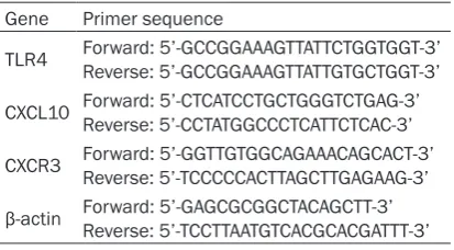
Primers for CXCL10, CXCR3 and TLR4 were list-ed in Table 1. PCR was performed under the following conditions: 50°C for 30 min, 95°C for 5 min, followed by 40 cycles each containing 95°C denature for 30 sec, 55°C annealing for 30 sec and 72°C elongation for 50 sec. The reaction was ended with 72°C elongation for 5 min. The relative expression level of target gene was determined by comparing its Ct value with internal reference gene (β-actin) using 2-ΔΔCt
method.
Western blotting
Pancreatic tissues were cut and mixed with lysis buffer (with PMSF). After homogenization, tissue lysate was incubated on ice for 10 min and centrifuged for 5 min. Total proteins were collected from the supernatant.
[image:2.629.99.306.92.206.2]Extracted protein solution was quantified by BCA and diluted to equal concentrations. After heat denature for 5 min, protein samples were loaded onto 10% SDS-PAGE for separation, and were transferred to PVDF membrane. The mem-brane was firstly blocked in 5% defatted milk powder for 1 hour at 37°C, and then was incu-bated with primary antibody against CXCL10,
Table 1. Specific primer for RT-PCR
Gene Primer sequence
TLR4 Forward: 5’-GCCGGAAAGTTATTCTGGTGGT-3’Reverse: 5’-GCCGGAAAGTTATTGTGCTGGT-3’
CXCL10 Forward: 5’-CTCATCCTGCTGGGTCTGAG-3’Reverse: 5’-CCTATGGCCCTCATTCTCAC-3’
CXCR3 Forward: 5’-GGTTGTGGCAGAAACAGCACT-3’Reverse: 5’-TCCCCCACTTAGCTTGAGAAG-3’
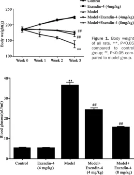
ing fur. Typical features of T1DM including poly-uria, polydipsia and polyphagia were observed, along with decreased body weight. After Exendin-4 intervention, the amplitude of body mass loss was decreased (Figure 1), suggest-ing the alleviation of T1DM after Exendin-4 treatment.
Blood glucose level
As shown in Figure 2, model group had signifi-cantly elevated blood glucose level compared to control group. The application of Exendin-4 remarkably decreased blood glucose level (P<0.05 compared to model group).
Serum levels of insulin, C-peptide and CXCL10
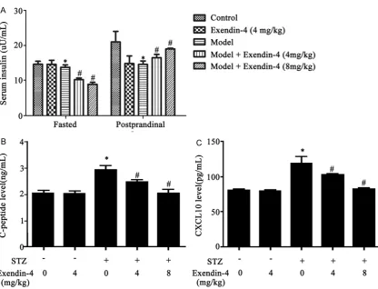
The insulin level in model rats was significantly depressed compared to control ones. After TLR4 or CXCR3 (1:1 000, Abcam, US) for
over-night incubation. After rinsing in TTBS for three times, secondary antibody (Proteintech, US) was added for 2-hour incubation. ECL chromo-genic substrates were used to visualize protein bands.
TUNEL assay
Pancreatic tissues were prepared for paraffin-based tissue blocks and were sectioned. After dewax and gradient hydration, tissues slices were processed in 20 mg/L proteinase K for 15 min to increase the tissue permeability. Endogenous peroxidase activity was then quenched by 0.3% hydrogen peroxide for 5 min. TUNEL reagent was prepared from TdT and bio-tinylated 16-dUTP, and was applied onto the tis-sue slice for 1-hour incubation. The slice was then incubated using 3% bovine serum albumin
(BSA) for 10 min, and was pro-cessed by novobiocin conju-gated with peroxidase for 30 min at room temperature. DAB substrate was used to develop the slice, which was then counter-stained by hematoxylin. 10 fields were randomly selected from each slice for counting number of apoptotic cells.
Statistical analysis
All data were presented by mean ± standard deviation (SD). Student t-test was used to compare means between two groups. One-way analysis of variance (ANOVA) com-pared the difference across groups, followed by SNK test in paired comparison. A sta-tistical significance was de- fined when P<0.05.
Results
General features
In control and Exendin-4 group, rats had normal body shape, acute response and good mental status. Model rats lacked spirit and acute response, along with brown-Figure 1. Body weight
of all rats. **, P<0.05 compared to control group; ##, P<0.05
com-pared to model group.
Figure 2. Blood glucose level. **, P<0.05 compared to control group; ##,
[image:3.629.103.381.87.449.2]Exendin-4 treatment, fasted insulin level was decreased while postprandial insulin was increased (Figure 3A), suggesting the improve-ment of islet functions. Serum peptide-C can reflect the secretion of insulin. We observed enhanced C-peptide secretion in model rats, and depressed level after Exendin-4 interven-tion (Figure 3B). As another indicator reflecting
islet function, CXCL10 showed similar patterns as C-peptide (Figure 3C).
CXCL10, CXCR3 and TLR4 protein expression
[image:4.629.107.524.77.395.2]Western blotting was used to quantify protein expression level of CXCL10, CXCR3 and TLR4 in pancreatic tissues. As shown in Figure 4, model Figure 3. Serum level of insulin (A), C-peptide (B) and CXCL10 (C) in all rats. *, P<0.05 compared to control group;
#, P<0.05 compared to model group.
Figure 4. Protein levels of CXCL10, CXCR3 and TLR4. *, P<0.05 compared to control group; #, P<0.05 compared to
[image:4.629.103.528.457.579.2]with T cell function. Both CXCL10 and T cell sur-face receptor CXCR3 participate in the recruit-ment of pancreatic specific T cells [15]. The study of CXCL10 and its receptor function is thus of critical importance for T1DM. In newly diagnosed T1DM patients, both CXCR3-positive lymphocytes infiltrated into pancreas and CXCL10 level were significantly up-regulated no matter the existence of enterovirus infection in islet [16]. The significantly lower incidence of diabetes in CXCR3-deficient mice [15] further suggested the importance of CXCR3 in islet function. The clinical study in patients with T1DM complicated with ketoacidosis found the existence of cascade immune reaction that may damage β cells [17]. This so called CXCL10-CXCR3 cycle is initiated by the co-expression of interferon-γ (IFN-γ) and CXCL10 in β cells after pancreatic injury. The secreted CXCL10 then activated autoimmune response via CXCR3 by activating local lymphocytes and recruiting for infiltration on islet tissues. Those lymphocytes infiltrated into islet tissues then released inflammatory cytokines to further aggravating the production of CXCL10 from remaining β cells. The consequently over-production of CXCL10 further accelerate activation of T cells and macrophage by CXCR3, further leading to the widely necrosis of β cells and diabetes. TLR4, as one novel receptor for CXCL10, is fre-quently expressed in myeloid cells, T cells, nat-ural killer cells, keratinocytes, endothelial cells rats had elevated expression of those proteins,
whose levels were depressed after Exendin-4 intervention.
mRNA level of CXCL10, CXCR3 and TLR4
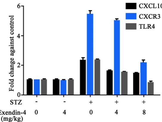
RT-PCR revealed consistent patterns of CXCL10, CXCR3 and TLR4 mRNA levels as those in protein expression: model rats had sig-nificantly elevated gene expression level, which was remarkably depressed after Exendin-4 treatment (Figure 5).
Islet cell apoptosis
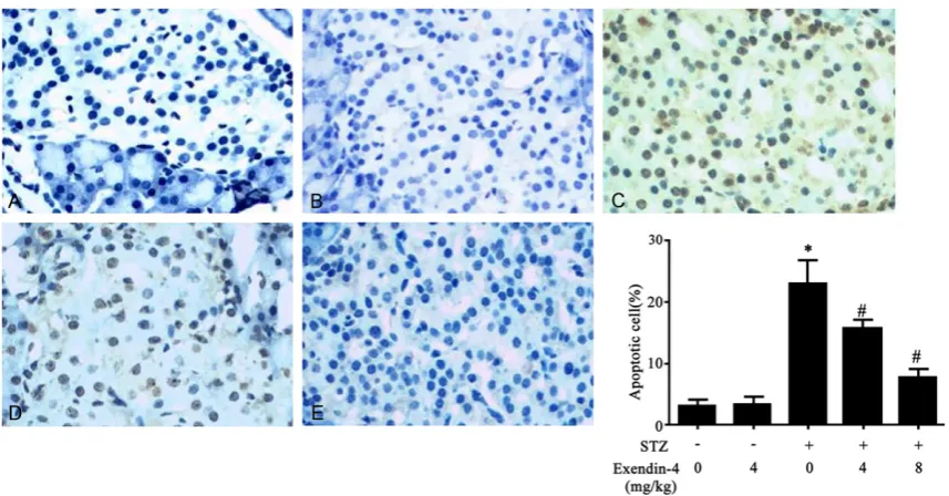
Using TUNEL assay, we found the decreased of islet cell apoptosis by Exendin-4 in a dose-dependent manner (Figure 6). This suggested the protective role of Exendin-4 on islet β cells via inhibiting apoptosis.
Discussion
The incidence of diabetes is increasing by years, with more severe complications includ-ing cardiovascular, kidney and retinal diseases. As one auto-immune disease, T1DM is com-monly believed to be related with immune-induced islet β cell damage [10]. Recent study has revealed CXCL10 as one indicator for islet β cell injury, thus making it one critical index in diagnosis and treatment of islet injury. Early study has suggested the gradual increase of CXCL10 in islet β cells at the early phase of
pancreatitis, followed by en- hancing level with disease aggravation [11]. Similarly, in T1DM patients, serum level of CXCL10 in early and sub-clinical phase T1DM patients was significantly potentiated [12]. In organ donors with T1DM, the production and secretion of CXCL10 was also elevated compared to normal individuals [13]. One study using cytokine signaling path-way inhibitior-1 gene trans-fection into islet β cells of NOD mice to suppress CXCL- 10 expression and show- ed the decreased of diabetes incidence [14].
[image:5.629.107.379.80.286.2]As one auto-immune disease, T1DM is closely correlated Figure 5. mRNA levels of CXCL10, CXCR3 and TLR4. *, P<0.05 compared to
and epithelial cells. TLR4 is also expressed in islet β cell. Via myeloid differentiation factor 88-dependent pathway, it can continuously activate PI3K-Akt and JNK signaling pathways to induce the cleavage of caspase-3 and PAK2, for down-regulating insulin expression and desensitizing glucose-dependent insulin secre-tion, in addition to cell apoptosis [13]. Moreover, TLR4 also participates in inflammation-induced apoptosis of islet β cells [18]. These studies all suggested CXCL10 and its receptor as critical mediators for inducing pancreatitis and partici-pating T cell-induced islet β cell damage. Some scholars have found the triple roles of GLP-1 after binding onto specific receptors on the membrane of islet β cell, including facilitat-ing insulin synthesis and secretion, inhibitfacilitat-ing islet β cell apoptosis and potentiating islet β cell mitosis [19]. GLP-1 was also known to mod-ulate both apoptotic and anti-apoptotic pro-teins including caspase-3, Bcl-2 and Bcl-xL via activating PI3K, PKB/Akt and PKA signaling molecules [20]. It can also inhibit cytokine or cytotoxicity induced islet β cell apoptosis via interfering with JNK signal pathway activation [7, 21].
All previous studies have suggested the critical role of CXCL10 and its receptor in T1DM. In this study, we measured the expressional pattern of CXCL10 and its receptors (CXCR3/TLR4) in T1DM rat islet via applying different dosages of
Exendin-4, to elucidate the role of Exendin-4 on the expression of CXCL10 (and its receptors) and cell apoptosis. Our results showed signifi-cant treatment efficacy of Exendin-4 on T1DM rats, including the modulation of blood glucose level, serum insulin, C peptide concentration, and the regulation of CXCL10 expression and secretion. The inhibition of CXCL10-CXCR3 cycle thus may provide further evidences for clinical treatment of T1DM and protecting islet β cells.
Acknowledgements
This study was supported by the Fujian medical innovation project (2015-CX-38).
Disclosure of conflict of interest
None.
Address correspondence to: Dr. Jinzhi Wu, Depart-
ment of Endocrine, Zhangzhou Hospital Affiliated to
Fujian Medical University, Zhangzhou 363000, Fujian, China. Tel: 596-2527451; Fax: +86-596-2527451; E-mail: [email protected]
References
[1] Dabelea D. The accelerating epidemic of child-hood diabetes. Lancet 2009; 373: 1999-2000.
[2] Corrado A, Ferrari SM, Ferri C, Ferrannini E, Antonelli A, Fallahi P. Type 1 diabetes and
Figure 6. Effect of Exendin-4 on islet cell apoptosis in T1DM rats. *, P<0.05 compared to control group; #, P<0.05
(C-X-C motif) ligand (CXCL) 10 chemokine. Clin Ter 2014; 165: e181-5.
[3] Pinto S, Martínez-Romero A, O’Connor JE, Gil-Benso R, San-Miguel T, Terrádez L, Monteagudo C, Callaghan RC. Intracellular coexpression of CXC- and CC- chemokine receptors and their ligands in human melanoma cell lines and dy-namic variations after xenotransplantation. BMC Cancer 2014; 14: 118.
[4] Novelli M, Beffy P, Menegazzi M, De Tata V, Martino L, Sgarbossa A, Porozov S, Pippa A, Masini M, Marchetti P, Masiello P. St. John's wort extract and hyperforin protect rat and hu-man pancreatic islets against cytokine toxicity. Acta Diabetol 2014; 51: 113-21.
[5] Richards P, Pais R, Habib AM, Brighton CA, Yeo GS, Reimann F, Gribble FM. High fat diet im-pairs the function of glucagon-like peptide-1
producing L-cells. Peptides 2015; [Epub ahead of print].
[6] Sherry NA, Chen W, Kushner JA, Glandt M, Tang Q, Tsai S, Santamaria P, Bluestone JA, Brillantes AM, Herold KC. Exendin-4 improves reversal of diabetes in NOD mice treated with anti-CD3 monoclonal antibody by enhancing recovery of beta-cells. Endocrinology 2007; 148: 5136-44.
[7] Kawasaki Y, Harashima S, Sasaki M, Mukai E, Nakamura Y, Harada N, Toyoda K, Hamasaki A, Yamane S, Yamada C, Yamada Y, Seino Y, Inagaki N. Exendin-4 protects pancreatic beta cells from the cytotoxic effect of rapamycin by inhibiting JNK and p38 phosphorylation. Horm Metab Res 2010; 42: 311-7.
[8] Perez-Arana G, Blandino-Rosano M, Prada-Oliveira A, Aguilar-Diosdado M, Segundo C. Decrease in {beta}-cell proliferation precedes apoptosis during diabetes development in
bio-breeding/worcester rat: beneficial role of
Exendin-4. Endocrinology 2010; 151: 2538-46.
[9] Sano H, Terasaki J, Mishiba Y, Imagawa A, Hanafusa T. Exendin-4, a glucagon-like pep-tide-1 receptor agonist, suppresses pancreatic beta-cell destruction induced by encephalo-myocarditis virus. Biochem Biophys Res Commun 2011; 404: 756-61.
[10] Mannering SI, Pathiraja V and Kay TW. The case for an autoimmune etiology of type 1
dia-betes. Clin Exp Immunol 2015; [Epub ahead of print].
[11] Shigihara T, Oikawa Y, Kanazawa Y, Okubo Y,
Narumi S, Saruta T, Shimada A. Significance of
serum CXCL10/IP-10 level in type 1 diabetes. J Autoimmun 2006; 26: 66-71.
[12] Thorand B, Kolb H, Baumert J, Koenig W, Chambless L, Meisinger C, Illig T, Martin S, Herder C. Elevated levels of interleukin-18 pdict the development of type 2 diabetes:
re-sults from the MONICA/KORA Augsburg Study, 1984-2002. Diabetes 2005; 54: 2932-8.
[13] Schulthess FT, Paroni F, Sauter NS, Shu L, Ribaux P, Haataja L, Strieter RM, Oberholzer J, King CC, Maedler K. CXCL10 impairs beta cell function and viability in diabetes through TLR4 signaling. Cell Metab 2009; 9: 125-39.
[14] Hultcrantz M, Jacobson S, Hill NJ, Santamaria P, Flodström-Tullberg M. The target cell re-sponse to cytokines governs the autoreactive T cell repertoire in the pancreas of NOD mice. Diabetologia 2009; 52: 299-305.
[15] Frigerio S, Junt T, Lu B, Gerard C, Zumsteg U, Holländer GA, Piali L. Beta cells are
responsi-ble for CXCR3-mediated T-cell infiltration in in -sulitis. Nat Med 2002; 8: 1414-20.
[16] Roep BO, Kleijwegt FS, van Halteren AG, Bonato V, Boggi U, Vendrame F, Marchetti P,
Dotta F. Islet inflammation and CXCL10 in re -cent-onset type 1 diabetes. Clin Exp Immunol 2010; 159: 338-43.
[17] Tanaka S, Nishida Y, Aida K, Maruyama T, Shimada A, Suzuki M, Shimura H, Takizawa S, Takahashi M, Akiyama D, Arai-Yamashita S, Furuya F, Kawaguchi A, Kaneshige M, Katoh R, Endo T, Kobayashi T. Enterovirus infection, CXC chemokine ligand 10 (CXCL10), and CXCR3 cir-cuit: a mechanism of accelerated beta-cell fail-ure in fulminant type 1 diabetes. Diabetes 2009; 58: 2285-91.
[18] Chang ZL. Important aspects of Toll-like recep-tors, ligands and their signaling pathways.
Inflamm Res 2010; 59: 791-808.
[19] Stoffers DA, Kieffer TJ, Hussain MA, Drucker DJ, Bonner-Weir S, Habener JF, Egan JM. Insulinotropic glucagon-like peptide 1 agonists stimulate expression of homeodomain protein IDX-1 and increase islet size in mouse pancre-as. Diabetes 2000; 49: 741-8.
[20] Farilla L, Bulotta A, Hirshberg B, Li Calzi S, Khoury N, Noushmehr H, Bertolotto C, Di Mario U, Harlan DM, Perfetti R. Glucagon-like peptide 1 inhibits cell apoptosis and improves glucose responsiveness of freshly isolated human is-lets. Endocrinology 2003; 144: 5149-58.