Original Article
Characterization and functional elucidation
of CD8+AT2+ lymphocytes in human
thoracic aortic aneurysm
Chenxi Wang, Weijun Wang, Mingjun Du, Xinyu Liu, Song Xue
Department of Cardiovascular Surgery, Renji Hospital, School of Medicine, Shanghai Jiaotong University, Shang-hai, China
Received December 26, 2016; Accepted March 14, 2017; Epub July 1, 2017; Published July 15, 2017
Abstract: Human thoracic aortic aneurysm (TAA) is one of the most fatal cardiovascular diseases. However, the underlying molecular mechanism behind this disease remains uncertain. Previous studies have proven the im-portance of angiotensin II receptor type 2 (AT2) and endothelial cells in aneurysm pathology. This study aimed to elucidate the effect of CD8+AT2+ lymphocytes in TAA pathogenesis. The expression levels of CD8, AT2, IL-2, and IFN-r were determined by real-time quantitative PCR. CD8+AT2+ lymphocytes were counted by semi-quantitative
im-munofluorescence and flow cytometric analysis. The wound-healing assay was used to detect endothelial cell migra
-tion. The expression levels of CD8 and AT2 in human nonsyndromic TAA tissues were significantly higher than those
of the control. TAA tissues have more CD8+AT2+ lymphocytes than the controls. Furthermore, circulating CD8+AT2+
lymphocytes were significantly elevated in TAA patients. The expression levels of IL-2 and IFN-r in CD8+AT2+ lym
-phocytes were significantly increased compared with those in CD8+AT2- lym-phocytes. The CD8+AT2+ lym-phocytes had a greater inhibitory effect on endothelial migration compared with CD8+AT2- cells. Our findings showed that the
increment of CD8+AT2+ lymphocytes in human TAA exhibited a protective effect by downregulating the release of
pro-inflammation cytokines and the inhibition of endothelial cell migration.
Keywords: Angiotensin II type 2 receptor, thoracic aortic aneurysm, endothelial cell, cytokine, inflammation
Introduction
Thoracic aortic aneurysm (TAA) is a life-threat-ening disease characterized by progressive dil-atation of the thoracic aorta. Usually, the dis-ease continues to develop without any symp-toms until the terminal stage, which is detect- ed by computed tomographic scans. Patients with TAA die because of aorta rupture [1]. Un- fortunately, the current standard therapeutic options are limited and include open surgical replacement or endovascular aortic repair [2]. Pharmaceutical treatment is only applied pre-operationally for surgical preparation [3]. There- fore, molecular pathogenesis is necessary for pharmacotherapy.
The pathogenesis of aneurysms is well known to involve tissue remodeling and chronic inflam -mation. Aneurysmal arteries are characterized by leukocyte infiltrates, particularly
tion, fibrosis, and apoptosis [16]. We previous-ly identified a new cell population, the CD4+ AT2+ T lymphocytes, in TAA and defined its functions in the inhibition of cell growth and MMP2 expression, and promotion of apoptosis in ECs. In the present study, we found another cell population, the CD8+AT2+ lymphocytes, in TAA tissues. Although CD8+AT2+ lymphocytes have been observed in ischemic heart injury and show cardioprotective functions [18], the role of this cell subpopulation in TAA has not been explored.
Materials and methods
Human aorta specimen
A total of 20 TAA patients participated in the present study. Patients with Marfan syn-drome or aortic dissection were excluded. In addition, 20 non-aneurysmal thoracic aortic samples were collected from patients with coronary heart disease undergoing coronary artery bypass surgery. The protocol was appro- ved by the Ethical Committee of Renji Hospi- tal. Informed consent was obtained from all in- cluded participants in the study. All the proce-dures performed in studies involving human participants agreed with the ethical standards of the institutional and/or national research committee, as well as the 1964 Helsinki decla -ration and its later amendments, or compara-ble ethical standards.
Immunofluorescence (IF) staining
Aorta tissues for IF staining were fetched from the operation room and immediately delivered to the laboratory. Tissues around the dilated region were cut and frozen for examination. Cryo-tissue section preparation and examina-tion was performed as previously described [10].
ctions. The following primer sequences were used: β-actin, 3’-AGAAAATCTGGCACCACACC-5’ (forward) and 3’-AGAGGCGTACAGGGATAG-CA-5’ (reverse); AT2, 3’-ACTGGCTCTTTGGACC-TGTG-5’ (forward) and 3’-GCCATACACCAAACA-AGGGG-5’ (reverse); CD8, 3’-GCCAGTGACCAT-TCCGGTA-5’ (forward) and 3’-GACCTAGCCTG-GACCTTGGA-5’ (reverse); IL-2, 3’-GAATCACGT-TACGTTCTGTCCT-5’ (forward) and 3’-CTTAGG-GTTTGAGTGGTCCTAC-5’ (reverse); IFN-r 3’-AC-CTGTAAGTTCAGTCAATGGCT-5’ (forward) and 3’-CGTCGATTTTGTCCCTTCGCT-5’ (reverse). The relative expression levels of CD8, AT2, IL-2, and IFN-r were calculated and normalized by the 2-ΔΔCt method relative to β-actin.
Flow cytometry analysis and sorting
Peripheral blood was collected prior to surgery. Mononuclear cell extraction from blood and its analysis was previously described [10]. After sorting, the cell subpopulations was kept in a mixed liquid for 30 min at 25°C and resuspend-ed for incubation.
Wound-healing assay
the wounded area. Three independent series of experiments were performed. The distance of cell migration from the initial wound was measured with the NIS-Elements D software (Nikon, Tokyo, Japan).
Statistical analyses
Data were expressed as mean ± SEM. Com- parison between two groups was performed by the two-tailed Student’s t-test. Multiple com-parisons were analyzed with one-way ANOVA
followed by the Bonferroni post hoc test. Dif- ferences were considered significant at P < 0.05.
Results
Increased CD8+AT2+ lymphocytes in TAA tis-sues
To investigate the presence of CD8+AT2+ lym-phocytes in TAA tissues, we first examined the expression levels of CD8 and AT2 in the aneu-Figure 1. The expression levels of CD8 and AT2 in human TAA tissues. Both CD8 (A) and AT2 (B) were significantly
[image:3.612.94.518.73.230.2]upregulated in TAA tissues. *P < 0.05, ***P < 0.001.
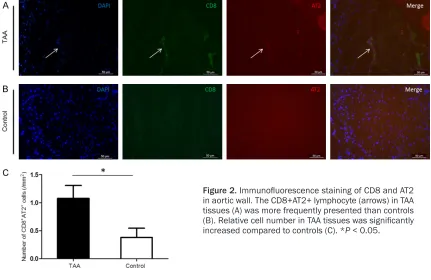
Figure 2. Immunofluorescence staining of CD8 and AT2
in aortic wall. The CD8+AT2+ lymphocyte (arrows) in TAA tissues (A) was more frequently presented than controls (B). Relative cell number in TAA tissues was significantly
[image:3.612.92.523.283.563.2]rysmal walls and controls. Both CD8 (P < 0.05) and AT2 (P < 0.001) were significantly upregu -lated in TAA tissues compared with the control (Figure 1). Furthermore, we examined the pro-tein levels of CD8 and AT2 in the aortic wall by immunofluorescence. CD8+AT2+ lymphocytes were rarely detected in the control. However, more cellular distribution was observed and the CD8+AT2+ lymphocytes had the tendency to cluster in TAA tissues (Figure 2). The density of CD8+AT2+ lymphocytes in TAA tissues (1.08 ± 0.23 cells/mm2, n = 16) was notably increas-
ed relative to the control (0.38 ± 0.16 cells/ mm2, P < 0.05, n = 16; Figure 2).
Increased circulation of CD8+AT2+ lympho-cytes in TAA patients
Alteration of peripheral lymphocyte subsets implies the local response of the immune sys-tem. The percentage of CD8+AT2+ lymphocy- tes from PBMCs was 8.87 ± 0.91% (n = 13) in TAA patients, which was significantly higher
than that in the control (4.70 ± 0.65%, P = 0.001, n = 13; Figure 3).
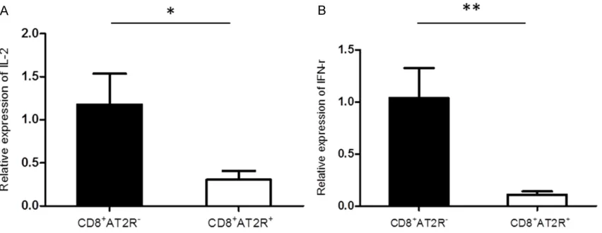
Downregulated IFN-r and IL-2 levels in CD8+AT2+ lymphocytes
Lymphocytes participate in local immune re- sponse by releasing pro-inflammation cytokin-es. We examined the expression levels of IFN-r and IL-2 in CD8+AT2+ and CD8+AT2- lympho-cytes. RT-qPCR revealed that the expression levels of IFN-r (n = 6, P < 0.01) and IL-2 (n = 6, P < 0.05) in CD8+AT2- lymphocytes were signifi -cantly higher than those in CD8+AT2+ lympho-cytes (Figure 4).
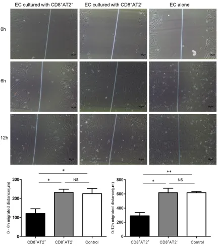
Effect of CD8+AT2+ lymphocytes on the migra-tion of aortic ECs
[image:4.612.92.520.76.186.2] [image:4.612.95.521.242.407.2]AT2- lymphocytes, and ECs alone. As shown in
Figure 5, the distance migrated by ECs with CD8+AT2+ lymphocytes (121 ± 25 μm, n = 3, 6 h; 289 ± 47, n = 3, 12 h) was shorter than either CD8+AT2- lymphocytes (232 ± 16 μm, 6 h, P < 0.05; 619 ± 60 μm, 12 h, P < 0.05) or ECs alone (225 ± 27 μm, 6 h, P < 0.05; 618 ± 17 μm, 12 h, P < 0.01) within 6 or 12 h. However, no statistically significant differences were
ob-served in the migration distance of ECs betw- een the CD8+AT2- lymphocyte and EC groups (n = 3 for each group).
Discussion
Previous studies have clarified the protective role of AT2 in aortic aneurysms of mice and humans [10, 19]. Similarly, our previous studies Figure 5. CD8+AT2+ lymphocytes inhibited ECs migration. There was significant difference in migration distance
between ECs cocultured with CD8+AT2+ lymphocytes and CD8+AT2- lymphocytes or ECs alone for 6 h and 12 h. *P
leading to inflammation cascades, cell infiltra -tion, and MMP production [20, 21]. IFN-r may activate NK cells and upregulate their killing ability to cause direct tissue damage. Com- pared with CD8+AT2- lymphocytes, CD8+AT2+ lymphocytes secreted low levels of IL-2 and IFN-r, thereby indicating that the anti-inflam-mation effect of CD8+AT2+ lymphocytes may be mediated by the AT2 pathway. Although the level of CD8+AT2+ lymphocytes in both peripheral blood circulation and aortic wall is increased, it is not clear where the cell subpop-ulation originate from. The underlying mecha-nism of elevated level of CD8+AT2+ lympho-cytes in TAA requires further investigation. Another important result of our study is the in- hibition of endothelial migration by CD8+AT2+ lymphocytes. Aortic ECs play an important role in the dilation of aneurysmal wall [10]. ECs can secrete MMP2 and inflammation cytokines to hydrolyze the aortic media and cause invasive migration [10, 11]. Endothelial cell migration within the vessels is marked by cell rearrange -ment, which was clearly demonstrated in our previous study [10, 22]. Therefore, EC-related neoangiogenesis could contribute to the final breakdown of the aortic wall [23]. The wound-healing assay reflects the motility and prolifera -tion of ECs [22]. Our study indicated that CD8+ AT2+ lymphocytes can dramatically inhibit EC migration. We concluded that the EC invasion into the aortic media could be significantly prevented by increasing the number of CD8+ AT2+ lymphocytes. EC disorder is one of the main causes of immune cell chemotaxis and aggregation. Therefore, the inflammatory reac -tion may be notably reduced by EC inhibi-tion. Although the underlying mechanism of inhibi-tion remains unclear, the effect of CD8+AT2+ lymphocytes on ECs may prevent media break
-Disclosure of conflict of interest
None.
Address correspondence to: Dr. Song Xue, Depart- ment of Cardiovascular Surgery, Renji Hospital, School of Medicine, Shanghai Jiaotong University, Shanghai, China. E-mail: [email protected]
References
[1] Guo DC, Papke CL, He R and Milewicz DM.
Pathogenesis of thoracic and abdominal aor- tic aneurysms. Ann N Y Acad Sci 2006; 1085: 339-352.
[2] Bacharach JM, Wood EA and Slovut DP. Man- agement of aortic aneurysms: is surgery of historic interest only? Curr Cardiol Rep 2015; 17: 105.
[3] Collins MJ and Elefteriades JA. Is losartan the true panacea for aneurysm disease? PRO. Cardiol Clin 2010; 28: 273-277.
[4] Tang PC, Yakimov AO, Teesdale MA, Coady MA, Dardik A, Elefteriades JA and Tellides G. Transmural inflammation by interferon-gam -ma-producing T cells correlates with outward vascular remodeling and intimal expansion of ascending thoracic aortic aneurysms. FASEB J 2005; 19: 1528-1530.
[5] Yang X, Chang Y and Wei W. Endothelial
dys-function and inflammation: immunity in rheu
-matoid arthritis. Mediators Inflamm 2016;
2016: 6813016.
[6] Saito T, Hasegawa Y, Ishigaki Y, Yamada T, Gao J, Imai J, Uno K, Kaneko K, Ogihara T, Shimo-sawa T, Asano T, Fujita T, Oka Y and Katagiri H. Importance of endothelial NF-kappaB sig -nalling in vascular remodelling and aortic an-eurysm formation. Cardiovasc Res 2013; 97: 106-114.
[8] Curci JA. Digging in the “soil” of the aorta to understand the growth of abdominal aortic an-eurysms. Vascular 2009; 17 Suppl 1: S21-29. [9] Swedenborg J, Mayranpaa MI and Kovanen PT.
Mast cells: important players in the orchestrat-ed pathogenesis of abdominal aortic aneu-rysms. Arterioscler Thromb Vasc Biol 2011; 31: 734-740.
[10] Wang C, Wu T, Hu X, Huang R, Lian F, Wang W,
Feng Y, Xie B, Hu Z, Zhai X, Liu J, Gu J, Chen Y, Li J and Xue S. Identification and characteriza -tion of CD4(+)AT2(+) T lymphocyte popula-tion in human thoracic aortic aneurysm. Am J Transl Res 2015; 7: 232-241.
[11] Newby AC. Matrix metalloproteinase inhibition therapy for vascular diseases. Vascul Pharma- col 2012; 56: 232-244.
[12] Daugherty A, Manning MW and Cassis LA. An- giotensin II promotes atherosclerotic lesions
and aneurysms in apolipoprotein E-deficient
mice. J Clin Invest 2000; 105: 1605-1612. [13] Daugherty A and Cassis L. Chronic angiotensin
II infusion promotes atherogenesis in low den-sity lipoprotein receptor -/- mice. Ann N Y Acad Sci 1999; 892: 108-118.
[14] Daugherty A, Rateri DL, Charo IF, Owens AP, Howatt DA and Cassis LA. Angiotensin II infu-sion promotes ascending aortic aneurysms:
attenuation by CCR2 deficiency in apoE-/-
mice. Clin Sci (Lond) 2010; 118: 681-689. [15] Rateri DL, Moorleghen JJ, Balakrishnan A,
Owens AP 3rd, Howatt DA, Subramanian V, Poduri A, Charnigo R, Cassis LA and Daugherty
A. Endothelial cell-specific deficiency of Ang II
type 1a receptors attenuates Ang II-induced ascending aortic aneurysms in LDL receptor-/- mice. Circ Res 2011; 108: 574-581.
[16] Steckelings UM, Rompe F, Kaschina E, Nam-solleck P, Grzesiak A, Funke-Kaiser H, Bader M
and Unger T. The past, present and future of angiotensin II type 2 receptor stimulation. J Renin Angiotensin Aldosterone Syst 2010; 11: 67-73.
[17] Altarche-Xifro W, Curato C, Kaschina E, Grze-siak A, Slavic S, Dong J, Kappert K,
Stecke-lings M, Imboden H, Unger T and Li J. Cardiac
c-kit+AT2+ cell population is increased in re -sponse to ischemic injury and supports cardio-myocyte performance. Stem Cells 2009; 27: 2488-2497.
[18] Curato C, Slavic S, Dong J, Skorska A,
Altarche-Xifro W, Miteva K, Kaschina E, Thiel A, Imboden
H, Wang J, Steckelings U, Steinhoff G, Unger T and Li J. Identification of noncytotoxic and
IL-10-producing CD8+AT2R+ T cell population in response to ischemic heart injury. J Immunol 2010; 185: 6286-6293.
[19] Habashi JP, Doyle JJ, Holm TM, Aziz H, Scho- enhoff F, Bedja D, Chen Y, Modiri AN, Judge DP and Dietz HC. Angiotensin II type 2 receptor signaling attenuates aortic aneurysm in mice through ERK antagonism. Science 2011; 332: 361-365.
[20] Lacraz S, Isler P, Vey E, Welgus HG and
Dayer JM. Direct contact between T lympho-cytes and monolympho-cytes is a major pathway for induction of metalloproteinase expression. J Biol Chem 1994; 269: 22027-22033.
[21] Malik N, Greenfield BW, Wahl AF and Kiener
PA. Activation of human monocytes through CD40 induces matrix metalloproteinases. J Im- munol 1996; 156: 3952-3960.
[22] Michaelis UR. Mechanisms of endothelial cell migration. Cell Mol Life Sci 2014; 71: 4131-4148.
[23] Kessler K, Borges LF, Ho-Tin-Noe B, Jondeau G, Michel JB and Vranckx R. Angiogenesis and