SYNTHESIS SILVER NANOPARTICLES AS ANTIMICROBIAL
AGAINST GRAM – NEGATIVE AND GRAM – POSITIVE
MULTIDRUG –RESISTANCE (MDR) BACTERIA
Anfal Ali Shakir1*, Iman Fadhil Abdul-Husin2 and Salih Abudl-Mahdi3
College of Biotechnology Al-Qasim Green University.
ABSTRACT
It is common phenomenon that microorganisms are developing their
resistance to many commercial drugs that is major cause of failure to treat bacteria infection diseases( Davies J. 1994), for which current antibiotic therapies are not effective which represent a growing problem, its well-known that the silver ion and silver nanoparticles have strong effect against microbial. In this study we prepare silver Np in chemical reduction methods and tested it on three types of multidrug resistance bacteria: Streptococcus fecalls, Klebsiella oxytoca, and Shigella flexneri The result showed that the sliver Np has a strong effect against gram-positive and gram-negative bacteria.
KEYWORD: Silver Np,antibacterial, Streptococcus fecalls, Klebsiella oxytoca, and Shigella flexneri.
INTRODUCTION
The field of nanotechnology is one of the most active research areas in modern materials
science. Nanoparticles exhibit new or improved properties based on specific characteristics such as size, distribution and morphology. There have been impressive developments in the field of nanotechnology in the recent past years, with numerous methodologies developed to synthesize nanoparticles of particular shape and size depending on specific requirements. New applications of nanoparticles and nano materials are increasing rapidly (mamun U.&et al 2013).
Nanotechnology can be termed as the synthesis, characterization, exploration and application
*Corresponding Author Anfal Ali Shakir
College of Biotechnology
Al-Qasim Green University. Article Received on
22 July 2016,
Revised on 11 August 2016, Accepted on 31 August 2016
www.wjpr.net
, 2016. Vol 5, Issue 9
1642
whose structures exhibit significantly novel and improved physical, chemical, and biological properties, phenomena, and functionality due to their nano scaled size. Because of their size, nanoparticles have a larger surface area than macro-sized materials. The intrinsic properties of metal nanoparticles are mainly determined by size, shape, composition, crystallinity and morphology. Nanoparticles, because of their small size, have distinct properties compared to the bulk form of the same material(Petr S. & et al2013), thus offering many new developments in the fields of biosensors, biomedicine, and bio nanotechnology
Silver nanoparticles are one of the promising products in the nanotechnology industry(Erieka R. & et al 2013). The development of consistent processes for the synthesis of silver
nanomaterials is an important aspect of current nanotechnology research. Silver nanoparticles can be synthesized by several physical, chemical and biological methods Silver nanoparticles (Ag-NPs) have been known for its inhibitory and bactericidal effects in the past decades.The applicability of silver nanoparticles as catalysis depends on the change of their size and stability ,The small surface area to the same ratio of volume means small nano size and that means larger active area of the catalyst . Due to high specific surface area , silver nanoparticles have high strong bacterial activity, against both gram positive and gram negative bacteria.There are many synthetic methods that have been developed to produce silver nanoparticles such as laser ablation, electrochemical method[Rodriguez etal.,2000] , chemical reduction methods[Shameli etal.,2011] etc,
Streptococcus fecalls, Klebsiella oxytoca, and Shigella flexneri. Those strains are responsible for several difficult –to- treat infections in humans also these strains have been also developed through the process of natural selection.[Emody etal.,2003]
In this study we prepare nano silver by chemical reduction methods , and we investigate the influence of silver nano particles against : Streptococcus fecalls, Klebsiella oxytoca, and Shigella flexneri.
MATERIALS AND METHODS
Silver nitrate (AgNo3), was purchased from Reagent World, tri sodium citrate,double –
Silver nano particles preparation Method
Silver Npa was prepared using chemical reduction methods by using tri sodium citrate as a reduction agent, 0.001 M of AgNo3 was heated to boil ,then 5 ml of tri sodium citrate was
added to the solution drop by drop, the solution were mixed and heated until color changed (pale-yellow), then it removed from heat device and it was stirred until cooled to the room temperature.
Below the mechanism of chemical reaction [Silva etal 2007and Hangxumx 2010] 4 Ag++C 6 H5D7 Na+2 H2O 4 Ag0 + C6H5O7H3 + 3Na+ +H+ +O2
Bacterial strains
Streptococcus fecalls, Klebsiella oxytoca, and Shigella flexneri bacterial cells were collected and selected from 45 patients (most founding strains were found samples 18 (40%)samples with Streptococcus fecalls, 17(37.7%) samples with Klebsiella oxytoca,10(22.2%) with samples Shigella flexneri, the bacteria isolates were collected from clinical samples of patient who were admit table in Al-Hilla teaching hospital, Babylon /Iraq / 2016. The Bacterial isolates were identified as Streptococcus fecalls, Klebsiella oxytoca, and Shigella flexneri
bacterial based on their morphology, Gram-staining. Vitek 2 system was performed to identify species of bacterial isolates.
Antimicrobial Susceptibility Test
The antimicrobial susceptibility patterns of isolates to different antimicrobial agents was determined and interpreted according to disk diffusion test that was used against 4 antibiotics, The following antimicrobial agents were obtained (from Oxoid, U.K) as standard reference disks as known potency for laboratory use:, trimethoprim-sulfamethoxazol (1.25/ 23.75mg), nitrofurantion (300mg), ciprofloxacin (5mg) and Ampicillin (l0mg)
Antibacterial activity
In vitro activity, silver Np against highly multi resistance strains gram – positive and gram- negative bacteria was evaluated by inhibition zone testing method by using (MHA ) Mueller – Hinton agar[CLSI],the inhibition zones were measured in mm after 24 hr of incubation at
35 C0.
www.wjpr.net
, 2016. Vol 5, Issue 9
1644
RESULT AND DISCUSSION
Silver Np was prepared by using chemical reduction method by adding Tri Sodium citrate to aqueous solution of AgNo3, the colorless solution change to pale- yellow color fig (1), this
indicates the formation of nano particles.
Fig (1): color of silver Np solution
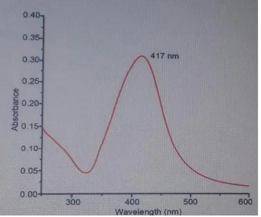
The formation and characterization of silver Np was determine by using UV-Vis absorption spectrometer with wave length band about 300-600 nm ,the band of surface plasmon
resonance (SPR) determined the morphology of the nanoparticles ,SPR bands are influenced by the morphology, size, and shape of the nanoparticles ,many studies have shown that the spherical silver Np contributes to the absorption band around 400 nm [Stamplecoskis K. 2010.] ,absorption band in this study was around (417) as shown in fig (2) wich strongly suggests that silver Np were spherical in the shape.
[image:4.595.175.420.167.317.2] [image:4.595.170.429.515.731.2]www.wjpr.net
, 2016. Vol 5, Issue 9
1645
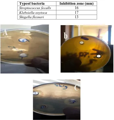
The size of nanoparticles determines its activity against bacteria because the binding of nanoparticles depends the surface area available for interaction ,when the particle is small this make large surface area available for interaction and this causes more activity of the nanoparticles against bacteria. table (1) and fig (3) show the inhibition zones of Bactria
Streptococcus fecalls, Klebsiella oxytoca, and Shigella flexneri respectively
Table (1): Inhibition zones of bacteria
Typeof bacteria Inhibition zone (mm)
Streptococcus fecalls 16
Klebsiella oxytoca 17
Shigella flexneri 13
Fig (3): Appearances of inhibitory zone with (A):Streptococcus fecalls (B): Klebsiella oxytoca, and (C):Shigella flexneri
The results shows that the nanoparticles have strong activity against gram negative and
gram positive bacteria and have more activity against Streptococcus fecalls and, Klebsiella oxytoca. the activity of the Nanoparticles against bacteria have been reported by many studies, but the mechanism of bactericidal effect of nanoparticles is not very well known, some studies have reported that the electrostatic attraction between positive charge of silver
[image:5.595.115.479.206.588.2]www.wjpr.net
, 2016. Vol 5, Issue 9
1646
Np and negative charged cell membrane of microorganism[Hamouda T2000, Dibror etal.,2002,Dragieva etal.,1999],other studies reported that the oxidation react at the surface of nanoparticles silver ion diffusion, thus causing structural change and finally bacterial will die. Melinte etal., 2011.
Other reason for bacteria death is that the silver Np may attach to surface membrane and disturb its power function such as permeability and respiration, Morible & et al G2009.
In conclusion silver Np has strong activity against bacteria and this activity is influenced by the size of nanoparticles, because of their size it can easily reach the nuclear content of bacteria and they present the large and impressive surface aria thus the contact with bacteria were the greatest, it can be reason behind the strong activity against bacteria and then more small particles make large inhibition zones
REFERENCES
1. Aslan K.,Lakowicz J., Geddes C.,2005" Plasmon light scattering in biology and medicine 'New sensing Approaches Visions and perspectives ",current opinions in chemical biology :analytical techniques ,9; 538.
2. Clinical and Laboratory Standards Institute (CLSI). (2012). Performance standards for Antimicrobial susceptibility testing. Approveds tandard M100-S20. 32(3); National Committee for Clinical Laboratory Standards, Wayne, PA.
3. Davies J.1994." inactivation of antibiotic and the dissemination of resistance genes), sci. 264: 375-382.
4. Dibror P.,Dzioba J.,Gosink K.,2002"chemiosmotic mechanism of antimicrobial activity of Ag (+) in vibrio cholera ,antimicrob agents chemo her ,46: 68-70..
5. Dragieva I.,Storea S.,Stoimeaov P.,1999" complex formation in solutions for chemical synthesis of nanoscaled particles prepared by borohydride reduction process ,Nano structure mater, 12: 267-270.
6. Emody, L.; Kerényi, M. and Nagy G. 2003"Virulence factors of uropathogenic
Escherichia coli International. J. Antim. Agent. 22: S29-S33.
8. Hamouda T.,Mys A.,Donovan B.,2000" Anovel surfactant nano emulsion with unique non-irnilat topical antimicrobial activity against bacteria enveloped viruses and fungi", Microbial Res., 156: 1-7.
9. Hangxumx U. and Kenmeths ,2010" water soluble fluorescent silver Nano clusters ,Adv.Mater, 22: 1078-1082.
10.Mamun U.,Khairl H.,EmranQ.2013." synthesis of silver nano particales (Ag-Nps) and their uses for Quantitative Analysis of Vitamin C tables )". Dhakaunir J.pharm. Sci.12(1). 11.Marible G., Guzma N.,jean D, etal ,2009"synthesis of silver nanoparticles by chemical
reduction method and their antibacterial activity", Int.J.chem.and bio.eng,2;104-111, Melinte V.,Burvana T.,Dmorarv I.,2011" silver – polymer composite materials with
antibacterial properties ,Digest J.of Nano materials and bio structures ,6: 213-223: 2011. 12.Petr S.,Ales P.,Robert P.et al, 2013" synthesis of small silver nanoparticles and their
catalytic activity in 4- nitrophenol reduction ".nanocon,16.18.
13.Rodriguez S.,Blanco M., Lopez Q.,2000" electro chemical synthesis of silver nanoparticles , J.,phy.,che.,104;: 9683-9688.
14.Sapsford K.,pons T.,Mediutz L.,etal,2006" Bio sensing with luminescent semiconductor quantum dots ",sensors ,6,925, Sepideh K.,Kamyar S., Zurina Z.," Rapid and green synthesis of silver nanoparticles Via sodium alginate media ",int.J.electrchem.sci, 2015; 10:486-497.
15.Shameli K., Ahmed M.,Zargar M., etal ,2011 "synthesis and characterization of silver /montmorillonite / chitosan bio nano composites by chemical reduction method and their antibacterial activity ,Int.J.Nano.Med., 6: 271-284.
16.Silva E.,Rivas J.,Leon L.2007," synthesis of silver –coated magnetite nanoparticles, J.Non crystalline solids , 352: 829-831.