Adaptive immune response in human Staphylococcus
aureus bloodstream infection
This thesis is submitted to the University of Dublin for the degree of
Doctor of Medicine (M.D.)
Dr Aisling F. Brown
MB BCh BAO MRCPI DipHIVMed School of Medicine
Trinity College Dublin March 2019
Advisors: Prof Rachel McLoughlin and Prof Thomas Rogers
iii
Declaration
I declare that this thesis has not been submitted as an exercise for a degree at this or any other university and it is entirely my own work. Where any of the content presented is the result of collaborative research, this is duly acknowledged in the text such that it is possible to ascertain how much of the work is my own.
I agree to deposit this thesis in the University’s open access institutional repository or allow the Library to do so on my behalf, subject to Irish Copyright Legislation and Trinity College Library conditions of use and acknowledgement.
I consent to the examiner retaining a copy of the thesis beyond the examining period, should they so wish (EU GDPR May 2018).
Signed: Aisling Brown 27th March 2019
v
Summary
Staphylococcus aureus bloodstream infection is a common and life-threatening condition. Treatment outcomes are poor, suboptimal clinical management is common, antimicrobial resistance continues to emerge, and there are few new agents in the drug discovery pipeline. There is an urgent need for novel methods of preventing and treating S. aureus infection. A lack of understanding of protective immunity or its correlates has greatly impeded progress in the development of rationally-designed anti-S. aureus vaccines. This project set out to characterise the pathogen, its clinical impact on patients, and the adaptive immune response it induced in humans, with a particular focus on T lymphocytes.
The clinical characteristics, management and outcomes of adult patients with S. aureus and E. coli bloodstream infection (SA-BSI and EC-BSI) were contrasted. The bulk of S. aureus disease was attributable to methicillin-susceptible S. aureus and infection was frequently community-acquired. S. aureus’s capacity to cause severe disease in otherwise healthy people
and to follow a complicated course distinguished it from E. coli. Failure, relapse or death was
seen in a large proportion of SA-BSI, whereas all E. coli infections were easily and rapidly cured. Healthcare resource utilisation was significantly greater in S. aureus infection. Of concern, clinical management of SA-BSI was suboptimal in over half of cases, and occurred when patients were not under the care of infection specialists. There was considerable genotypic heterogeneity, particularly in MSSA, in invasive S. aureus isolates. Despite this variety in lineage, genes for several cell-wall antigens were present and conserved on next-generation genome sequencing, which may be promising for vaccine development.
to S. aureus during recovery from bloodstream infection. This response was primarily Th1-mediated, with a lesser contribution from Th17 cells. Th1 immune memory was formed or re-activated during systemic infection. Similar pathogen-specific T helper cell activation was not seen in patients with E. coli bloodstream infection, implying a substantial difference between the role of T cells in these two bacterial infections. A S. aureus antigen-specific Th1/17 signature thus seems particular to recovery from invasive staphylococcal disease and, similar to animal models, may be a correlate of protective immunity.
The final part of this work successfully developed a model assay to measure T cell immunogenicity of specific S. aureus antigens (and T cell-directed adjuvants) using blood donor buffy coats and clumping factor A as a model antigen. This assay can be used to map T helper cell responses over the course of infection in future SA-BSI patients to correlate relative potency of T cell activation with clinical outcome.
vii
Acknowledgement
There are many people that deserve my thanks for their contribution to this work.
I would particularly like to thank Professor Rachel McLoughlin for taking a chance on a medic as a novice scientist and for her insightful and encouraging ideas, feedback and constant support. I owe a debt of gratitude too to Professor Tom Rogers for his kind and perceptive advice throughout the years.
To all my colleagues in the McLoughlin Lab Host-Pathogen Interactions group, and in our allied 3.12 labs and its reading room for teaching me how to do everything. Their experience and ability meant that my mistakes were usually caught in time and if not, at least both commiserations and pizza were shared. Dr Micheál Mac Aogáin and Dr Dara O’Halloran are colleagues from other departments who deserve special mention for their assistance with particular experiments.
To the medical scientists and clinical microbiologists in St. James’, Tallaght and Beaumont Hospitals, thank you for letting me wander in and out to check the bacteraemia books, sub my plates and for all your courtesy and assistance in this project.
The years between taking my first tentative steps in the lab to completing this thesis have seen many changes in my life – completion of specialist training, moving country (twice), my first appointment as a consultant physician and the addition of a new member of our family. My friends and family have provided a constant quiet support without which none of that could have gone as smoothly as it did.
ix
Abbreviations
AD Atopic dermatitis
AD-HIES Autosomal dominant hyper-IgE syndrome
AMR Antimicrobial resistance
APC Antigen-presenting cell
BSI Bloodstream infection
CA Community-acquired
CA-MRSA Community-acquired methicillin-resistant S. aureus
CC Clonal complex
CD Cluster of differentiation
CFU Colony-forming units
CGD Chronic granulomatous disease
ClfA Clumping factor A
CTL Cytotoxic T lymphocyte
E. coli Escherichia coli
EC-BSI Escherichia coli bloodstream infection
HA Hospital-acquired
HCA Healthcare-associated
HKEC Heat-killed E. coli
HKSA Heat-killed S. aureus
HLA Human leukocyte antigen
IFNγ Interferon gamma
IL-10 Interleukin-10
IL-17 Interleukin-17
MDSC Myeloid suppressor dendritic cells
MHC Major histocompatibility complex
MLST Multilocus sequence typing
MRSA Methicillin-resistant S. aureus
MSSA Methicillin-susceptible S. aureus
OD Optical density
RC Research clinician
ROS Reactive oxygen species
S. aureus Staphylococcus aureus
SA-BSI Staphylococcus aureus bloodstream infection
SEA Staphylococcal enterotoxin A
SpA Staphylococcal protein A
ST Sequence type
TCR T cell receptor
Th T helper cell
Th1 T helper cell type 1
Th17 T helper cell 17 (producing IL-17)
Th2 T helper cell type 2
TNFα Tumour necrosis factor alpha
xi
Table of Contents
Chapter 1. Introduction ...1
1.1. The pathogen, the host and infectious disease ...3
1.2. Staphylococcus aureus...5
1.2.1. Clinical significance...5
1.2.2. Epidemiology ...7
1.2.3. Colonisation...9
1.2.4. Transmission and Infection...9
1.2.5. Genome...10
1.2.6. Antimicrobial resistance...10
1.2.7. Virulence Factors ...13
1.3. Immune response to Staphylococcus. aureus infection ...14
1.3.1. Innate immune response to S. aureus infection ...14
1.3.2. Adaptive immune response to S. aureus infection ...18
1.3.2.1. B Lymphocytes and S. aureus...18
1.3.2.2. T lymphocytes and S. aureus ...18
1.3.2.1.1. αβ T cells (CD4+ T helper cells and CD8+ cytotoxic T cells)..18
1.3.2.1.2. γδ T cells and S. aureus ...21
1.4. Immune Regulation ...25
1.5. Why Immunity Fails ...26
1.5.1. Immune Evasion ...26
1.5.2. Host Immunodeficiency and Susceptibility to S. aureus infection ...27
1.6. Immunisation and Immune memory ...29
1.6.1. Clinical experience with S. aureus vaccines ...30
1.6.2 Challenges in S. aureus vaccine development ...36
1.7. Project aims ...38
Chapter 2. Materials & Methods ...39
2.1. Materials ...41
2.1.1. General reagents ...41
2.1.2. Custom (mixed) reagents ...41
2.1.3. Protein Assay Reagents ...42
2.1.4. FACS Antibodies and Reagents ...42
2.1.5. ELISA kits ...43
2.2. Clinical Study Methods ...45
2.2.1. Study Design and Recruitment ...45
2.2.2. Clinical Definitions ...48
2.2.3. Sample Handling ...49
2.2.4. Ethical approval ...49
2.2.5. Blood Donors ...50
2.2.6. Funding ...50
2.3. Microbiological Methods ...51
2.3.1. Bacterial identification and Antimicrobial Susceptibility Testing ...51
2.3.2. Genetic characterisation of S. aureus strains ...51
2.3.3. Virulence and immune evasion genes in S. aureus isolates ...52
2.3.4. Preparation of heat-killed S. aureus and Escherichia coli ...55
2.3.5. Protein Quantification ...57
2.3.6. Expression of Clumping factor A in S. aureus before and after heat-inactivation ...57
2.3.7. Bacterial adherence to fibrinogen before and after heat-inactivation ...57
2.3.8. Purification of Clumping factor A ...58
2.4. Immunological Methods ...59
2.4.1. Isolation of sera and cells ...59
2.4.2. Short ‘ex vivo’ re-stimulation with heat-killed bacteria ...59
2.4.3. CFSE staining ...59
2.4.4. Lymphocyte proliferation assay ...60
2.4.5. T cell-enriched lymphocyte proliferation assay ...64
2.4.6. Surface and intracellular cytokine staining ...67
2.4.7. Flow cytometry ...67
2.4.8. Enzyme-linked immunosorbent assay (ELISA) ...71
2.4.9. Antibody Assays ...71
2.5. Statistical analysis ...72
Chapter 3. Clinical and Microbiological characterisation of Staphylococcus
aureus bloodstream infection ...73
3.1 Introduction ...75
3.2 Results ... .78
3.2.1 Recruitment and clinical course of patients with S. aureus and E. coli bloodstream infections ...78
xiii
3.2.1.2 Clinical Characteristics, Management and Outcomes ...81 3.2.2. Genotypic and phenotypic characterisation of S. aureus bloodstream
isolates...96 3.2.2.1 Multilocus sequence typing (MLST) and clonal complex (CC) distribution ...96 3.2.2.2 Prevalence of Virulence and Immune Evasion Factor genes in S. aureus bloodstream isolates ...104 3.2.2.3 Clumping factor A illustrates the limited antigenic variability of candidate vaccine antigens ...106 3.2.2.4 Antimicrobial resistance in S. aureus bloodstream isolates ...108 3.3 Discussion ...110
Chapter 4. Adaptive immune responses in human Staphylococcus aureus
bloodstream infection ...115
4.1 Introduction ...117 4.2 Results ...120Chapter 5. General Discussion ...163
5.1 Summary of Results ...165
5.2 Host-Targeted Therapeutics for invasive S. aureus infection ...169
5.2.1 Immunomodulatory Therapy for S. aureus infection ...169
5.2.2 The Future for next-generation S. aureus vaccines ...170
5.2.3 A Roadmap for S. aureus Vaccine Clinical Trial Design ...172
5.3 Future Directions ...173
References ...175
xv
List of Figures
Chapter 1
Figure 1.1. Host-pathogen interactions determine disease severity ... 4 Figure 1.2. National S. aureus bloodstream infection rates (2008-2017) for Ireland including methicillin resistance rates ...6 Figure 1.3. National S. aureus bloodstream infection rates (2017-2018) for England by age group and gender ... 8 Figure 1.4. Emergence of antibiotic-resistant S. aureus and variation in MRSA rates across European countries ... 12 Figure 1.5. Immune cell lineage ... 17 Figure 1.6. T lymphocyte activation ... 23 Figure 1.7. CD4+ T cell differentiation depends on the cytokine environment (Signal 3) ... 24
Chapter 2
Figure 2.1. Gram staining and microscopy of S. aureus before and after heat inactivation ...56 Figure 2.2. PBMC assay for investigation of S. aureus antigen-induced T cell proliferation and cytokine production in bloodstream infection patients ... 62 Figure 2.3. Plate map PBMC S. aureus antigen-specific proliferation assay ... 63 Figure 2.4. CD4+ T cell-enriched assay for investigation of antigen-specific T helper
proliferation ... 66 Figure 2.5. Fluorescence-activated cell sorting (FACS) gating strategy for proliferation assay ... 69
Chapter 3
Figure 3.7. MLST type and clonal complex distributions of S. aureus bloodstream isolates ...99 Figure 3.8. Whole genome sequencing demonstrates significant genetic diversity among invasive S. aureus isolates ... 100 Figure 3.9. Lineages of bloodstream S. aureus strains do not cluster by site ... 101 Figure 3.10. Lineages of bloodstream S. aureus strains do not cluster by acquisition risk ... 102 Figure 3.11. Lineages of bloodstream S. aureus strains do not cluster by complexity of infection... 103 Figure 3.12. Presence of virulence and immune evasion genes in clinical and laboratory S. aureus isolates ... 105 Figure 3.13. Allelic variation in clumping factor A (clfA) genes of clinical and laboratory S. aureus isolates ... 107 Figure 3.14. Methicillin resistance in S. aureus bloodstream isolates from recruited adult patients ... 109
Chapter 4
Figure 4.1. Non-specific inflammatory marker C-reactive protein is similar in initial and early recovery periods of S. aureus or E. coli bloodstream infection ... 122 Figure 4.2. Absolute neutrophil count and proportion are similar in initial and early recovery periods of S. aureus or E. coli bloodstream infection. ... 123 Figure 4.3. Lymphocyte recovery differs in S. aureus and E. coli bloodstream infection ...
... 124 Figure 4.4. T lymphocyte subset proportions remain within normal ranges during early recovery from S. aureus or E. coli bloodstream infection ... 126 Figure 4.5. S. aureus bloodstream infection is associated with elevated serum IFNγ and Th1-skewed response ... 128 Figure 4.6. IFNγ responses from S. aureus antigen-specific CD4+ and CD8+ T lymphocytes are evident in S. aureus bloodstream infection ... 131 Figure 4.7. E. coli bloodstream infection induces pathogen-specific IFNγ production in
xvii
bloodstream infection patients, and this proliferation is patient-specific rather than strain-specific ... 137 Figure 4.10. CD4+ T lymphocytes from S. aureus bloodstream infection patients proliferate significantly more than CD4+ cells from E. coli bloodstream infection patients in response to S. aureus antigens ... 138 Figure 4.11. S. aureus-specific CD8+ proliferation and E. coli-specific CD4+ proliferation is not significantly different between S. aureus and E. coli bloodstream infection patients ... 139 Figure 4.12. S. aureus antigen-specific Th1 expansion is evident only in CD4+ T lymphocytes from S. aureus bloodstream infection patients, and does not differ between bacterial strains ... 141 Figure 4.13. S. aureus antigen-specific Th1 expansion is the major T lymphocyte response in S. aureus bloodstream infection patients ... 142 Figure 4.14. S. aureus bloodstream infection induces significant S. aureus antigen-specific IFNγ response exclusively from CD4+CD45RO+ memory T lymphocytes ...145 Figure 4.15. Anti-staphylococcal antibodies in health and disease ... 147 Figure 4.16. Anti-staphylococcal antibodies that discriminate active S. aureus infection ... 148 Figure 4.17. Anti-Clumping factor A IgG antibodies are detectable in S. aureus bloodstream infection patients and healthy volunteers ... 151 Figure 4.18. Expression of ClfA is conserved across strains and remains present on the cell surface after heat-killing ... 152 Figure 4.19. Heat inactivation of S. aureus significantly impairs binding to fibrinogen ..
List of Tables
Chapter 1
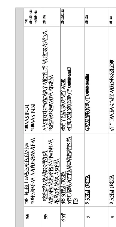
Table 1.1. Clinical trials of anti-S. aureus vaccines ... 34
Chapter 2
Table 2.1. General Reagents ... 41Table 2.2. Custom (Mixed) Reagents ... 41
Table 2.3. Protein Assay Reagents ... 42
Table 2.4. FACS antibodies and Reagents ... 42
Table 2.5. ELISA Ready-SET-Go!® kits ... 43
Table 2.6. ELISA Reagents ...44
Table 2.7. Inclusion and Exclusion Criteria ... 47
Table 2.8 Genotypic analysis sources for selected S. aureus antigen genes ... 53
Chapter 3
Table 3.1. Clinical characteristics and outcomes of S. aureus bloodstream infection patients ... 85Table 3.2. Clinical characteristics and outcomes of E. coli bloodstream infection patients ...89
Table 3.3. Comparison of S. aureus and E. coli bloodstream infection patient demographics and infection source ... 90
Table 3.4. Antimicrobial consumption in S. aureus bloodstream infection patients ... 95
Table 3.5. Characteristics of invasive S. aureus isolates ...98
Chapter 1
1.1. The pathogen, the host and infectious disease
In 1881, Alexander Ogston, the surgeon who first described Staphylococcus aureus, wrote that the bacterium “has been held to be the most potent factor in producing blood-poisoning and has also been asserted to be an innocent organism” [1]. This paradox persists today. S. aureus colonises a substantial proportion of us. Colonised individuals establish an equilibrium with bacteria and most never develop staphylococcal infection. In apparent contrast, S. aureus is also an opportunistic pathogen which frequently causes life-threatening community- and healthcare-acquired infections. Ogston found that experimental infection with S. aureus produced “varying effects”: “blood-poisoning… suppuration…” or “may be resisted by strong individuals under favouring circumstances” [1]. What factors conspire to determine these outcomes remain largely unknown to this day, and S. aureus’s dual identity as innocent bystander and guilty party endures.
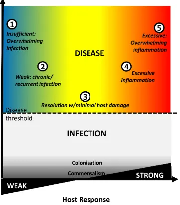
The traditional pathogen-centred view, with microbes as the sole perpetrators of infectious diseases, has changed. Increasingly, the concept of a healthy host incorporates a diverse microbiome, including S. aureus and other pathobionts. The mere presence of potentially pathogenic bacteria does not inevitably herald clinical infection, largely due to a successful and balanced immune response. Possible outcomes of host-microbe contact are shaped by a complex dance between both parties (Fig. 1.1). Commensalism or colonisation occurs when interaction with a microbe, though it can elicit an immune response, results in no (or clinically inapparent) damage to the host. Infectious disease is a clinical manifestation of damage, which may be pathogen-mediated, host-mediated, or both, and evolves differently among individuals [2]. Ideally, it is resolved with minimal host damage and lasting protective immunity. Alternatively, the pathogen directly damages host tissues, or leads to overwhelming infection. Conversely, an excessive, dysregulated, or misplaced host immune response can lead to overwhelming inflammation.
Figure 1.1. Host-pathogen interactions determine disease severity
Commensalism/colonisation occurs when host-microbe contact results in no apparent damage, though it can elicit an immune response. Infectious disease is a clinical manifestation of damage, which may be pathogen-mediated, host-pathogen-mediated, or both. If immune response is insufficient, overwhelming infection may result (1). Where response is weak, recurrent or chronic infection occurs (2). An ideal response easily resolves infection with minimal host damage (3). A persistently dysregulated immune response leads to chronic inflammation (4). Finally, where host response is entirely excessive, overwhelming inflammation may cause death (5).
1.2. Staphylococcus aureus
1.2.1. Clinical Significance
Staphylococcus aureus is a species of Gram-positive, coagulase-positive bacteria. A major human pathogen, it causes diverse disease: superficial cutaneous infection (e.g., skin abscess, surgical site infections); systemic disease (e.g., bloodstream infection, endocarditis); serious deep-seated infection (e.g., osteomyelitis, septic arthritis, necrotising pneumonia, meningitis); and toxinoses (e.g., toxic shock syndrome, scalded skin syndrome and food poisoning). Cutaneous infection accounts for the majority of staphylococcal disease, but is rarely life-threatening. Serious invasive S. aureus infections, on the other hand, have an enormous impact.
Figure 1.2. National S. aureus bloodstream infection rates (2008-2017) for Ireland including methicillin resistance rates
1.2.2. Epidemiology
Age is a powerful determinant of invasive disease with the highest incidence of SA-BSI occurring at the extremes of life (Fig. 1.3). This is not unique to SA-BSI, but rather a universal finding among all bloodstream infections [14]. Age remains a predictor of increased S. aureus-attributable mortality even after correction for co-morbities and other variables [3,15]. Male gender is also consistently associated with increased rates for unknown reasons [16]. While some studies have sugggested females have a higher mortality due to hormonal or health-seeking behaviour differences, larger age-adjusted studies have not borne this out [3]. Lower socioeconomic status is also independently associated with increased incidence of SA-BSI, particularly CA-MRSA. Invasive infection is also seen more frequently among certain ethnicities (including African-Americans, indigenous Australians, Maoris) as compared to Caucasian populations [16]. This finding is not fully explained by socioeconomic disparities between ethnic groups and is not associated with an increased mortality. It may, however, reflect the much greater burden of staphylococcal and other skin disease among indigenous populations [17]. The genetic or immunological basis for any of these host susceptibility differences has not yet been investigated.
Figure 1.3. National S. aureus bloodstream infection rates (2017-2018) for England by age group and gender
Population rates by age group and gender for SA-BSI in England demonstrate the burden of MSSA disease as compared to MRSA infection, as well as an increased incidence in tnfants and older adults. There is also a consistently higher rate ratio for males to females. Figure adapted from Public Health England [22].
1.2.3. Colonisation
The primary S. aureus reservoir in humans is the anterior nares, although colonisation at other sites is common. Extra-nasal sites include skin, throat, perineum, vagina and gastrointestinal tract [23-25]. Approximately 20% of adults are persistently colonised with S. aureus, and the remainder intermittently carry fewer bacteria [26]. Certain populations tend to have higher rates of colonisation, e.g., almost all adults with atopic dermatitis are S. aureus carriers [27]. Defining differences between carrier and non-carrier groups has proved difficult. Particular strains of S. aureus may be superior colonisers, but predilection for S. aureus colonisation seems largely host-determined [19]. Experimental nasal inoculation results in most volunteers returning to their original non-carrier or persistent carrier state [26]. Nasal carriage of S. aureus is strongly associated with infection. Carriers have up to a 20-fold relative risk for bloodstream infection [28,29]. Despite this, the vast majority will never develop invasive disease.
Bacterial carriage may also ‘prime’ the immune system, changing its behaviour – either favourably or unfavourably – during subsequent infection [30-32]. Intriguingly, some data suggest that nasal carriage, while strongly predictive of disease, may be partially protective in invasive infection [30,33]. Among those who developed SA-BSI, carriers had lower all-cause and S. aureus-attributable mortality than non-carriers [30]. There were signiciant clinical confounders which may have influenced this difference in outcome – carriers were significantly younger, had fewer cardiac issues but were non-specifically ‘immunocompromised’. It is possible that immunocompromise may have dampened detrimental excessive inflammation. Alternatively, a degree of immune tolerance to S. aureus may have created a better orchestrated response to invasive disease. Although the mechanism is not known, examining the immune imprint of S. aureus colonisation is critical to predicting and influencing response to infection and immunisation in future patients.
1.2.4. Transmission and Infection
infection) [37]. Determinants of whether a new acquisition will end in eradication, colonisation or infection are unknown.
1.2.5. Genome
S. aureus population structure is largely clonal with infrequent spontaneous mutations giving rise to new lineages. The distribution of these lineages differs geographicallly. S. aureus has core and accessory genomes [38]. The core genome contains ~ 75% of genes that encode for essential proteins. Most are highly conserved, and thus a reliable method of inferring evolutionary relationships. ‘Core variable’ genes – often encoding regulators of virulence genes or surface proteins involved in host interactions – vary between lineages or may even be missing. The accessory genome is largely comprised of horizontally exchanged mobile genetic elements (fragments of DNA encoding toxins, virulence factors, antimicrobial resistance and genes involved in host adaptation).
1.2.6. Antimicrobial resistance (AMR)
The advent of anti-staphylococcal antibiotics revolutionised outcomes for patients with S. aureus BSI, reducing mortality by two-thirds [10,39]. This panacea was short-lived. Immense quantities of (often inappropriate) antimicrobials introduced into the clinical and wider environments since the 1940s applied enough selective pressure to bring resistance mechanisms already present in bacteria to the fore. This condensed the emergence and global spread of AMR in staphylococci into a remarkably short timeframe.
confined to hospitals, are now acquired in the community. Community-acquired MRSA (CA-MRSA) was first described in remote indigenous communities in Western Australia as genetically and phenotypically distinct from healthcare-associated strains, but is now prevalent in many parts of the world [43]. MRSA has added to, rather than replaced, the overall burden of SA-BSI. The problem of beta-lactam resistance is further amplified by S. aureus having also demonstrated resistance to every other current anti-staphylococcal drug [40,44,45].
Figure 1.4. Emergence of antibiotic-resistant S. aureus and variation in MRSA rates across European countries.
Timeline of the emergence of penicillin, methicillin-, and vancomycin resistant S. aureus in the USA (A) [49]. Proportion of invasive S. aureus isolates with resistance to methicillin (MRSA) by EU/EEA country 2017 (B). Data from European Centre for Disease Prevention and Control Surveillance Atlas (Antimicrobial Resistance). Available at https://ecdc.europa.eu/en/antimicrobial-resistance/surveillance-and-disease-data/data-ecdc. [Accessed Oct 2018]
PRSA = penicillin-resistant S. aureus; MRSA = methicillin-resistant S. aureus; VRSA = vancomycin-resistant S.
[image:30.595.65.500.125.530.2]1.2.7. Virulence Factors
1.3. Immune response to S. aureus infection
The immune system comprises cells and molecules responsible for protection against infection. It can be divided into two arms which work in synergy. The innate immune system provides an immediate, non-specific response to neutralise or eliminate a pathogen. The adaptive immune system provides a delayed acquired response to specific pathogen elements that, crucially, can form immunological memory. Memory results in faster and more precise attacks on subsequent encounters with the same pathogen, and forms the basis of immunisation.
1.3.1. Innate Immune Response to S. aureus
Recognition and initial inflammatory response
Phagocytosis and Antigen Presentation
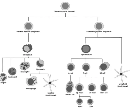
The professional phagocytes – neutrophils, macrophages and dendritic cells (Fig. 1.5) – are richly endowed with PRRs. They are the most important effector cells of the innate immune system. Phagocytes utilise chemical cocktails to kill engulfed micro-organisms. Dead or dying neutrophils are the major component of the pus so characteristic of S. aureus infection. Neutrophils are equipped with three major methods of killing bacteria – production of reactive oxygen species (ROS), release of packaged enzymes via degranulation and NETosis [55,56]. Monocytes leave the circulation to mature into macrophages, the major tissue-resident phagocytes. Activated macrophages act as scavengers, ingesting pathogens and dying cells, to clear infection and limit inadvertent tissue damage [57]. Although neutrophils are conventionally thought of as the primary protective phagocyte in S. aureus infection, human macrophages show comparable intracellular killing of S. aureus [58]. Phagocytes recruit other inflammatory cells via chemokine signalling. Many are also professional antigen-presenting cells (APCs) that serve as the crucial link between innate and adaptive immunity. These migrate to regional lymph nodes and present fragments of digested bacteria (peptide:MHC complexes) along with co-stimulatory molecules to expand and polarise antigen-specific T lymphocytes and trigger adaptive immunity (Fig 1.6). Dendritic cells (DCs) are the primary APCs of the immune system. A new antigenic challenge cannot induce a T cell response without DC involvement.
Figure 1.5. Immune cell lineage.
1.3.2. Adaptive immune response to S. aureus infection
The adaptive immune system is essential for eliminating pathogens that have not been cleared by more primitive defences. It constructs receptors specific to individual pathogen elements, allowing them to be targeted with much greater precision and, critically, generating memory. The antigen-specific cells formed during primary infection persist as memory cells that react more readily and effectively on re-infection. It comprises humoral (antibodies) and cellular (lymphocytes) components.
1.3.2.1. B lymphocytes and S. aureus infection
B lymphocytes recognise extracellular antigens and become efficient antibody-producing cells, with assistance from helper T lymphocytes. Antibodies may have neutralising or opsonising functions. Immunoglobulin (Ig) M is the first class of antibody produced, followed by IgG. IgG-opsonised bacteria engage phagocyte receptors to mediate phagocytosis. B cell cytokines also polarise T helper cells towards specific functions (Fig. 1.7).
There is little evidence that B cell deficiency states in humans (or in mice) result in greater incidence or severity of invasive S. aureus disease [15,64]. Healthy children and adults already have a multitude of anti-S. aureus antibodies [65]. During S. aureus infection, B cells readily generate antibodies against multiple S. aureus components. These antibodies appear functional and potentiate opsonophagocytosis in vitro. However, it has proved difficult to consistently correlate presence or titre of anti-staphylococcal antibodies with improved clinical outcomes [66-68]. The role of B cells in defence against S. aureus is further complicated by staphylococcal protein A, which so effectively disables the humoral immune response during infection (see 1.5.1). Finally, neither passive immunisation, nor active immunisation with vaccines that successfully produced robust humoral responses, have prevented or attenuated the course of infection in clinical trials (Table 1.1).
1.3.2.2. T Lymphocytes and S. aureus infection
T cells are identified by the surface marker CD3, and are categorised on the basis of T cell receptor (TCR; e.g., αβ or γδ). Intravenous S. aureus challenge in T cell knockout mice is rapidly fatal [64].
1.3.2.2.1. αβ T lymphocytes
lymphocytes are helper cells which produce cytokines to promote other immune cell functions. CD8+ cells can directly kill compromised or infected cells. T cell response is
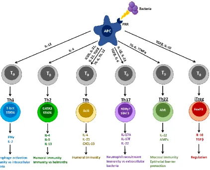
orchestrated by the APCs of the innate immune system by three signals – processing and presenting of antigens (signal 1), co-stimulation (signal 2), and secretion of cytokines to direct lymphocyte differentiation (signal 3) (Fig. 1.6). Subsets of T helper cells and their roles in S. aureus infection are reviewed below.
CD4+ T cells and S. aureus
The primary function of CD4+ T cells is to produce cytokines to help other immune cells.
CD4+ subsets are characterised by expression of specific transcription factors, signal
transduction pathways and cytokine secretion profiles (Fig. 1.7).
Th1 cells
Th1 cells produce IFNγ as their signature cytokine. IFNγ activates macrophage phagocytosis and ROS formation. It enhances antigen presentation by upregulating expression of MHC. Finally, IFNγ promotes immunoglobulin switching to IgG1/3, which bind avidly to phagocyte Fc receptors and activate opsonophagocytosis [69]. There is accumulating clinical and experimental evidence that Th1 cells play important roles in protection against S. aureus, particularly in systemic infection. Patients with acquired impairment of Th1 function (e.g., HIV infection, end-stage renal disease and diabetes mellitus) have the greatest relative risk for S. aureus bloodstream infection [18,20,21]. Murine studies also imply a protective role for these cells. Th1-skewed C57BL/6 mice were significantly more likely than Th2-skewed Balb/c to survive i.v. inoculation [70]. Adoptive transfer of S. aureus-specific Th1 cells led to accelerated clearance of bacteria [32]. Th1 cells are a dominant source of IFNγ, and exogenous IFNγ administration increased phagocytosis, bacterial clearance and survival in systemic infection [70,71]. Deficiency in IFNγ or its receptor was associated with worse survival [72,73]. Th1 response seems most beneficial early in the course of systemic infection, by aiding phagocytic clearance of extracellular S. aureus, and activating phagocyte killing of intracellular staphylococci.
the site and stage of infection, and needs regulation to avoid counterproductive inflammation (see 1.4. Immune Regulation).
Th2 cells
Th2 cells produce IL-4 as their signature cytokine. They promote B lymphocyte proliferation, antibody production, isotype switching to IgG2/4 and IgE, and activation of eosinophils to control large extracellular pathogens like helminths and other parasites. Th1 and Th2 lymphocytes are antagonistic. Th1-derived IFNγ directly suppresses IL-4 secretion, inhibiting differentiation of naïve T cells into Th2 cells. Th2-derived IL-4 suppresses secretion of IL-12 and IFNγ, blocking Th1 development [69]. Th2 cells drive antibody production in S. aureus infection.
Th17 cells
Th17 cells produce IL-17 as their signature cytokine and are important in mucosal protection against extracellular bacterial and fungal infections [75]. IL-17 recruits neutrophils to sites of infection. It may prevent haematogenous dissemination from mucosal S. aureus infection [76]. Mice deficient in IL-17A/F are more susceptible to mucocutaneous, but not to systemic, S. aureus infection than wild type mice [77,78]. Adoptive T cell transfer from Th1-deficient S. aureus-exposed mice lessened severity of S. aureus skin infection – an effect thought due to transfer of IL-17-producing T cells. Blocking of IL-17 abrogated this protection [79]. Gamma-delta T cells (discussed below) seem the predominant source of IL-17 in mice [31,80,81].
Data from the clinic supports the hypothesis that IL-17-producing T cells are essential to mucocutaneous immunity, although CD4+ (Th17), not γδ+, T cells are the primary source.
CD8+ T cells and S. aureus
CD8+ T cells are cytotoxic T lymphocytes (CTLs) that interact with MHC I molecules on the
surface of compromised host cells, such as those infected or transformed by malignancy. CTLs produce cytokines (e.g., IFNγ and TNFα) and/or release of cytotoxic granules to stimulate apoptosis. CTLs are particularly effective in defence against viruses, but also play a role in some, particularly intracellular, bacterial infections (e.g., Mycobacterium tuberculosis). CD8+ T cells have not been demonstrated to play a significant role in defence
against S. aureus infection. Adoptive transfer of vaccine-induced S. aureus-specific CD8+ T
cells did not demonstrate protection in murine experiments and a CTL response was not generated to any of four S. aureus antigens in a human vaccine study [88,89] .
1.3.2.2.2. Gamma-delta (γδ) T cells and S. aureus
Gamma delta T cells are at a crossroads between adaptive and innate cells, and express TCRs composed of γ and δ chains. They are a minority population in the blood, but are abundant at epithelial sites, where they contribute to mucosal immunity. In addition to their γδ TCR, they can also be activated by PRR ligation, cytokine stimulation and/or specific antigens [90]. Gamma-delta cells can be directly cytotoxic, or may act as phagocytes, cytokine-secretors or APCs. There are three major human γδ subsets. (Vγ9) Vδ1 cells are primarily distributed in skin and mucosa, Vδ2 in peripheral blood, and Vδ3 are a scanty population found in gut and liver. Human γδ cells have significant plasticity, and can readily assume Th1-, Th2-, Th17- or Treg-like functions [91].
Staphylococci can activate murine γδ+ cells. In cutaneous and pulmonary S. aureus infection,
γδ+ cell IL-17 production is important in early containment and clearance of staphylococci,
presumably by recruiting and activating neutrophils [81,92]. In a peritonitis/systemic infection model, γδ+ lymphocytes producing IL-17 (but not IFNγ) accelerated bacterial
clearance, and adoptive transfer of these S. aureus-primed cells was similarly protective [31]. This IL-17+γδ+ signal has only been described animals. Murine γδ subsets are not directly
analogous to human subsets, which limits the utility of comparative studies (e.g., Vδ2+ cells
are only found in primates) [93]. Human Vδ2+ cells were not activated by supernatants from
neutrophils that had phagocytosed S. aureus [94]. Interestingly, transfer of pamidronate-activated Vδ2+ cells improved bacterial clearance and survival in humanised mice after i.v. S.
Figure 1.6. T lymphocyte activation.
Complete activation of naïve T lymphocytes requires 3 signals delivered by the APC. Signal 1 (antigen presentation) is delivered via the TCR when it binds a matching peptide–MHC complex on the APC surface. MHC class I molecules are constitutively expressed by all nucleated cells, and report on intracellular events, such as infection, damage or malignant transformation. Surface peptide:MHC I complexes act as alarm ‘flags’ or ‘self-destruct’ signals to attract and direct CD8+ cytotoxic T cells (CTLs) to destroy compromised cells. Expression of MHC II molecules is restricted to professional APCs. MHC II presents processed pathogen fragments to CD4+ helper T cells (Th). Signal 2 (co-stimulation) is an accessory signal (typically CD28:CD80/CD86 binding) that induces clonal expansion. Signal 3 (T cell differentiation) comprises the cytokine signals delivered from APC (principally DCs) to T cell that determine its differentiation into an effector cell subset (e.g., Th1, Th2, Th17, CTL).
Figure 1.7. CD4+ T cell differentiation depends on the cytokine environment (Signal 3)
1.4. Immune Regulation in S. aureus infection
While the immune response is critical to pathogen clearance and host survival, it must also be under tight control to limit inflammatory collateral damage. The ability of immunity to self-regulate is essential, and accomplished by a repertoire of innate and adaptive regulatory cells: tolerogenic DCs, alternatively activated (M2) macrophages, myeloid suppressor dendritic cells (MDSCs), regulatory T (Tregs) and B lymphocytes (Bregs) [96-98]. Their mechanisms include direct cell-contact and production of anti-inflammatory cytokines (e.g., IL-10 and TGFβ). S. aureus can facilitate its own survival by inhibiting expansion of pro-inflammatory cells [99]. IL-10 was beneficial in localised S. aureus infection by constraining excessive inflammation and tissue destruction, without affecting bacterial clearance [100,101]. However, in systemic infection, limiting inflammation may be detrimental. During persistent intravenous challenge, murine T cells became irreversibly hyporesponsive (anergic) to staphylococcal antigens, largely due to the inhibitory effect of MDSCs [102,103].
1.5. Why Immunity Fails
1.5.1. Immune Evasion by S. aureus
Many virulence factors not only directly damage human cells (see 1.2.7), but also impair or subvert host pathways designed to clear infection. S. aureus is a master manipulator of both innate and adaptive immunity, and its primary danger may lie in this ‘anti-immunity’. A striking feature is that S. aureus often has multiple mechanisms to subvert the same defence.
S. aureus evades innate defences by lysing immune cells with toxins, avoiding phagocytic killing, and persisting in metabolically inactive biofilms. Ideally, neutrophils ingest and kill bacteria before undergoing apoptosis to be cleared by macrophages. S. aureus subverts this at multiple steps. Up to half of phagocytosed S. aureus can survive and replicate in neutrophils, macrophages or dendritic cells, and later orchestrate cell lysis to ensure bacterial release [58,63,105]. Viable bacteria may then be trafficked to distant sites inside these ‘Trojan horse’ phagocytes to cause metastatic infection [106]. Staphylococci may also invade and persist in non-immune cells, particularly vascular endothelial cells [107]. Intracellular survival protects the organism from host defence and antimicrobial drugs, and has been proposed as a major contributor to metastatic disease and persistent or recrudescent infection requiring prolonged therapy. The particular problem of S. aureus biofilms deserves special mention. Staphylococci in biofilms are buried in matrix, making them inaccessible for phagocytosis and other host immune responses. Conventional antibiotic therapy has little activity against quiescent non-dividing S. aureus in biofilm phase. In device-related infections, biofilms pose such a significant problem they usually require device removal to achieve cure.
completely unresponsive to staphylococcal antigens other than SpA itself [109,110]. Neutralisation of SpA would enable an effective humoral response to many staphylococcal antigens, and is a target for future vaccines, but wild-type SpA is not a suitable vaccine component due to its B cell superantigen effect.
Superantigens (SAgs) are secreted toxins which activate T lymphocytes in a pathological way, independent of antigen processing and MHC-dependent presentation. They can activate 10-30% of the T lymphocyte pool by directly cross-linking the TCR and MHC II. This precipitates a massive polyclonal proliferation and release of pro-inflammatory cytokines. The primary function of staphylococcal SAgs may be to prevent normal development of S. aureus antigen-specific effector and memory T cells.
1.5.2. Host immunodeficiency and susceptibility to S. aureus infection
Immunodeficiency is a congenital or acquired quantitative or qualitative deficiency of element(s) of the immune response. Certain immunodeficiencies are associated with increased acquisition or severity of S. aureus infection, and may give clues as to which arms of the immune system to target for future immunisation and therapeutic strategies. While much research focus to date has been on pathogen attributes, there is compelling clinical evidence that the outcome of human S. aureus infection may, in fact, be more dependent on host than microbial factors [3,4,111].
(i.e., potentiate opsonophagocytosis) in the healthy population, but boosting their titres has not prevented or attenuated the course of infection [65-68].
1.6. Immunisation and Immune Memory
Immunisation is the administration of specific, but relatively harmless, antigenic components to induce protective immunity against an infectious agent. It is one of the greatest triumphs of modern medicine, and a single human disease (smallpox) has been eradicated in this way. Throughout the mid-20th century, it led to widespread control of diphtheria, tetanus, pertussis, Haemophilus influenzae type b, poliomyelitis, measles, mumps, rubella and typhoid. In the past 30 years, significant reductions in death and disease attributable to meningococcal and pneumococcal disease, hepatitis A and B, influenza and rotavirus have resulted from vaccine development. Finally, vaccines against oncogenic viruses (hepatitis B and human papilloma virus) have heralded a new era of immunisation to prevent cancer.
Effective vaccines for many diseases remain elusive. Pathogen factors such as antigenic drift, serotype variability, redundancy of virulence factors and immune evasion pose ongoing challenges. Human factors, such as immunocompromise due to extremes of age or medical therapy remain difficult barriers. In addition, there is an ongoing battle for public opinion to keep vaccination rates sufficiently high to maintain population-level protection. Vaccinology has also been challenged by organisms which do not produce disease via a single mechanism (e.g., toxin that can be easily neutralised), which persist intracellularly, or where natural infection does not produce sterile immunity. The failure to develop vaccines against diseases such as human immunodeficiency virus (HIV) and malaria despite decades of effort has underscored the need to better understand mechanisms of protective immunity. It is now clear that the immune system has evolved different responses to protect against different pathogens, and these are informing the movement from empiric to rational vaccine design.
antigen, and direct the quality of that response. Aluminium-based adjuvants are most common and preferentially induce type 2 responses. TLR agonists induce mainly type 1 responses [114]. Novel delivery systems such as attenuated viral vectors have also entered the field. Viral-vectored vaccines with replication-deficient viruses can express complete antigens, eliminating the requirement to define epitopes for multiple HLA types. They also produce sustained T cell responses [115]. Route of administration (intramuscular, subcutaneous, intradermal, mucosal) may also play a role in the character of immune response and its concentration in certain tissue compartments.
Knowledge of the most effective immune response against a pathogen enables selection of vaccine antigens, adjuvants and delivery systems that best invoke those responses. With improved immunological methods we now have an unprecedented opportunity to obtain a more comprehensive understanding of adaptive immunity in invasive S. aureus infection, to avoid repeating the disappointments of past vaccine efforts.
1.6.1. Clinical experience with anti-S. aureus vaccines
Surface Polysaccharides:
Capsular polysaccharides (CP) inhibit opsonophagocytosis and increase lethality in animal models. Active or passive immunisation of mice with CP 5 and 8, or anti-CP IgG resulted in reduced bacterial burden and improved survival in systemic infection [116]. However, adjunctive infusion of polyclonal anti-CP 5 and 8 IgG failed to improve outcome in adult S. aureus bloodstream infection [117]. It also failed to prevent bloodstream infection in very low birth weight neonates [118]. Active immunisation with CP 5 and 8 (StaphVax®) was tested in adult haemodialysis patients. Despite high antibody titres (and 80% of S. aureus isolates expressing vaccine-type CP) a reduction in BSI was not observed [119]. More recently, these polysaccharides have been included in multivalent candidate vaccines, SA4Ag and GSK2392103A.
Clumping factor A and other adhesins:
IsdB immunisation:
Antibody titres against the highly conserved iron-scavenging protein iron-regulated surface determinant (IsdB) increase significantly in patients recovering from SA-BSI, suggesting it is expressed in vivo [127]. Immunisation with IsdB improved survival in a murine i.v. S. aureus challenge, where protection correlated with anti-IsdB antibody titres [128]. Immunisation of mice also expanded an IsdB-specific Th17 cell population, adoptive transfer of which was protective [88]. This dual activation of humoral and cellular immunity made it a highly attractive molecule. In the phase III trial, ~8000 pre-cardiothoracic surgery patients were vaccinated with IsdB (V710) without adjuvant. Anti-IsdB titres and opsonophagocytic activity increased, but the vaccine failed to reduce S. aureus BSI, deep sternal wound infections, or all-cause mortality [129]. Moreover, the trial was terminated early due to a potential safety signal. Although overall mortality did not differ between groups, the risk of death in patients who developed post-operative S. aureus infection was five-fold higher in vaccine than in placebo recipients (23 vs 4 per 100 person-years, p=0.001). Although a causal relationship was not established, concerns were raised about vaccine-induced ‘immune priming’ being detrimental to outcome of subsequent infection (see Chapter 5) [130]. Development of V710 was halted.
Protein A
Staphylococcal protein A (SpA) disables B cells and antibodies. 514G3 is an investigational IgG3 monoclonal antibody against SpA [108]. 514G3 binds to and opsonises S. aureus, to facilitate immune-mediated clearance. Both as monotherapy and in combination with vancomycin, it reduced mortality from S. aureus BSI in mice, and is currently being evaluated as adjunctive therapy in humans [131].
Multivalent antigens
Whole killed organism:
Killed whole S. aureus cells provide a multitude of antigens. Repeated intradermal vaccination with formalin-killed bacteria did not influence antibody production or disease in a small sample of chronic furunculosis patients [133]. Most of these were nasally colonised, and already had pre-vaccination anti-S. aureus antibodies. Protection was not seen in mice using irradiated whole S. aureus cells [134].
Anti-toxin strategies
Ta
b
le
1.
1.
(c
on
ti
nu
ed
)
Ac ti v e I m m u ni sa ti o n S ta p h V ax ® C P 5 , C P 8 ( co n jug at ed to p se u d o m o n al ex o to xin A ) – III (1 ) A d ul t H D , p re ven tiv e vs BS I. (2 ) R ed uc ti o n in n asa l co lo n isa tio n (1 ) N o ef fic ac y (2 ) N o ef fic acy (1 ) [119 ,1 4 0 ] (2 ) [1 4 1] V 7 10 Isd B – III A d ul t ca rdi o th o ra cic surg er y p at ien ts , p re ven tiv e vs BS I an d /o r st er n al w o un d in fe ct io n N o ef fic ac y, in cre ase d m o rta lit y am o n g va cc in es w h o d ev elo p ed S . a u re u s in fe ct io n [129 ] SA 4 Ag C P 5 , C P 8 , C lfA , Mn tC co n jug at ed to C R M 19 7 – I/ II b I: H ea lt h y ad u lt s II b : P re -sp in al fus io n , p re ven tiv e vs SSI I: I m m un o gen ic ( h um o ra l) II b : U n pu b li she d ( N CT 0 2 38 8 16 5) [1 32 ,1 4 2 ] R S A V (re co m b in an t S . a u re u s va cc in e) Isd B, rA T , S p A 5 , S E B, Mn tC A lum I H ea lt h y ad ul ts U n pu b li she d ( N CT 0 2 8 0 4 71 1) [1 4 3 ] G S K 2 39 2 10 3 A C P 5 , C P 8 ( co n jug at ed to T T ), rA T , C lfA A S0 3B I H ea lt h y ad ul ts Im m un o gen ic ( h um o ra l an d c ell ul ar) [89 ] T o xo id s rL uk s-P V , rA T A lum I H ea lt h y ad ul ts Im m un o gen ic ( h um o ra l) [1 4 4 ] N D V -3 rA ls3 p ( C lf A a n al o gue) A lum I – Im m un o gen ic ( h um o ra l an d c ell ul ar) [126 ] S T E BV ax SEB A lum I H ea lt h y ad ul ts Im m un o gen ic ( h um o ra l) [1 36 ] S A 7 5 C h lo ro fo rm -in ac tiv at ed w h o le S . a u re u s cell s – I H ea lt h y ad ul ts Im m un o gen ic ( h um o ra l) [1 4 5 ] A d j. = a d juv a n t; G rf A = A B C t ra n sp o rt er c o m p o n en t; m A b = m o n o cl o n a l A b ; B S I = b lo o d st re a m i n fec ti o n ; C P = ca p su la r p o ly sa cc h a ri d e; C lf A = c lu m p in g f a ct o r A ; Fn b P = fi b ri n o gen -b in d in g p ro tei n ; S d rG = ser in e-asp a rt a te r ep ea t-co n ta in in g p ro tei n G p re cu rso r; H L A = α -h a em o ly si n ; L T A = li p o tei ch o ic a ci d ; S p A = st a phy lo co cc a l p ro tei n A ; I sd B = ir o n -r eg u la te d s u rf a ce d et er m in a n t; Mnt C = m a n ga n es e tr a n sp o rt p ro tei n C ; (r )A T = (r ec om b in a n t) a lp h a t o xo id ; SE B = m on o cl o n a l st ap hy lo co cc a l en te ro to xi n B ; T T = t et a n u s t o xo id ; A S03 = a
[image:53.842.113.496.43.787.2]1.6.2. Challenges in anti-S. aureus vaccine development
Vaccine triumphs of the past – where antibodies neutralise dangerous toxins (e.g., diphtheria or tetanus) or opsonise bacteria for phagocyte or complement clearance (e.g., pneumococcus or H. Influenzae type b) – have seduced vaccinologists into a continued focus on humoral immunity. Replication of this strategy has failed so far in the case of S. aureus. The scientific challenges to development of an anti-S. aureus vaccine are daunting. Namely, the organism’s multiple mechanisms of pathogenesis, its status as a commensal, difficulties with translational immunology and lack of known correlates of human immunity.
In contrast to many other bacteria, the elements essential to pathogenesis in S. aureus infection are unknown – there is no single toxin, adhesin or immune evasion factor that causes disease when present, and fails to cause disease when absent. Attempts to identify a limited range of particularly ‘virulent’ strains associated with invasive infections have largely failed [146,147]. Its multiple redundant virulence and immune evasion factors means that a vaccine that neutralises a single one is unlikely to impact on bacterial replication, invasion or evasion.
S. aureus’s status as a commensal organism complicates vaccine design and testing. Whatever their colonisation status at the time of enrolment in a clinical study, human participants, unlike their laboratory animal counterparts, are not immunologically naïve to S. aureus [65,148]. A lifetime’s S. aureus exposure may alter the immune system’s response to subsequent infection or immunisation. A vaccine must have a clear purpose. Would it aim to eliminate colonisation or simply protect against invasion? What effect would a vaccine influencing colonisation (by design or by accident) have on the balance of the microbiome? There has been almost no examination of these issues to date.
epitope-based vaccines. The mouse breed used (e.g., Th1-skewed C57BL/6 or Th2-skewed BALB/c) influences experimental findings and clouds translation to a balanced human immune system [70]. These mice require large bacterial inocula to establish infection with S. aureus. In contrast, bacterial concentration in the blood in human S. aureus BSI is typically < 10 CFU/ml and even in endovascular infections is < 500 CFU/ml [154,155]. As such, the reduction in organ bacterial loads reported as efficacious in vaccinated animals may be completely irrelevant for human infections, and even in murine experiments bacterial load does not necessarily correlate with mortality [155-157]. Direct extrapolation of protection from animals to humans cannot be expected and there must be renewed focus on human immunology.
1.5. Project aims
Chapter 2
2.1. Materials
2.1.1. General Reagents
Table 2.1. General Reagents
Reagent Supplier
10X RBC Lysis Buffer eBioscience
Brefeldin A Sigma Aldrich
Columbia blood agar plates St James’s Hospital Microbiology
CpG Oligos Etc.
Fibrinogen Enzyme Research Laboratories
Ionomycin calcium Sigma Aldrich
Lymphoprep Axis-Shield
Phorbol 12-myristate 13-acetate (PMA) Sigma Aldrich Phosphate Buffered Saline (PBS) Sigma Aldrich Protect Microorganism Preservation
System
Technical Service Consultants Ltd
RPMI Sigma Aldrich
Staphylococcal Enterotoxin A Sigma Aldrich
Vybrant® CFDA SE Cell Tracer Kit
[Carboxyfluorescein diacetate succinimidyl ester (CFSE)]
Invitrogen
2.1.2. Custom Reagents
Table 2.2. Custom (Mixed) Reagents
Reagent Components
1% Bovine Serum Albumin 2.5 g Bovine Serum Albumin lyophilised powder (Sigma Aldrich)
250 mL 1X PBS
10X PBS 80g NaCl (Sigma Aldrich)
11.6g Na2HPO4 (Sigma Aldrich)
2g KH2PO4 (Sigma Aldrich)
Table 2.2. (continued)
cRPMI Media 500 ml RPMI (Sigma Aldrich)
10% (v/v) Fetal Bovine Serum (FBS; Biosera); heat-inactivated at 56°C x 1 h 100mM L-Glutamine (Gibco)
100μg/ml penicillin/streptomycin (Gibco)
FACS Buffer 1X PBS (Sigma Aldrich)
MACS Buffer 0.5% (w/v) BSA
2mM EDTA (Sigma) 1X PBS
pH 7.2
2.1.3. Protein Assay Reagents
Table 2.3. Protein Assay Reagents
Pierce BCA Protein Assay Kit (Thermo Scientific)
Standard 2mg/mL bovine serum albumin in 0.9%
saline and 0.05% sodium azide
Working Reagent 50 mL Reagent A
1 mL Reagent B
BCA Reagent A 500mL (sodium carbonate, sodium
bicarbonate, bicinchoninic acid and
sodium tartrate in 0.1M sodium hydroxide)
BCA Reagent B 25mL (4% cupric sulfate)
2.1.4. FACS antibodies and Reagents
Table 2.4. FACS antibodies and Reagents
Marker Clone Supplier
Extracellular
CD3 OKT3 eBioscience
CD4 RPA-T4 eBioscience
CD8a RPA-T8 eBioscience
CD45RO UCHL1 eBioscience
Table 2.4 (continued)
Gamma delta TCR B1.1 eBioscience
TCR Vδ2 B6 eBioscience
Intracellular
IFN gamma 4S.B3 eBioscience
IL-10 JES3-9D7 eBioscience
IL-17AF 20LJS09 eBioscience
TNF alpha MAb11 eBioscience
Permeabilisation Kit DakoCytomation Intrastain Kit
Alere
Viability Dyes
Live/Dead Aqua Invitrogen
Proliferation Tracker Dye Vybrant® CFDA SE Cell Tracer Kit (CFSE)
Invitrogen
Compensation
OneComp eBeads eBioscience
2.1.5. ELISA kits
Table 2.5. ELISA Ready-SET-Go!® kits
Cytokine Supplier Capture Ab
Dilution
Standard cytokine dilution
Detection Ab dilution
IFNγ eBioscience 1:250 1:2000 1:250
IL-5 eBioscience 1:250 1:2000 1:250
IL-10 eBioscience 1:250 1:3000 1:250
2.1.6. ELISA Reagents
Table 2.6. ELISA Reagents
Components
Coating Buffer 10mL 10X PBS ELISA Coating buffer
(eBioscience) 90mL dH20
Assay Diluent (PBS/Fetal bovine serum/1% ProClin preservative)
10mL 5X Assay Diluent (eBioscience) 40mL dH20
Wash Buffer 1L 10X PBS
9L dH20
5ml Tween-20 (Sigma Aldrich)
Substrate solution Tetramethylbenzidine (1X TMB,
eBioscience)
Stop Reagent (1M H3PO4) 34mL 85% phosphoric acid (Sigma Aldrich) 466mL dH20
Detection enzyme Streptavidin-HRP (Sigma Aldrich) diluted
2.2. Clinical Study Methods
2.2.1. Study Design and Recruitment
Adult inpatients from three acute tertiary care centres in Dublin with bloodstream infection due to S. aureus or E. coli who met defined inclusion criteria (Table 2.7) were invited to participate. These criteria were designed to collect data on the ‘normal’ immune response to infection without confounding by iatrogenic immunosuppression. As such, groups of clearly immunosuppressed patients (particularly those with severe functional lymphocyte deficits) were excluded. Prior to commencing recruitment at each site, an email was sent to all clinical consultants explaining the study and inviting any objections to patient inclusion (none were received). New cases of bloodstream infection were prospectively identified by checking blood culture bench or clinical bacteraemia logbooks during regular visits to the participating clinical microbiology laboratories by the research clinician (RC).
If potentially suitable for inclusion, inpatients were approached by the RC for further assessment. During this visit the study was explained, Patient Information Leaflet (PIL, Appendix A) provided for review and any questions were answered. PILs were tailored to each hospital site so that on-site clinical contact names and numbers were correct, but other text remained the same. At a single study site, assent could be obtained from next-of-kin, who received a modified Patient Representative Information Leaflet (Appendix B), for cases where patients were unable to consent themselves (e.g. critical care patients). Patients willing to participate provided a sample on the same day (rarely) or more often within the following 1-2 days, to allow a period of reflection. Consent forms (Appendix C) or assent forms (Appendix D) were signed on the day of sampling. On recruitment, participants were assigned a unique identifier (UI). Patient medical record numbers were recorded alongside this study UI on the Recruitment Record (Appendix E). Clinical isolates of S. aureus and E. coli were coded numerically to correspond with source patient numbering. The recruitment record was kept in the study file. Consent forms and the recruitment record were the only items with identifiable data (patient signatures and medical record numbers) and will be retained for 10 years before being destroyed in line with standard practice.
of symptom onset. On day 7 ± 2 post-onset of bacteraemia, 40ml venous blood was collected by the RC. Relevant clinical data on all patients was collected by history-taking during the initial RC visit and accessing the medical record. This was recorded by the RC on a standardised Case Report Form (CRF, Appendix F) and subsequently entered on a password-protected database. At the end of the study, electronic medical records were accessed once more by the RC to determine longer-term clinical outcomes. Despite the Irish acute hospital system depending mostly on paper-based records for contemporaneous clinical entries, re-admissions, deaths, subsequent microbiology or radiology results, and summaries of clinical information in subsequent discharge or outpatient letters are captured electronically. These would generally be expected to capture significant longer-term patient outcomes, including relapses and deaths, as long as patients re-attend the same hospital. Due to the absence of national-level patient unique identifiers and recording of multi-centre patient episodes, mining these records at the recruitment sites only, may not accurately track outcomes for patients who subsequently present at other healthcare institutions. Healthy volunteers were recruited from staff of participating institutions (School of Biochemistry & Immunology, Trinity College Dublin and Departments of Microbiology at clinical sites) by email notice and word of mouth. Two healthy family members of recruited patients also volunteered to donate blood samples. Healthy volunteers also received the information leaflet and formally consented to participate.
Table 2.7. Inclusion and Exclusion Criteria Inclusion Criteria
Patients must satisfy all the following criteria to