www.wjpr.net Vol 6, Issue 8, 2017. 2022 Senthilkumar et al. World Journal of Pharmaceutical Research
POTENT ANTIDIABETIC ACTIVITY OF AQUEOUS EXTRACT OF
BROWN SEAWEED
PADINA BOERGESENII
IN
STREPTOZOTOCIN-INDUCED DIABETIC RATS
Senthilkumar P.1*, Ranjith Santhosh Kumar D. S.1, Surendral L.1, Sudahar B.1,
Nazir Hazza Abdul Karim Taha, Harshini J.1 and Sathya Priya N.1
1
PG and Research Department of Biotechnology, Kongunadu Arts and Science College,
Coimbatore - 641 029, Tamilnadu, India.
ABSTRACT
Identification of new drugs from marine has major focus of
researchers. In the present study, the effect of aqueous extract of brown
algae Padina boergesenii in streptozotocin (STZ) induced diabetic rats.
Diabetes mellitus was induced by a single intraperitoneal injection of
STZ (60 mg/kg bw). Oral administration of the effective dose of P.
boergesenii to the diabetic rats for 20 days showed abridged effects on
protein, hemoglobin and glycosylated hemoglobin levels. Significant
difference was observed serum urea, uric acid and creatinine levels in
diabetic rats after P. boergesenii extract treatment (p<0.005). The
extract of P. boergesenii significantly increased the activities of
protein, hemoglobin in liver and kidney of control and experimental
rats. The effect of urea, uric acid and creatinine was more pronounced
in serum, liver and kidney lipid profile treatment with the P. boergesenii extract. The present
study clearly indicated significant antidiabetic effect with the P. boergesenii shows to have a
potential value for the development of an effective phytomedicine for diabetes and lends
support for its traditional usage.
KEYWORDS: Seaweed, aqueous extract, Padina boergesenii, antidiabetic activity,
Streptozotocin.
World Journal of Pharmaceutical Research
SJIF Impact Factor 7.523
Volume 6, Issue 8, 2022-2035. Research Article ISSN 2277– 7105
*Corresponding Author
Dr. Senthilkumar P.
PG and Research
Department of
Biotechnology, Kongunadu
Arts and Science College,
Coimbatore - 641 029,
Tamilnadu, India.
Article Received on 14 June 2017,
Revised on 03 July 2017, Accepted on 24 July 2017
www.wjpr.net Vol 6, Issue 8, 2017. 2023 Senthilkumar et al. World Journal of Pharmaceutical Research
INTRODUCTION
Type 2 diabetes is a clinically and heritably heterogeneous metabolic disorder expressed
through unusually high levels of glucose in the blood.[1] It is a severe communal health problem, all over the humanity, with a predictable worldwide occurrence of 422 million
people in 2014, compared to 108 million in 1980.The prevalence of diabetes has closely
doubled since 1980, rising from 4.7% to 8.5% in the adult population.[2] It is boosting the search for novel methods and targets for the treatment of this incurable disease. The
researchers are developing indigenous natural resources in order to for controlling diabetes.[3] The use of natural medicines for treatment of diabetes has been attempted for their
antidiabetic potential by the various examinations in experimental animals.[4] In this present investigation is undertaken to explore the antidiabetic potential of a marine algae, P.
boergesenii in type 2 diabetic animals.
In our earlier reports, P. boergesenii Allender & Kraft (Dictyotaceae), brown seaweed
lavishly growing in Gulf of Mannar, Southeast coast of Tamilnadu, India was found to have
on blood glucose, insulin, carbohydrate metabolism such as hexokinase,
glucose-6-phosphatase and fructose- 1, 6-diglucose-6-phosphatase and lipid profile in STZ-induced diabetic rats.[5] Antioxidant effect.[6] Recently we have reported on the synthesis and characterization and antifungal activity of silver nanoparticles synthesized from P. boergesenii.[7] And potent α-glucosidase inhibitory activity of green synthesized gold nanoparticles[8] and it has also been reported for hepatoprotective activity.[9] Chemo preventive affects[10] and herbivory effects.[11]
Except the initial report, there is no report of systematic investigation of antidiabetic activity
of aqueous extract of P. boergesenii STZ-induced diabetic rats. Thus, the aim of the present
study was to perform detailed studies on the antidiabetic activity of aqueous extract of P.
boergesenii.
MATERIALS AND METHODS
Drugs and chemicals
Streptozotocin (STZ) was purchased from Sigma–Aldrich Co, USA. All the biochemical used
in this experiment were obtained from Sigma Chemical Company (St. Louis MO, USA). All
other chemicals utilized were obtained either from Hi Media (Mumbai) or SD-Fine
Chemicals (Mumbai). Reagents and chemicals used in the present study were of analytical
www.wjpr.net Vol 6, Issue 8, 2017. 2024 Senthilkumar et al. World Journal of Pharmaceutical Research
Animals and diet
Adult wistar (albino) rats weighed between 150 to 200 g were obtained from animal house of
Kongunadu arts and science college, Coimbatore, India. The animal tests were carried out
according to the guidelines of Committee for the Purpose of Control and Supervision of
Experiments on Animals (CPCSEA). The Institutional AnimalEthical Committee approved
added experimental design performed in this study for the use of wistar albino rats as an
animal model for antidiabetic activity. The animals were fed with standard pellet diet
(Hindustan Lever Limited, Mumbai, India) and water freely available throughout the
experimental period and replenished daily. The animals were housed in well ventilated large
polypropylene cages under controlled conditions of light (12 hr light/12 hr dark), humidity
(50-55 %) and ambient temperature (25 ± 2°C).
Seaweed material
Padina boergesenii (Allender & Kraft) was collected from Mandapam coastal region
(78°8’E, 9°17’N), in Gulf of Mannar, Tamilnadu, South India on low tide during October
2015, The seaweed material was taxonomically identifiedand authenticated by the Botanical
Survey of India and the voucherspecimen (No.BSI/SRC/5/23//TECH486) was retained in our
laboratory for future reference.
Preparation of seaweed extract
The collected algae were instantlytransported to the laboratory in polythene bags with
seawater and washed numerous times with seawater to remove sand, mud and attached fauna.
The seaweedwas cleaned using brush for the removal of epiphytes with distilled water. After
cleaning, seaweedwas dried in shade at room temperature for one week. The dried
seaweedmaterials were homogenized to fine powder and further subjected to extraction.
About 50 g of powdered seaweed material was mixed with 250 ml of double distilled water in
a 500 ml conical flask and was placed in shaker for 16 h. The solution was then extracted
using a separating funnel and was concentrated by lyophilizer. A brownish-black powdered
material was obtained (12 g) and stored in a dessicator and used for further experiments. The
pH was adjusted to 7.5-8 and osmolarity was adjusted to 290-300 mOsm respectively.
Induction of experimental diabetes
Type 2diabetes was induced by single intraperitoneal injection of 60 mg/kg of Streptozotocin
www.wjpr.net Vol 6, Issue 8, 2017. 2025 Senthilkumar et al. World Journal of Pharmaceutical Research
Benedict’s qualitative test, were used for the study. No adverse effect was observed at the
tested concentration throughout the study.
Experimental design
Five groups of 6 rats each were used in this experiment. Group 1, normal control (the animals
were given normal saline only). Group 2, diabetic group induced by streptozotocin (60 mg/kg
bw). Group 3, treatment group diabetic animals treated with P. boergesenii aqueous extract at
400 mg/kg bw (effective dose of the extract). Group 4, Positive control (the diabetic
ratstreated with glibenclamide at 2 mg/kg bw). Group 5, control (animals were treated with P.
boergesenii at 400 mg/kg bw.[13] The animals were weighed and dose was given through oral intragastric tube every day. The test sample and reference standard drugs were given orally
and the experiment was terminated in overnight fasted rats at the end of 30 days. After the
experimental regimen, the animals were sacrificed by cervical dislocation after giving mild
anesthesia using chloroform. Blood was collected using EDTA as the anticoagulant and
serum was separated bycentrifugation at 2500 rpm. Liver and kidneys were
immediatelydissected out, washed and stored in 0.9 % ice cold saline and weight was
recorded. A 10 % homogenate of the liver and kidney tissue were prepared with 0.1 M Tris -
HCL buffer, pH 7.4. The homogenates were used to analyze the enzyme activities and
biochemical parameters.
Analytical procedure
The protein, urea, uric acid and creatinine were assayed according to the methods of Lowry et
al., 1951,[14] Natelson et al., 1951,[15]Caraway et al., 1963[16] and Brod et al., 1964[17] respectively. Estimation of hemoglobin andglycosylated hemoglobin were estimated
according to the standard methods (Saibene et al., 1979 and Drabkin et al., 1932).[18,19] The lipid profile oftotal cholesterol, free cholesterol, phospholipids, free fatty acids and
triglycerides were calculated by the standard methods of Parekh and Jung, 1970,[20]Rouser et al., 1970,[21]Horn and Mehanan, 1981[22]and Rice, 1970[23] respectively.
Histopathological analysis
All the group ofanimals were sacrificed at the close of the experiment and pancreases, live
and kidney tissue of rats was exposed to histological studies. The study achieved by cutting
5µm sections of the tissues using a microtome and fixing in 10% formalin and staining with
www.wjpr.net Vol 6, Issue 8, 2017. 2026 Senthilkumar et al. World Journal of Pharmaceutical Research
Statistical analysis
The results obtained were expressed as mean ± SD. The statistical comparison among the
groups were performed with one-way ANOVA and DMRT using statistical package (SPSS
version 10.0) at p<0.05.
RESULTS
The present study exhibited a sharp decline in total proteins in uncontrolled diabetic rats. In
P. boergesenii aqueous extract and glibenclamide treated rats, there was an improvement in
protein profile. Treatment with aqueous extract P. boergesenii showed significant increase.
Table 1: Changes in the levels of protein, hemoglobin and glycosylated hemoglobin in
STZ induced diabetic rats.
Groups Protein
*
(g/100 ml)
Hemoglobin* (g/100 ml)
Glycosylated hemoglobin#
(mg fructose/ hemoglobin) Group I 6.22 ± 0.07 c 12.54 ±0.14 c 0.15 ± 0.01 a
Group II 4.90 ± 0.07 a 9.10 ± 0.39 a 0.26 ± 0.01 c Group III 6.22 ± 0.38 c 12.46 ± 0.28 c 0.15 ± 0.01 a Group IV 5.76 ± 0.24 b 11.93 ± 0.37 b 0.18 ± 0.01 b Group V 5.74 ± 0.20 b 11.93 ± 0.58 b 0.17 ± 0.01 b
Values are expressed as mean ± SD (n=6); values not sharing a common letter; differ
significantly at < 0.05 by DMRT. (*g/100 ml # mg fructose/ hemoglobin).
Table 2: Changes in the levels of serum urea, uric acid and creatinine in STZ induced
diabetic rats.
Groups Urea
*
(mg /100 ml)
Uric acid* (mg /100 ml)
Creatinine* (mg /100 ml) Group I 47.97 ± 2.70 a b 1.37 ± 0.10 a 0.75 ± 0.04a Group II 61.90 ± 2.56 d 2.03 ± 0.10 c 1.79 ± 0.09 d Group III 47.94 ± 3.01 a 1.37 ± 0.06 a 0.70 ± 0.02 a Group IV 51.34 ± 2.57 c 1.53 ± 0.08 b 1.03 ± 0.04 c Group V 51.35 ± 1.56 b 1.54 ± 0.10 b 0.95 ± 0.04 b
Values are expressed as mean ± SD (n = 6); values not sharing a common letter; differ
significantly at < 0.05 by DMRT. (* mg/ 100 ml).
In the hemoglobin level in diabetic rats. Mean elevation of hemoglobin observed was 12.46 ±
0.28in treated group (Table 1). Serum contents of renal markers, like urea, uric acid, and
www.wjpr.net Vol 6, Issue 8, 2017. 2027 Senthilkumar et al. World Journal of Pharmaceutical Research
control rats (Table 2). Diabetic rats treated withP. boergesenii aqueous extract showed
markedly decreased renal markers, which is closer to normal levels. However, non-significant
[image:6.595.48.554.165.287.2]decreases were observed in P. boergesenii aqueous extract alone treated rats (P<0.05).
Table 3. Changes in the serum lipid profile levels in control and experimental rats.
Groups Total
cholesterol *
Free
cholesterol * Phospholipids
* Free fatty
acids*
Triglycerides
*
Group I 77.86 ± 2.98 a 8.57 ± 0.25 a 98.83 ± 1.43 a 95.99 ± 1.95 a 59.51 ± 1.39 a Group II 148.16 ± 15.3 d 15.70 ±0.62 d 140.71 ±2.11 c 174.32 ± 10.9 c 106.65 ± 4.01 c Group III 76.26 ± 2.18 a 8.42 ± 0.58 a 97.42 ± 2.69 a 96.98 ± 6.10 a 60.23 ± 1.78 a Group IV 97.06 ± 5.10 b 9.50 ± 0.54 b 113.18 ±2.19 b 118.35 ± 3.21 b 71.74 ± 3.33 b Group V 99.44 ± 4.68 c 10.12 ±0.43 b 112.63 ± 2.1 b 122.17 ± 6.90 b 73.43 ± 2.16 b
Values are mean ± SD (n=6), values not sharing a common letter differ significantly at <0.05
by DMRT. (* mg/100m).
The levels of serum total cholesterol, free cholesterol, ester cholesterol, free fatty acids,
triglycerides and phospholipids in normal and experimental animals are summarized in
(Table 3). A significant increase in the levels of serum cholesterol, free cholesterol, free fatty
acids, triglycerides and phospholipids were observed in diabetic rats. Treatment with the P.
boergesenii extract and glibenclamide significantly reduced the levels of lipids, when
compared to the control rats (P <0.05).
Table 4: Changes in the liver lipid profile levels in control and experimental rats
Values are mean ± SD (n=6), values not sharing a common letter differ significantly at
<0.05 by DMRT. (* mg/100ml).
Groups Total
cholesterol *
Free
cholesterol * Phospholipids
* Free fatty
acids * Triglycerides
*
www.wjpr.net Vol 6, Issue 8, 2017. 2028 Senthilkumar et al. World Journal of Pharmaceutical Research
Table 5: Changes in the kidney lipid profile levels in control and experimental rats.
Groups Total
cholesterol *
Free
cholesterol * Phospholipids
* Free fatty
acids* Triglycerides
*
Group I 3.37 ± 0.11 a 0.96 ± 0.03 a 15.40 ± 0.21 a 6.54 ± 0.2 a 3.69 ± 0.09 a Group II 5.95 ± 0.18 c 1.70 ± 0.02 d 17.30 ± 0.69 c 13.05 ± 0.53 c 5.27 ± 0.12 c Group III 3.36 ± 0.17 a 0.96 ± 0.05 a 15.38 ± 0.38 a 6.62 ± 0.30 a 3.66 ± 0.10 a Group IV 3.84 ± 0.13 b 1.16 ± 0.68 b 16.14 ± 0.40 b 7.26 ± 0.43 b 4.15 ± 0.09 b Group V 3.72 ± 0.40 b 1.23 ± 0.06 c 16.09 ± 0.31 b 7.36 ± 0.37 b 4.26 ± 0.08 b
Values are expressed ± SD (n=6), values not sharing a common letter differ significantly at <
0.05 by DMRT. (* mg/tissue).
(Table 4 and 5) represents the levels of total cholesterol, free cholesterol, free fatty acids,
triglycerides and phospholipids in liver and kidney of normal and experimental rats. Oral
administration of P. boergesenii extract significantly lowered the levels of total cholesterol,
free cholesterol, ester cholesterol, free fatty acids, triglycerides and phospholipids as
compared to Group II animals (P <0.05). Treatment of diabetic rats with Glibenclamide also
lowered the tissue level of total cholesterol, free cholesterol, ester cholesterol, free fatty acids,
triglycerides and phospholipids.
Fig. 1: Liver histology in experimental rats.
(A) Normal liver showing the central vein (CV),with radiating cords of hepatocytes; (B)
Diabetic liver shows Macro vesicular steatiosis (MVS); (C) Conjugated and edementous
Poratal vein (PV); (D) Poratal tract showing normal features with mild hemorrage ; (E)
[image:7.595.83.521.416.675.2]www.wjpr.net Vol 6, Issue 8, 2017. 2029 Senthilkumar et al. World Journal of Pharmaceutical Research
The histopathological examination of liver in normal portal tracts of all three zones. The
hepatic sinusoids and kuppfer cells showed preserved cytoplasm, nucleus and central veins
whereas the streptozotocin induced rat liver tissue section shows distortion in the
arrangement of cells around the central vein-periportal fatty infiltration with focal necrosis of
hepatocytes (Fig. 1A and 1B). The P. boergesenii extract and glibenclamide treatment
brought back the cellular arrangement around the central vein and reduced necrosis. It also
helped to bring the blood vessels to normal condition (Fig. 1C and 1D). The Group V did not
show any significant change of liver, when compared with Group I (Fig. 1E).
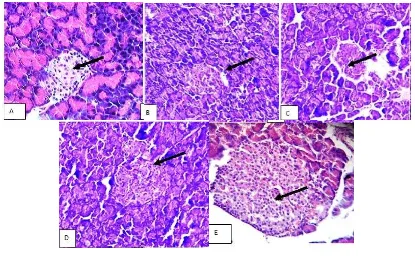
Histopathology of pancreas in normal tissues section shows lobules of exocrine acini,
interlobular ducts and occasional islets of langerhan which is not observed in STZ induced
diabetic pancreas (Fig. 2A and 2B). In P. boergesenii extract treated pancreas, the cells seem
to have gathered together and small preserved islets similar to the normal (Fig. 2C and 2D).
The Group V did not show any significant change of pancreas, when compared with normal
[image:8.595.91.506.387.643.2]pancreas (Fig. 2E).
Fig. 2: Pancreatic histology in experimental rats.
(A) Pancreas shows exocrine acini and endocrineIslets (IL); (B) Shows depleted isletes
(DIL); (C) Shows exocrine acini and small preserved islets; (D) Shows exocrine acini; (E)
www.wjpr.net Vol 6, Issue 8, 2017. 2030 Senthilkumar et al. World Journal of Pharmaceutical Research
Histological examination of renal section of the control rats showed (Fig. 3) the glomeruli,
tubules, interstitium and blood vessels appear normal. Kidney sections of rats treated with
streptozotocin shows tubular damage proteinurea and haemorrhage. In the case of P.
[image:9.595.84.514.165.423.2]boergeseniiextract treated diabetic rats and diabetic rats treated with glibenclamide
Fig. 3: Kidney histology in experimental rats.
(A) Normal kidney shows glomorulin (GM) andProximal convoluted tubules;
(B) Shows tubular damage proteinurea and homorrage;
(C) Shows glomoruli, tubules interstitium and blood vessels appear normal;
(D) Shows glomoruli, tubules without proteiurea and homorrage;
(E) Shows normal structure, shows the glomeruli, tubules, interstitium and blood vessels
appear normal. The P. boergesenii extracts alone treated group showed normal structure
comparable to the control group.
DISCUSSIONS
In P. boergesenii extract and Glibenclamide treated rats, there was an improvement in protein
profile which may be due to marked change in circulating amino acid level, hepatic amino
acid uptake and muscle output of amino acid concentrations.[25]This may be due to decline in either albumin or globulin or both, hypoalbuminemia is a common problem in diabetic
animals and it’s generally attributed to the presence of nephropathy.[26]
Hemoglobin leads to
www.wjpr.net Vol 6, Issue 8, 2017. 2031 Senthilkumar et al. World Journal of Pharmaceutical Research
to N-terminal of hemoglobin beta chain. This process is non-enzymatic and reflects the
average exposure of hemoglobin to glucose over an extended period.[27]
Treatment with P. boergesenii extract showed significant increase in the hemoglobin level in
diabetic rats. Mean elevation of hemoglobin observed was 12.46 ± 0.28 in P. boergesenii
treated group. The hemoglobin level was decreased in diabetic rats that may increase the
formation of glycosylated hemoglobin. Glycosylated hemoglobin was found to increase in
diabetic mellitus and the amount of increase is directly proportional to that of fasting blood
glucose level.[28]
In the present study, administration of P. boergesenii extracts to diabetic rats reduced the
elevated levels of urea, uric acid and creatinine to normal; this shows the normalizing effect
of the seaweed extract on urea and creatinine synthesis.The increase in serum creatinine and
urea levels in Streptozotocin induced diabetic rats may be due to hyperglycemia that causes
osmatic diuresis and depletion of extracellular fluid volume. Several studies have shown an
increased correlation between serum creatinine and urea in diabetic patients.[29]
Insulin plays an important role in metabolism of lipids; insulin is potent inhibitor of lipolysis.
It inhibits the activities of hormone sensitive lipases in adipose tissue and suppresses the
release of free fatty acids.[30] The antihyperglycemic effect of P. boergesenii extract may be due to the down regulation of NADPH and NADH, a cofactor in the fat metabolism. Higher
activity of Glucose-6-phosphatase provides H+ which binds with NADP+ in the synthesis of
fats from carbohydrates. When glycolysis slows down because of cellular activity, the
pentose phosphate pathway still remains active in live to breakdown glucose that
continuously provides NADPH which converts acetyl radicals into long fatty acid chains. The
P. boergesenii extract might be the capable of oxidizing NADPH. Abnormalities in lipid
profile are one of the most common complications in diabetes mellitus and cardiomyopathy.
Acute insulin deficiency initially causes an increase in free fatty acid mobilization from
adipose tissue.[31] Excess of fatty acids in serum of the Streptozotocin induced diabetes promotes conversion of excess fatty acids into phospholipids and cholesterol in liver. These
two substances along with excess triglycerides formed in liver may be discharged into blood
in the form of lipoproteins.[32]
The therapeutic effect of P. boergesenii was confirmed by histopathological study of the
www.wjpr.net Vol 6, Issue 8, 2017. 2032 Senthilkumar et al. World Journal of Pharmaceutical Research
The histopathological changes in the liver, pancreases and kidney in diabetic rats treated with
aqueous extract of P. boergesenii restore the normal structure. Consequently, seaweed P.
boergesenii accomplished of reduction oxidative stress and restore tissue damage and
increase activities of endogenous antioxidant enzymes in diabetic animals. Therefore, P.
boergesenii may have therapeutic benefits effect. The outcome of the histopathological study
was agreement with Balasee et al., 1972[33], reported that P. boergesenii have protective effect with reduced histopathological changes.
CONCLUSION
In conclusion, the results of the present study show that P. boergeseniiextract showed potent
antidiabetic activity in STZ induced diabetic rats. The effective dose of P. boergeseniiextract
was found to be 400 mg/kg body weight. The action of P. boergesenii was comparable with
antidiabetic drug glibenclamide. Results of this experimental study indicated that P.
boergesenii has potent antidiabetic activity in STZ-induced experimental diabetes in rats. P.
boergesenii appears to have a favorable significance for the increase of a potent
phytomedicine from marine flora for diabetes, however more inclusive pharmacological
studies are required to elucidate the exact mechanism of actionof the P. boergeseniiextract.
ACKNOWLEDGEMENT
The authors are grateful to the authorities of Kongunadu Arts and Science College,
Coimbatore, Tamilnadu, India for providing facilities and for their encouragement.The
authors would like to acknowledge Karpagam University, Coimbatore, Tamilnadu, India for
providing animal house facilities and for their support. Authors also thank Botanical Survey
of IndiaSouthern Circle TNAU Campus, Coimbatore, Tamilnadu, India for the species
identification.
REFERENCES
1. American Diabetes Association. Diagnosis and classification of diabetes mellitus. Diab
Care, 2010; 34: S62–S69.
2. Global report on diabetes. World Health Organization. Geneva, 2016.
3. Lautamaki R, Airaksinen KEJ, Seppa M, Toikka J, Luotolahti M, Ball E. Rosiglitazone
improves myocardial glucose uptake in patients with type 2 diabetes and coronary artery
www.wjpr.net Vol 6, Issue 8, 2017. 2033 Senthilkumar et al. World Journal of Pharmaceutical Research
4. Sharma B, Balomajumder C, Roy P. Hypoglycemic and hypolipidemic effects of
flavonoid rich extract from Eugenia jambolana seeds on Streptozotocin induced diabetic
rats. Food Chem Toxicol, 2008; 46: 2376–83.
5. Senthilkumar P, Prakash S, Sudha S. Antidiabetic activity of aqueous extract of Padina
boergesenii in streptozotocin-induced diabetic rats. Int J Pharm Sci, 2014; 6(5): 418-22.
6. Senthilkumar P, Sudha S. Evaluation of Antioxidant activity and Total Phenolic content
of Padina boergesenii from Gulf of Mannar. Drug Invention Today, 2012; 4(12): 635-9.
7. Senthilkumar P, Bhuvaneshwari, Janani, Prakash, Lakshmi Priya and Ranjith Santhosh
Kumar. Green synthesis and characterization of silver nanoparticles from aqueous extract
brown seaweed of Padina boergesenii and its antifungal activity. World J Pharm Sci,
2015; 410: 1858-70.
8. Senthilkumar P, Bhuvaneshwari, Janani, Prakash, Lakshmi Priya and Ranjith Santhosh
Kumar. Potent α-glucosidase inhibitory activity of green synthesized gold nanoparticles
from the brown seaweed padina boergesenii. Int J adv multidiscip Res. 2015; 2(11):
0917-23.
9. Vasanti HR, Jaswanth A, Saraswathy GV, Rajamanickam. Control of urinary risk factors
of stones by Padina boergesenii (Allender and Kraft), brown algae in experimental
hyperoxaluria. J Nat Rem, 2003; 3(2): 189-194.
10.Rajamani KT, Manivasagam P, Ananatharaman TST. Chemopreventive effect of Padina
boergesenii on ferric nitrilotriacetate (Fe-NTA) induced oxidative damage in Wistar rats.
J Appl Phycol, 2010; 257(2): 257–63.
11.Guillermo D, Luisa Villamil, Viviana Almanza. Herbivory effects on the morphology of
the brown alga Padina boergesenii (Phaeophyta). Phycologia, 2007; 46(2): 131-6.
12.Pandit R, Jagtap A. Antidiabetic effect of Ficus religiosa extract instreptozotocin-induced
diabetic rats. J Ethnopharmacol, 2010; 128: 462–6.
13.Arokiyaraj S, Balamurugan R, Augustian P. Antihyperglycemic effect of Hypericum
perforatum ethyl acetate extract on streptozotocin-induced diabetic rats. Asian Pacific J
Trop Biomed, 2011; 386–90.
14.Lowry OH, Rosenbrough NJ, Farr AL, Randall RJ. Protein measurement with the Folin’s
Phenol reagent. J Biol Chem, 1951; 193: 265-75.
15.Natelson S, Scott ML, Beffa CA. Rapid method for the estimation of urea in biologic
fluids. Am J Clin Pathol 1951; 21:275-81.
16.Caraway WI. Uricacid. In: Standard methods of clinical chemistry. Seligson D, editor.
www.wjpr.net Vol 6, Issue 8, 2017. 2034 Senthilkumar et al. World Journal of Pharmaceutical Research
17.Saibene V, Brembilla L, Bertoletti L, Bolognani L, Pozza G. Chromatographic and
colorimetric detection of glycosylated hemoglobins: a comparative analysis of two
different methods. Clin Chim Acta. 1979; 93: 199.
18.Drabkin DL, Austin JH. Spectrophotometric constants for common hemoglobin
derivatives in human, dog, rabbit blood. J Biol Chem. 1932; 98: 719-68.
19.Parekh AC, Jung DH. Cholesterol determinutesation with ferric acetate-uranyl acetate
sulphuric acid, ferrous sulphate reagents. Anal Chem, 1970; 42: 1423-7.
20.Rouser G, Fleisher S, Yamanoto A. Two dimensional thin layer chromatographic
separation of polar lipids and determinutesation of phospholipids by phosphorus analysis
of spots. Lipids, 1970; 5: 494-6.
21.Horn WT, Menahan LA. Sensitive method for determinutesation of free fatty acids in
plasma. J Lipid Res, 1981; 122: 377-81.
22.Rice EC. Triglycerides in serum:Standard methods of clinical chemistry. Vol.VI. Ceds
Roberict Pand Medorald, editors.New York.Academic press, 1970; 215-222.
23.El-Khatib AS. Biological active free radicals and their scavengers: A review. Saudi
Pharm J, 1997; 5: 79-95.
24.CotgreaveI A, Moldeus P, Orrenius S. Host biochemical defense mechanisms against
pro-oxidants. Ann Rev Pharmacol Toxicol, 1988; 28: 189-212.
25.Mallikarjuna Rao C, Parameshwar S, Srinivasan KK. Oral antidiabetic activities of
different extracts of Caesalpinia bonducella seed kernels. Pharmaceutical biology, 2002;
40(8): 590-8.
26.Soon YY, Tan BKH. Evaluation of the hypoglycemic and antioxidant activities of
Morinda officinalis in streptozotocin induced diabetic rats. Singapore med J, 2002; 43:
077-85.
27.Mohammadi J, Naik Prakash R. Evaluation of hypoglycemic effects of Morus albain an
animal model. Indian J Pharmacol, 2008; 40(1): 15-8.
28.Sellamuthu P, Balu Periamalli-patti M, Sathiya Moorthi P, Murugesan K.
Antihyperglycemic effects of Mangiferin in Streptozotocin induced Diabetic rats. J
Health Sci, 2009; 55(2): 206-14.
29.Meister. New aspects of glutathione biochemistry and transport selective alterations of
glutathione metabolism. Nutr Rev, 1984; 42: 397-410.
30.Bays H, Mandarino L, DeFronzo RA. Role of the adipocyte, free fatty acids, and ectopic
www.wjpr.net Vol 6, Issue 8, 2017. 2035 Senthilkumar et al. World Journal of Pharmaceutical Research
agonsits provide a rational therapeutic approach. J Clin Endocrinol Metab, 2004; 89:
463-78.
31.Balasee EO, Bier DM, Hanel RJ. Early effects of anti-insulin serum on hepatitis