Pre-processing and Segmentation of Brain Image
for Tumor Detection
Prof.(Dr.) Samir Kumar Bandyopadhyay Advisor to Chancellor, JIS University, India
Abstract: In today's world, computer aided technology is touching every sphere of human life ranging from communication, smart systems and even medical diagnosis. One of the broadest and upcoming area of research is biomedical image processing that includes biomedical signal gathering, picture processing, image forming, and display to medical diagnosis based on various features extracted from the images. One of the most challenging and complex area of research in biomedical image processing is the segmentation and analysis of brain tumor. This paper proposed method for pre-processing and segmentation of brain image for tumor detection.
Keywords: FCM, Images Registration, Image Fusion, Segmentation
I. INTRODUCTION
In the day-to-day life, computational applications are gaining much importance. Specially, the use of the Computer Aided Diagnosis (CAD) tool for application in the field of
computational biomedical analysis is being studied to a greater extent. In today's health care arena, detection and analysis of Brain tumour is one of the most commonly occurring fatality. According to the the report of the National Cancer Institute statistics (NCIS), over the last two decades, the overall cases of carcinogen, that includes brain cancer, has increased more than10%. The National Brain Tumour Foundation (NBTF) for research in USA had estimated that approximately 29,000 patients in the United States are diagnosed with primary brain tumours every year, moreover, out of them 13,000 patients die. Specially among kids, brain tumours causes one quarter of all deaths related to cancer. The average yearly incidence of primary brain tumours in the United States is11 to 12 for every 100,000 people for primary malignant brain tumours, that rate is 6 to 7 for every 1,00,000. In the United Kingdom, over 4,200 people are diagnosed with brain tumour each year (2007 estimates). There are nearly 200 other categories of tumours diagnosed in United Kingdom every year. Approximately 16 out of every 1,000 cancers detected in the United Kingdom are in the brain (or 1.6%). 80,271 people are affected in India by various types of tumour.
One of the specific medical image analysis methodologies is fully computerized brain
disorder diagnosis like brain tumour detection from MRI. Brain tumour is the uncontrolled growth of the tissue cell in the brain. The cells that supplies blood in the arteries are bounded tightly together which makes general laboratory test inadequate to analyze the chemistry of human brain. The various modalities of bio-medical imaging that allows the doctor and researchers to analyze the brain anatomy by studying the brain without surgical invasion are computed tomography, Magnetic resonance imaging and Positron emission tomography.Magnetic Resonance Imaging (MRI) is a bio-medical imaging technique used by radiologist for viewing the anatomical structures. MRI provides detailed information about soft tissue structural anatomy of human. MRI assist in diagnosis of the brain tumour. MR images are used for analyzing and studying the anatomy of the brain. One of the most challenging task in today's medical image analysis is automated brain tumor detection from MRI images. MR produces images of the anatomy of soft human tissue. It is used to study the human anatomy without invasive surgery [1-3].
Brain image segmentation, that defines the process of creating partition and analyzing the image into visibly and anatomically different regions, is among the most vital and critical
aspect of computer aided clinical diagnostic of tumor or other anatomical abnormalities. various types of noise that are found in the Brain MR images are multiplicative in nature and reductions of these noises are critical. From the clinical aspect, it is very essential to ensure that the sensitive anatomical details are not removed by the noise reduction algorithms. Thus precise segmentation of brain MR images is a challenging research area. Hence, highly precise segmentation of the MRI images is very critical for proper diagnosis by computer aided clinical tools. A wide variety of procedures for segmentation of MR images had been proposed till date.
recognition, mobile robot navigation, estimation of 3D surface area from 2D images, contour based image retrieval and many more. Multiple algorithms or mathematical modeling methods have been proposed for texture analysis, and they can be classified into three major categories: statistical methods, model based methods and structural methods. Each method has its own merits and demerits. This paper focused on MR Image segmentation, tumor detection and texture analysis of tumor. We had proposed an algorithm on spatial domain segmentation of brain tumor using multiple images of MRI. Another algorithm of brain tumor segmentation in the frequency domain using wavelet transformation is also proposed. The proposed algorithm is compared with various other algorithm over different performance metrics to show the effectiveness of our proposed algorithm.
Brain is the most complicated and delicate anatomical structure in human body. Statistics proves that, among various brain ailments, brain tumor is the most fatal and in many cases those tumors become carcinogenic i.e. brain cancer. Brain tumor is characterized by an abnormal and uncontrolled growth of brain cells, and takes up space within the cranial cavity. It varies in shape, size, position and characteristics and can be benign or malignant, or even spread to different parts of brain and body, which makes the detection of brain tumor very critical and challenging. The most vital information a neurologist or neurosurgeon needs to have is the precise size and location of the tumor in the brain and also whether it is causing any swelling or compression of the brain that may need urgent attention. Moreover, this information is also necessary for planning of surgery or post-operative care that may include radiology. Medical imaging plays a critical role in detection of brain tumor. Magnetic Resonance Imaging (MRI) is important to provide detailed and very precise information about tumor size, location and compression of adjacent brain structures. MR Images are of very high resolution that can be analyzed using Computer aided tool for automatic segmentation and analysis of tumor. Computer aided systems are preferred over conventional manual segmentation because automated segmentation is highly accurate and precise, free from human error and much faster than manual segmentation. So, there is a lot of research on the design of efficient algorithms for segmentation and analysis of Brain MR Image.
In addition to Brain tumor segmentation, the detection of brain tumor surface texture
is challenging for researchers. Normally, MRI datasets have very low resolution. Image enhancement technique based on wavelet is used.
To give a complete analysis of brain MR image segmentation and surface texture analysis, the paper primarily throws light on four aspects, namely,
1) Brief discussion about different brain imaging modalities
2) Review of different existing segmentation techniques and texture analysis of Brain MRI. 3) Spatial Domain Segmentation of Brain Tumor using Multiple Images of Brain MR
4) Discrete Wavelet Transform Algorithm for Brain MR image Segmentation based on Frame Theory 5) Brain Tumor Texture Analysis using Wavelets
6) A detailed comparative study of the different techniques have been discussed
This paper aims at providing a 360-degree insight into the recent research areas of biomedical image processing on MR Images that involves understanding of various imaging modalities, proposed technologies of MR Image segmentation and also latest hybrid procedures that involves different aspect of signal processing with spatial image operation. It also provides an analysis of surface texture of brain tumor using wavelets and fractal geometry.
II. LITERATURE REVIEW
minute anatomical variations which requires very precise and exact segmentation for doing diagnosis clinically [5]. Segmentation of brain from MR image is very critical and challenging but extremely precise and accurate segmentation is required for various clinical diagnosis like, detection, analysis and classifying various tumor categories, such as, edema, haemorage detection and necrotic tissues etc. Unlike CT scan, Magnetic Resonance image acquisition parameters are greatly adjustable for creating high contrast image having distinct gray levels for different cases of neuropathology [6]. Hence, segmenting MR images is the recent research focus in biomedical image processing domain. In neuro-science segmenting of Magnetic Resonance image is required in diagnosis of neuro-degenerative and also various psychiatric disorders [7].
Recent work by [8] used a combination of mathematical morphology, wavelet based segmentation and K-means to achieve tumor detection. Another novel approach using color based feature extraction using wavelet decomposition can be found in [9]. Authors used Berkeley wavelet transform for feature extraction and a Support Vector Machine for classification of features [10]. A paper introduced frame and Gabor systems and its use in wavelet analysis for brain segmentation [11]. The use of frame theory and wavelet is used for image restoration [12]. Thus, it signifies that frame theory and wavelets can be used effectively in image processing. Authors also employed techniques of discrete wavelet transform (DWT) and principal component analysis (PCA), and used K-nearest neighbor (KNN) for segmentation [13]. Some employed stationary wavelet transforms to take place of traditional Discrete Wavelet Transform [14]. Afterwards, to train the classifier, they introduced a novel algorithm that is a hybridization of PSO and ABC. Researchers combined discrete wavelet transform with principal component analysis for segmentation [15]. Although those above mentioned algorithms give good results, still, segmentation accuracy could be improved further. Here we design an algorithm based on frame theoretic methods and using discrete wavelet transforms mirror and apply it to brain MRI images and compare its performance with other segmentation algorithms. Significant gains in performance are observed, although the execution time is traded off.
III. PROPOSED METHOD
Thresholding, region growing, statistical models, active control models and clustering had been implemented for segmenting images. As the intensity distribution in biomedicalimages is complex, thresholding becomes a critical task and fails in most cases. Fuzzy C means is a widely used technology for biomedical image segmentation but it considers only the image intensity thereby giving unsatisfactory output in noisy images [16]. A number of algorithms are proposed to make FCM robust against noise and in homogeneity but it's still far from perfect. In probabilistic classification, a very accurate estimation of the probability density function (PDF) is necessary. Non-parametric approach does not take any assumption in getting the parameters of PDF, hence makes it precise but costly. In parametric approach, a function is considered to be a PDF function. It is relatively simple to implement but at certain cases it lacks in preciseness and do not correlate with real data distribution.
for all types of registration. The algorithm must take into consideration not only the type of geometric deformations among the image but also various radiometric deformations and noise corruption effects that required precision of registration process and data characteristics which are dependent on applications. Most registration algorithms consist of the following steps:
1) Feature Detection: Distinct and salient objects such as contours, boundary regions, line intersections, edge, corner, etc. that are automatically found by analysis of image histogram and filtering by spatial mask of 9 X 9 matrix across the Region of Interest. Then, those feature is designated by the point representative, here centre of gravity, known as control point (CPs). Minimum three control points are designated for this case.
2) Feature Matching: Here, correlation between feature found in sensed image and the feature observed in referenced image are established.
3) Transform Model Estimation: The sensed image is aligned with reference image based on the mapping function parameters that are calculated by the establishing feature correspondence. Here, rigid geometric transformation is implemented. Rotation and translation of the image is performed for aligning with base image.
4) Image Transformation and Re-Sampling: Image values in non-integer coordinates. The values are calculated by interpolation. Here, transformation is done using nearest neighbour interpolation.
The critical aspect of fusion images is determining the process of combining sensor images. The naive image fusion technique is to simply take the average pixel gray level value of source image. The algorithm fuse two or more pre-registered images in one high
quality image. Gray and Colour Image is supported. Here, Factor is changed to alter the degree of fusion of every constituent
images. Having = 0:5, both images are fused in equal proportion. Having < 0:5, background image will have greater impact.
Having > 0:5, foreground image will have higher impact.
The scull bone is removed by generating skull-mask from the MRI. Otsu's algorithm is implemented for image histogram shape-based thresholding[19-22] i.e., reducing the gray level image to binary image. OTSU algorithm for thresholding defines that images that needs thresholding had two class of pixel value i.e., foreground and background, after that the estimation of optimum threshold separating these two classes id one such that intra-class variance is minimum. (weight of foreground * variance of foreground) + (weight of background *variance of background) is calculated for the image and the minimum value is considered as thresholding. After doing complete thresholding, the scull is extracted by subtraction the binary image of scull from the original image.
A three step refining segmentation algorithm is discussed here. The steps are as follows:
a) Initial segmentation using k-means algorithm
b) Grid based coarse grain localization using local standard deviation
c) Grid based _ne grain localization using local standard deviation
It is the clustering problem in which image gray levels is clustered in pre-defined number of gray levels that are clustered. The most used unsupervised learning methodologies which explains well-known clustering problem is the K-means [20]. The algorithm uses simple and efficient method to classify a set of data using finite number of cluster, assuming k clusters, that is pre-defined. Those centroids should be placed randomly and separated from one another. The challenge lies in labeling k centroids, for each cluster. Next for each point the location is calculated for a given dataset and correlated with adjacent centroid. After this step k new centroids are recalculated for those cluster resulting from previously. After calculating those k new centroids, a new binding is done among those equivalent data set point and their neighboring new centroids. As an effect of iteration, it is concluded that those k centroids change their position at each iteration until no further changes are done in subsequent iteration, i.e. centroids do not shift any more [21-22].
Lastly, the algorithms tend to minimize the objective function, using squared error function in which the square is calculated as the distance between a data point and cluster center.
The algorithm executes through those steps:
i) Place K distinct points in original image which are characterized by objects which are getting clustered. Initial group centroids are signified by those points.
ii) Every object are assigned to a group by the closest centroid.
iii) After the assignment of every object, recalculation of position of K centroids is executed.
iv) Repeat Steps 2 and 3 until the centroids stop movement.
that will be executed after having done segmentation using K - means. The local standard deviation of output of the k - means segmented image is calculated. This image is projected onto big grid, ideally such grid is 8 by 8 pixel. Local standard deviation of every pixel is generated depending on the pixel values of these 64 pixels of the grid. After that histogram of every grid is generated and depending on this local standard deviation and histogram in every grid, the boundary of segmentation in those grids are re-calculated to give better result.
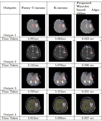
Selecting a larger grid dimension helps to reduce noise in segmentation. But, on the contrary, the larger grid dimension removes the fine anatomic variations like sharp curves in boundary region of tumour or overlapping area of gray and white matters of the brain. Hence, to achieve segmentation with optimal result, we re-process the output segmented image using the method of grid based fine grain localization using local standard deviation. Grid size of 3 by 3 pixel is chosen for the purpose. Hence, selecting a smaller grid helps to focus on fine anatomical variations of Magnetic Resonance image which requires proper preservation. It restores the minute details of the tumor border and fine analysis of the overlapping region of gray and white matter. The results are shown in the following figure 1 to figure 4. Table 1 shows the result of comparison with other methods.
[image:5.612.159.486.226.717.2]Figure 1 Input and Enhanced Image
Figure 2 Registered Image
Figure 4 Skull Mask, Skull Removed and K -Means Segmented Image
Table 1 Traditional segmentation algorithms are compared with the proposed method
IV. CONCLUSIONS
[image:6.612.135.482.215.625.2]REFERENCES [1] W. Gonzalez, Image processing. PHI, India.
[2] S. Bandhyopadhyay and T. Paul, “Segmentation of Brain MRI Image - A Review," IJARCSSE, 2012.
[3] S. Bandhyopadhyay and T. Paul, “Automatic Segmentation of Brain Tumour from Multiple Images of Brain MRI," IJAIEM, 2013.
[4] C. P.L. and T. W.G., “Exploiting the self-organizing map for medical image segmentation," in Twentieth IEEE international symposium on computer-based medical systems, IEEE, 2007.
[5] H. L.O., B. A.M., C. L.P., V. R.P., S. M.S., and B. J., “A comparison of neural network and fuzzy clustering techniques in segmenting magnetic resonance images of the brain," IEEE Trans Neural Network, 1992.
[6] T. D. and F. L., “A brain MR images segmentation method based on som neural network," in The 1st international conference on bioinformatics and biomedical engineering, 2007.
[7] B. M. et al, “New multi-scale medical image segmentation based on fuzzy c-mean (fcm)," in IEEE conference on innovative technologies in intelligent systems and industrial applications, 2008.
[8] Kharrat, Benamrane, Messaoud, and Abid, “Detection of brain tumor in medical images," Signals, Circuits and Systems, 2009.
[9] Karkanis, Iakovidis, Maroulis, Karras, and Tzivras, “Computer-aided tumor detection in endoscopic video using color wavelet features," IEEE Transactions on Information Technology in Biomedicine, 2003.
[10] Bahadure, Ray, and Thethi, “Image analysis for MRI based brain tumor detection and feature extraction using biologically inspired bwt and svm," International Journal of Biomedical Imaging, 2017.
[11] O. Christensen, “A short introduction to frames, gabor systems, and wavelet systems.," Azerbaijan Journal of Mathematics, 2014. [12] Z. Shen, “Wavelet frames and image restorations," Proceedings of the International Congress of Mathematicians, 2010.
[13] E.-D. ESA, T. Hosny, and A. Salem, “Hybrid intelligent techniques for MRI brain images classification," Digital Signal Processing, 2010.
[14] S. Wang, Z. Dong, and S. Du, “Feed-forward neural network optimized by hybridization of pso and abc for abnormal brain detection," International Journal of Imaging Systems and Technology, 2015.
[15] D. Nayak, R. Dash, and B. Majhi, “Brain MR image classification using two dimensional discrete wavelet transforms and adaboost with random forests," Neuro-computing, 2016.
[16] L. P.L., M. J.M., and C. T., “Image selective smoothing and edge detection by nonlinear diffusion," SIAMJNA, 1992. [17] L. G. Shapiro and G. C. Stockman, Computer Vision. New Jersey, Prentice-Hall, 2001.
[18] W. S. et al, “Automatic identification of grey matter structures from MRIto improve the segmentation of white matter lesions," Journal of Image Guided Surgery, 1995.
[19] M. J.B., “Some methods for classification and analysis of multivariate observations," in Proceedings of 5-th Berkeley Symposium on Mathematical Statistics and Probability, University of California Press, 1967.
[20] K. A., S. G., and S. P., “Different Techniques of Edge Detection in Digital Image Processing," IJERA, 2013.
[21] A. A. et al, “A modified fuzzy C-means algorithm for bias field estimation and segmentation of MRI data," IEEE Transactions on Medical Imaging, 2002. [22] V. K. B. et al, “MRI brain image segmentation using modified fuzzy c-means clustering algorithm," in International Conference on Communication Systems