Original Article
Correlation of serum vitamin D levels with ovarian
reserve markers in patients with primary
ovarian insufficiency
Zhengfen Xu1,2*, Quanmei Wang1,3*, Lianhong Zhu4, Linjuan Ma1, Xiaoqing Ye5, Chunming Li1, Yibing Lan1, Yizhou Huang6, Jing Liu5, Jianhong Zhou1
1Department of Gynecology, Women’s Hospital, School of Medicine, Zhejiang University, Hangzhou 310006, Zhejiang, China; 2Department of Gynecology, Jiaxing Maternity and Child Care Hospital, Jiaxing 314000, Zhejiang, China; 3The First People’s Hospital of Tonglu, Hangzhou 311500, Zhejiang, China; 4Tongxiang First People’s Hospital, Jiaxing 314500, Zhejiang, China; 5MOE Key Laboratory of Environmental Remediation and Ecosystem Health, College of Environmental and Resource Sciences, Research Center for Air Pollution and Health, College of Environmental and Resource Sciences, School of Public Health, Zhejiang University, Hangzhou 310006, Zhejiang, China; 6School of Medicine, Zhejiang University, Hangzhou 310006, Zhejiang, China. *Equal contributors.
Received April 17, 2018; Accepted November 9, 2018; Epub April 15, 2019; Published April 30, 2019
Abstract: The cause of primary ovarian insufficiency (POI) is unknown for many women. Previous studies have
suggested that vitamin D plays an important role in reproduction, but there have been few studies to investigate
possible correlation between serum vitamin D levels and ovarian reserve markers in patients with POI. In this study, 33 women with POI and 72 women with normal menstrual cycles were recruited. Serum levels of follicle stimulat
-ing hormone (FSH), anti-Müllerian hormone (AMH), 25-hydroxyvitamin D (25(OH)D and total cholesterol (TC) were measured. Correlations of 25(OH)D with AMH and FSH levels were assessed using regression models. There was a statistically significant difference in 25(OH)D levels between the POI group and the control group. The 25(OH)
D levels were inversely correlated with log-transformed FSH and positively correlated with log-transformed AMH.
However, no statistical significance was found. The results show low levels of vitamin D may contribute to decreased
ovarian reserve. Additional nationwide studies should be conducted to clarify whether appropriate vitamin D levels
reduce the risk of developing POI.
Keywords: 25(OH)D, FSH, AMH, primary ovarian insufficiency
Introduction
Primary ovarian insufficiency (POI) is a clinical syndrome defined by loss of ovarian activities before the age of 40. POI is one of the major causes of female infertility, and the prevalence is approximately 1%. POI is characterized by menstrual disturbance (amenorrhea or oli-gomenorrhea) with raised gonadotrophins and low oestradiol [1]. Anti-Müllerian hormone (AMH) is a protein hormone and a well-recog-nized biomarker of ovarian reserve. AMH is mainly expressed in granulosa cells of growing antral and pre-antral follicles [2]. AMH regu-lates follicle growth by inhibiting their sensitivi-ty to follicle-stimulating hormone (FSH) [3]. Thus, reduced AMH serum levels in patients is an important marker of POI [2]. Several causes
autoimmune, metabolic, toxic, infectious and iatrogenic factors [1, 4]. However, the precise cause of most POI cases has not been eluci -dated [4].
role of vitamin D in the ovarian reserve [7-9]. A prior study confirmed that vitamin D stimulated the production of progesterone, oestrone and oestradiol in cultured human ovarian cells [10]. Serum 25(OH)D is the major circulating form of vitamin D and is used by clinicians to determine vitamin D status [11]. A previous study found that low levels of 25(OH)D were associated with POI patients [12]. However, another study showed no difference in 25(OH)D levels bet-ween control and POI patients [13].
This study sought to investigate the correlation between serum levels of vitamin D and ovarian reserve markers (such as AHM and FSH) in patients with POI because the relationship between vitamin D and POI is unclear. This case-control study recruited 33 women with POI and 72 healthy women as controls. The serum levels of FSH, AMH, 25(OH)D and total cholesterol (TC) were measured. The correla-tions of 25(OH)D with AMH and FSH levels in women were assessed using binary logistic regression models.
Materials and methods
Subjects
The initial sample consisted of 110 women with POI and 179 healthy women of whom 174 were excluded (Figure S1). This case-control study recruited 33 women with POI from the Gyna-ecology Endocrinology Outpatient Clinic at Wo-men’s Hospital, School of Medicine, Zhejiang University, Hangzhou, China from September 2015 to April 2016. There were 72 healthy women recruited as the control group during the same study period. This study was approved by the Ethics Committee of Women’s Hospital of Zhejiang University. All patients included in the study provided informed consent before participating in the study. The following inclu-sion criteria were used: 1) diagnosis of primary ovarian insufficiency: (i) oligo/amenorrhea for at least 4 months and (ii) an elevated FSH level > 25 IU/l on two occasions > 4 weeks apart [1]; 2) no iatrogenic cause or known chromosomal abnormality; 3) age between 18 and 40; 4) no hormone therapy for at least 6 months; and 5) the control group consisted of individuals with regular menstruation cycles and without history of infertility. The study exclusion criteria includ-ed the following: 1) taking vitamin D supple -ments or using medication likely to affect the
levels of vitamin D and ovarian reserve determi-nants; 2) history of hysterectomy, oophorecto-my, ovarian surgery, chemotherapy and/or radi-otherapy; 3) cigarette smoking and 4) women with autoimmune diseases (e.g., autoimmune thyroid disease, autoimmune liver diseases, systemic lupus erythaematosus).
Blood collection
Each patient in the POI group provided a blood sample on the day of their first consultation. The control group blood samples were collect-ed from the first day to the fifth day of sponta -neous bleeding episodes during the menstrual cycle. The participants completed question-naires addressing socio-demographic charac-teristics, gynecological, and medical histories, and lifestyle factors.
Hormones and 25(OH)D measurement
Serum samples were separated by centrifuga-tion at 4,000 rpm for 10 minutes within 30 minutes of blood sampling. The samples were then stored in polypropylene tubes at -80°C until the time of analysis. The serum levels of FSH and AMH were determined using an elec-tro-chemiluminescence immunoassay (cobas e602, Roche). Serum 25(OH)D levels were measured with mass spectrometry (API 3200, SCIEX). The serum total cholesterol (TC) levels were measured with enzymatic colorimetric methods with commercial kits (cobas c701, Roche). Pro-vitamin D3 is a precursor in the cholesterol biosynthetic pathway, and there is a significant association between serum 25(OH) D and serum lipids [14]. Therefore, the plasma levels of 25(OH)D were adjusted by TC. The intra- and inter-assay coefficients of variation (CVs) were both < 10% based on laboratory controls.
Statistical analysis
The odds ratios (OR) for POI associated with 25(OH)D were examined by binary logistic regression analysis before and after adjusting for potential confounders. The log transforma-tion of AMH and FSH was performed to assess the plausibility of linear regression. Correlations for 25(OH)D, transformed FSH, and log-transformed AMH levels were examined by mul-tivariate linear regression before and after adjusting for potential confounders. All covari-ates (age, BMI, education, household income) were simultaneously entered into the multivari-ate linear regression model to identify regres-sion coefficients for factors related to the log-transformed AMH and log-log-transformed FSH levels. All P-values < 0.05 were considered sta-tistically significant.
ly lower than those in the control group (3.27 ± 2.33 ng/ml) (P < 0.001). The 25(OH)D levels did not differ between the POI (92.38 ± 31.07 nmol/L) and the control groups (96.76 ± 33.12 nmol/L) (P = 0.523). However, the adjusted 25(OH)D levels were significantly lower in POI (18.47 ± 6.03 nmol/mmol) group compared to controls (22.79 ± 7.90 nmol/mmol) (P = 0.006) (Table 2).
Odds ratio (OR) for POI associated with 25(OH) D
The logistic regression models were used to assess the OR and the corresponding 95% CI for the relationship between the adjusted 25(OH)D levels and POI risk. There was a signifi
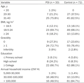
-Table 1. Demographic characteristics of the POI group and the control group
Variable POI (n = 33) Control (n = 72)
Age, y
< 20 1 (3%) 0
20-30 7 (21.2%) 27 (37.5%)
31-40 25 (75.8%) 45 (62.5%)
BMI, kg/m2
< 18.5 4 (12.1%) 13 (18.1%)
18.5-24 23 (69.7%) 49 (68.1%)
≥ 24 6 (18.2%) 10 (13.8%)
Gravidity
0 9 (27.3%) 17 (23.6%)
≥ 1 24 (72.7%) 55 (76.4%)
Infertility 3 (9%) 2 (2.8%)
Education
≤ Primary school 3 (9.1%) 4 (5.6%)
High school 8 (24.2%) 6 (8.3%)
≥ College 22 (66.7%) 62 (86.1%) Annual household income (CNY ¥)
5,000-30,000 1 (3%) 1 (1.4%)
30.000-100,000 16 (48.5%) 10 (13.9%)
[image:3.612.90.370.96.374.2]> 100,000 16 (48.5%) 61 (84.7%)
Table 2. Biochemical parameters of the POI group and the control group
Variable POI (n = 33) Control (n = 72) P
FSH (IU/L) 59.56 ± 32.67 7.05 ± 2.90 < 0.001
AMH (ng/ml) 0.02 ± 0.004 3.27 ± 2.33 < 0.001
25(OH)D (nmol/L) 92.38 ± 31.07 96.76 ±33.12 0.523
TC (mmol/L) 5.04 ± 0.838 4.30 ± 0.732 < 0.00
25(OH)D/TC (nmol/mmol) 18.47 ± 6.03 22.79 ± 7.90 0.006
Values are expressed as mean ± SD. Testing for differences used student’s t test.
Results
Demographic characteristics
The demographic characteris-tics of the women included in this study are presented in
Table 1. The majority of wo- men were older than 30 years of age (POI 75.8%, control 62.5%). Additionally, more than half (POI 69.7%, control 68.1%) of the women had BMI values ≥ 18.5 and < 24 kg/m2. The majority of women in this study had been pregnant once (POI 72.7%, control 76.4%). There were 3 infertile women in the POI group but only 2 in the con -trol group. The participants were generally highly educat-ed, and 66.7% of cases in POI group and 86.1% of subjects in the control group had at least a college education. Participants in the control group (84.7%) had higher annual household income than in the POI group (48.5%).
Biochemical parameters
[image:3.612.89.373.418.498.2]significant-cant negative association ob- served between POI risk and 25(OH)D levels in the unadjust -ed analysis (OR = 0.915, CI: 0.857-0.978, P = 0.009). After adjusting for potential con-founders (age, BMI, education, household income) the nega-tive correlation between POI risk and 25(OH)D levels was similar (OR = 0.924, CI: 0.861-0.991, P = 0.026) (Table 3).
The relationship between 25(OH)D and ovarian reserve markers
A multiple linear regression analysis was conducted to evaluate the relationships between the 25(OH)D levels and the log-transformed FSH or log-transformed AMH. The 25(OH)D level was inversely correlated with log-transfor- med FSH (r = -0.192, P = 0.05) (Figure 1) and positively corre-lated with log-transformed AMH (r = 0.175, P = 0.075) (Figure 2). However, there was no statistically significant dif -ference. Additional adjustmen- ts for age, BMI, education, and household income did not yield a significantly statistical difference between 25(OH)D and log-transformed FSH (r = -0.175, P = 0.071) or log-trans-formed AMH (r = 0.153, P = 0.120) (Table 4).
Discussion
[image:4.612.90.374.97.137.2]The cause of POI is unknown for many women, and these cases are described as having unexplained or idiopathic POI [4]. Chromosome abnormali-ties were discovered in several POI patients. Previous studies also reported that autoimmun-ity, metabolism, infection, sm- oking, and iatrogenic causes may contribute to the develop-ment of POI [17].
Table 3. Odds Ratio (OR) (95% CI) for POI Associated with 25(OH) D
Variable Unadjusted Model Adjusted Modela
95% CL (0.857, 0.978) 95% CL (0.861, 0.991)
25(OH)Db OR = 0.915 P = 0.009 OR = 0.924 P = 0.026
[image:4.612.92.372.177.404.2]aThe adjusted model included age, BMI, annual household income and education. bSerum 25(OH)D levels were adjusted for TC (nmol/mmol).
Figure 1. Correlation between 25(OH)D/TC ratio and log-transformed FSH
in all participants.
[image:4.612.91.371.456.683.2]Serum 25(OH)D is the major circulating form of vitamin D. According to the US Preventive Services Task Force guidelines [18] and the Endocrine Society guidelines [19] vitamin D deficiency is generally recognized as a 25(OH)D level below 20 ng/mL and vitamin D insufficien -cy has been defined as a serum 25(OH)D level of 21-29 ng/mL. In our study, the mean 25(OH) D levels was (92.38 ± 31.07 nmol/L, 26.40 ± 8.88 ng/mL) in POI cases and (96.76 ± 33.12 nmol/L, 27.65 ± 9.46 ng/ml) in the control groups. The results indicate that most of the women in the study had inadequate 25(OH)D levels. Our study also found the infertility rate in the POI group was very high (9%) and the adjusted 25(OH)D levels were significantly lower in POI. All these findings improved the power of the study to detect an association between low vitamin D and ovarian reserve markers.
The biological actions of vitamin D are mediat-ed through VDR, which is expressmediat-ed in many reproductive organs such as ovary, placenta and the uterus [6]. Previous studies found the promoter region of the AMH gene contained a vitamin D response element [20]. This discov-ery prompted researchers to focus on the rela-tionship between vitamin D and ovarian reserve markers. Recent evidence from animal and human studies suggests vitamin D is involved in many functions of the reproductive system. Vitamin D deficiency reduced overall fertility by 75% in female rats when diet interventions reduced levels [21]. Furthermore, VDR null mutant mice showed gonadal insufficiencies [22]. Uterine hypoplasia and impaired follicu- logenesis were observed in female VDR-defe- ctive mice [22]. In a previous human study involving 35 women with POI and 28 control women, there were significantly lower levels of serum vitamin D found in the POI group com
-pared with the healthy control group with nor-mal menstrual cycles [12]. This study found an association of lower 25(OH)D levels with increased risk of POI in women, which is con -sistent with prior observations.
The occurrence of POI is accompanied by high levels of FSH, and the FSH elevation is usually used as a diagnostic basis for POI. Several studies showed a link between vitamin D levels and FSH. Vitamin D levels were found to be inversely correlated with FSH levels in POI women [12]. Another study demonstrated that vitamin D was inversely related to urinary levels of FSH in older premenopausal women [23]. In this study, 25(OH)D was negatively correlated with log-transformed FSH which confirmed that low levels of vitamin D play a role in the aetiol-ogy of POI.
AMH is an important marker of ovarian reserve, and multiple studies have reported a relation-ship between vitamin D and AMH in women. In a study of 33 premenopausal women, the change in AMH level was correlated with the magnitude of change in vitamin D levels [24]. In this study, a similar trend for the positive cor-relation of 25(OH)D and AMH levels. However, there was no significant statistical difference.
Although these previous studies and our obser-vations suggest that low vitamin D levels might have an adverse effect on ovarian reserve, sev-eral previous studies failed to identify the asso-ciation between vitamin D and ovarian reserve markers. For example, a recent study showed that 25(OH)D levels in follicular fluid were nega -tively correlated with AMH mRNA levels in human granulosa cells of small follicles [25]. A prospective cross-sectional study showed that vitamin D was not associated with the ovarian reserve markers such as AMH and antral folli -cle count in infertile women [26]. Another study of 130 women reported that 25(OH)D3 levels did not differ between the premature ovarian failure (POF) group and the control group [13]. There are several limitations of our study. First, the women we recruited were mainly residing in Zhejiang province, whose sun-light exposure was similar. Another limitation is the small study sample. Future studies should be rand-omized studies with larger samples and have patients with various concentrations of serum vitamin D.
Table 4. Relationship between 25(OH)D and log-transformed FSH or log-transformed AMH after adjusting for potential confounders (age, BMI, education and annual household income) in all participants
Variable 25(OH)Da
r p
Log-transformed FSH -0.175 0.071
Log-transformed AMH 0.153 0.120
aSerum 25(OH)D levels were adjusted for TC (nmol/
In conclusion, this study reveals that low levels of vitamin D may contribute to decreased ovarian reserve and induce POI. Further investi -gations in a larger population are needed to corroborate these observations and determine the mechanism(s) by which vitamin D interacts with POI.
Acknowledgements
The authors would like to thank the women who agreed to participate. This work was sup -ported by the National Nature Science Foun- dation of China (grant No. 81703236) and Project for Zhejiang Medical Technology Pro- gram (grant No. 2018KY437, 2016KYA049 and WKJ-ZJ-1621) and the Zhejiang provincial Nature Science Foundation of China (grant No. Y18H040007).
Disclosure of conflict of interest
None.
Address correspondence to: Dr. Jianhong Zhou,De-
partment of Gynecology, Women’s Hospital, School
of Medicine, Zhejiang University, Hangzhou 310006, China. Tel: 87061501; Fax: 0086-571-87061878; E-mail: zhoujh1117@zju.edu.cn
References
[1] European Society for Human Reproduction
and Embryology (ESHRE) Guideline Group on POI, Webber L, Davies M, Anderson R, Bartlett J, Braat D, Cartwright B, Cifkova R, de Muinck
Keizer-Schrama S, Hogervorst E, Janse F, Liao
L, Vlaisavljevic V, Zillikens C and Vermeulen N. ESHRE Guideline: management of women with premature ovarian insufficiency. Hum Reprod
2016; 31: 926-937.
[2] Visser JA, de Jong FH, Laven JS and Themmen
AP. Anti-Mullerian hormone: a new marker for
ovarian function. Reproduction 2005; 131: 1-9.
[3] Pellatt L, Rice S, Dilaver N, Heshri A, Galea R,
Brincat M, Brown K, Simpson ER and Mason HD. Anti-Mullerian hormone reduces follicle sensitivity to follicle-stimulating hormone in human granulosa cells. Fertil Steril 2011; 96: 1246-1251 e1241.
[4] Shestakova IG, Radzinsky VE and Khamoshina MB. Occult form of premature ovarian insuffi
-ciency. Gynecol Endocrinol 2016; 32: 30-32.
[5] Pfotenhauer KM and Shubrook JH. Vitamin D deficiency, its role in health and disease, and
current supplementation recommendations. J
Am Osteopath Assoc 2017; 117: 301-305.
[6] Johnson LE, DeLuca HF. vitamin D receptor null mutant mice fed high levels of calcium are fertile. J Nutr 2001; 131: 1787-1791.
[7] Franasiak JM, Molinaro TA, Dubell EK, Scott
KL, Ruiz AR, Forman EJ, Werner MD, Hong KH and Scott RT Jr. Vitamin D levels do not affect IVF outcomes following the transfer of euploid
blastocysts. Am J Obstet Gynecol 2015; 212:
315 e311-316.
[8] van de Vijver A, Drakopoulos P, Van Landuyt L, Vaiarelli A, Blockeel C, Santos-Ribeiro S, Tour
-naye H and Polyzos NP. Vitamin D deficiency
and pregnancy rates following frozen-thawed embryo transfer: a prospective cohort study. Hum Reprod 2016; 31: 1749-1754.
[9] Fabris A, Pacheco A, Cruz M, Puente JM,
Fate-mi H and Garcia-Velasco JA. Impact of circulat -ing levels of total and bioavailable serum vita-min D on pregnancy rate in egg donation recipients. Fertil Steril 2014; 102: 1608-1612. [10] Parikh G, Varadinova M, Suwandhi P, Araki T,
Rosenwaks Z, Poretsky L and Seto-Young D.
Vitamin D regulates steroidogenesis and
insu-lin-like growth factor binding protein-1 (IGFBP-1) production in human ovarian cells.
Horm Metab Res 2010; 42: 754-757.
[11] Holick MF. Vitamin D deficiency. N Engl J Med
2007; 357: 266-281.
[12] Kebapcilar AG, Kulaksizoglu M, Kebapcilar L, Gonen MS, Unlu A, Topcu A, Demirci F and Taner CE. Is there a link between premature
ovarian failure and serum concentrations of vitamin D, zinc, and copper? Menopause 2013; 20: 94-99.
[13] Ersoy E, Ersoy AO, Yildirim G, Buyukkagnici U, Tokmak A and Yilmaz N. Vitamin D levels in pa
-tients with premature ovarian failure. Ginekol
Pol 2016; 87: 32-36.
[14] Jorde R, Figenschau Y, Hutchinson M, Emaus
N and Grimnes G. High serum 25-hydroxyvita -min D concentrations are associated with a
favorable serum lipid profile. Eur J Clin Nutr
2010; 64: 1457-1464.
[15] Ye X, Pan W, Zhao S, Zhao Y, Zhu Y, Liu J, Liu W. Relationships of pyrethroid exposure with go-nadotropin levels and pubertal development in chinese boys. Environ Sci Technol 2017; 51: 6379-6386.
[16] Ye X, Liu J. Effects of pyrethroid insecticides on hypothalamic-pituitary-gonadal axis: a re-productive health perspective. Environ Pollut 2019; 245: 590-599.
[17] LM N. Primary ovarian insufficiency. N Engl J
Med 2009; 360: 606-614.
[18] Chung M LJ, Terasawa T, Lau J, Trikalinos TA.
Vitamin D with or without calcium supplemen-tation for prevention of cancer and fractures: an updated meta-analysis for the U.S. Pre-
ventive services task force. Ann Intern Med
[19] Holick MF, Binkley NC, Bischoff-Ferrari HA, Gor -don CM, Hanley DA, Heaney RP, Murad MH, Weaver CM and Endocrine S. Evaluation,
treat-ment, and prevention of vitamin D deficiency:
an endocrine society clinical practice guide-line. J Clin Endocrinol Metab 2011; 96: 1911-1930.
[20] Malloy PJ, Peng L, Wang J, Feldman D. Interac-tion of the vitamin D receptor with a vitamin D response element in the Mullerian-inhibiting substance (MIS) promoter: regulation of MIS expression by calcitriol in prostate cancer cells. Endocrinology 2009; 150: 1580-1587. [21] Halloran BP, DeLuca HF. Effect of vitamin D
de-ficiency on fertility and reproductive capacity in
the female rat. J Nutr 1980; 110: 1573-1580. [22] Yoshizawa T, Handa Y, Uematsu Y, Takeda S,
Sekine K, Yoshihara Y, Kawakami T, Arioka K, Sato H, Uchiyama Y, Masushige S, Fukamizu A, Matsumoto T, Kato S. Mice lacking the vitamin
D receptor exhibit impaired bone formation uterine hypoplasia and growth retardation
af-ter weaning. Nat Genet 1997; 16: 391-396.
[23] Jukic AM, Steiner AZ and Baird DD. Association
between serum 25-hydroxyvitamin D and ovar-ian reserve in premenopausal women. Meno- pause 2015; 22: 312-316.
[24] Dennis NA, Houghton LA, Jones GT, van Rij AM,
Morgan K and McLennan IS. The level of se-rum anti-Mullerian hormone correlates with vi-tamin D status in men and women but not in boys. J Clin Endocrinol Metab 2012; 97: 2450-2455.
[25] Merhi Z, Doswell A, Krebs K and Cipolla M. Vi-tamin D alters genes involved in follicular de-velopment and steroidogenesis in human cu-mulus granulosa cells. J Clin Endocrinol Metab 2014; 99: E1137-1145.
[26] Drakopoulos P, van de Vijver A, Schutyser V, Milatovic S, Anckaert E, Schiettecatte J, Block -eel C, Camus M, Tournaye H and Polyzos NP. The effect of serum vitamin D levels on ovarian
reserve markers: a prospective cross-sectional