2-(2-Hydroxy-2-phenylethyl)-1-methyl-cyclopropan-1-ol
Hui Mao,* Ya-Wei Tu, Shi-Kun Li, Xiao-Juan Wang and Peng-Peng Wang
College of Chemistry and Life Science, Zhejiang Normal University, Jinhua 321004, Zhejiang, People’s Republic of China
Correspondence e-mail: maohui2011@zjnu.cn
Received 17 December 2012; accepted 23 December 2012
Key indicators: single-crystal X-ray study;T= 296 K; mean(C–C) = 0.004 A˚;
Rfactor = 0.070;wRfactor = 0.249; data-to-parameter ratio = 20.3.
The asymmetric unit of the title compound, C12H16O2,
contains two independent molecules in which the dihedral angles between the benzene and cyclopropane rings are 75.9 (3) and 76.3 (3). In the crystal, the molecules are connected by O—H O hydrogen bonds into a three dimensional supramolecular structure.
Related literature
For applications of cyclopropane derivatives, see: Pietruszka (2003); Heleneet al.(2003); Wessjohannet al.(2003); Charette & Marcoux (1995).
Experimental
Crystal data
C12H16O2 Mr= 192.25
Triclinic,P1 a= 9.1700 (8) A˚ b= 10.3863 (10) A˚ c= 11.9412 (11) A˚ = 98.133 (7)
= 90.854 (6)
= 91.841 (7)
V= 1125.07 (18) A˚3
Z= 4
MoKradiation = 0.08 mm1
T= 296 K
0.130.100.08 mm
Data collection
Bruker SMART APEXII area-detector diffractometer 16925 measured reflections
5146 independent reflections 2391 reflections withI> 2(I) Rint= 0.038
Refinement
R[F2> 2(F2)] = 0.070
wR(F2) = 0.249
S= 1.04 5146 reflections 253 parameters
4 restraints
H-atom parameters constrained max= 0.37 e A˚
3 min=0.39 e A˚
3
Table 1
Hydrogen-bond geometry (A˚ ,).
D—H A D—H H A D A D—H A
O1—H1A O2i 0.82 1.94 2.745 (2) 167
O1A—H1AA O2Aii
0.82 1.95 2.757 (2) 167
O2—H2B O1A 0.82 1.96 2.768 (3) 167
O2A—H2AB O1iii
0.82 1.98 2.778 (2) 165
Symmetry codes: (i)x;yþ1;z; (ii)xþ1;yþ1;zþ1; (iii)x;y;zþ1.
Data collection:APEX2(Bruker, 2006); cell refinement:SAINT (Bruker, 2006); data reduction:SAINT; program(s) used to solve structure:SHELXS97(Sheldrick, 2008); program(s) used to refine structure: SHELXL97 (Sheldrick, 2008); molecular graphics: SHELXTL(Sheldrick, 2008); software used to prepare material for publication:SHELXL97.
Supplementary data and figures for this paper are available from the IUCr electronic archives (Reference: XU5667).
References
Bruker (2006).SAINTandAPEX2. Bruker AXS Inc., Madison, Wisconsin, USA.
Charette, A. B. & Marcoux, J. C. (1995).Synlett,12, 1197–1207.
Helene, L., Marcoux, J. C., Molinaro, C. & Charette, A. B. (2003).Chem. Rev. 103, 977–1050.
Pietruszka, J. (2003).Chem. Rev.103, 1051–1070. Sheldrick, G. M. (2008).Acta Cryst.A64, 112–122.
Wessjohann, L. A., Brandt, W. & Thiemann, T. (2003).Chem. Rev.103, 1625– 1648.
Acta Crystallographica Section E
Structure Reports Online
supporting information
Acta Cryst. (2013). E69, o189 [doi:10.1107/S1600536812051768]
2-(2-Hydroxy-2-phenylethyl)-1-methylcyclopropan-1-ol
Hui Mao, Ya-Wei Tu, Shi-Kun Li, Xiao-Juan Wang and Peng-Peng Wang
S1. Comment
Due to the special structure and versatile biologically activity of the chiral cyclopropanes their medicinal properties and
synthetic utility have inspired numerous chemists to fascinate (Pietruszka, 2003; Helene et al., 2003; Wessjohann et al.,
2003; Charette & Marcoux, 1995). In this work, we reported the synthesis and crystal structure of cis
-2-(2-hydroxy-2-phenyl-ethyl)-1-methyl-cyclopropanol.
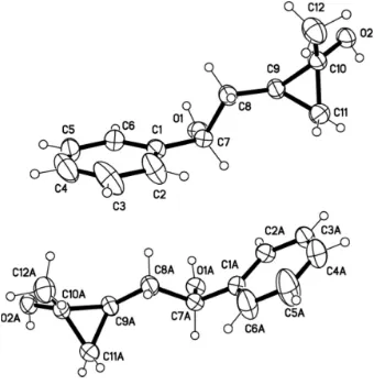
X-ray crystallography confirmed the molecular structure and the atom connectivity for the title compound(I), as
illustrated in Fig. 1. A view on the crystal structure of the title compound, the angle of (C9—C10—C11) is 60.8 (2)°, and
the angle of (C10—C9—C11) is 59.4 (2)°, and the angle of (C9—C11—C10) is 59.8 (2)°. It can be speculated that the
structure of the three ring was similar equilateral triangle. The dihedral angle between the benzene ring and the
cyclo-propane ring is 75.9 (3) and 76.3 (3)°. The structure is more stable by intramolecular hydrogen bond (O2—H2B···O1A).
The intermolecular hydrogen (O1A—H1AA···O2A; O1—H1A0···O2; O2A—H2AB···O1) results in the formation of a
three-dimensional structure in the crystal.
S2. Experimental
To a two-necked flask containing samarium powder (2.5 mmol), was added THF (18 ml) and ally bromide (2.2 mmol)
under nitrogen. The mixture was allowed to stir at room temperature for 1 h (the color would turn into purple). HMPA
(2.0 ml) and H2O (1.0 mmol) was then added in sequence via a syringe. A solution of 4-Acetoxy-4-phenyl-1-butene (1.0
mmol) in THF (5.0 ml) was subsequently added. The color would fade out in 3 h (monitored by TLC). After treatment,
afford the solid products. Recrystallization condition: Petrol/EtOAc (5/1, v:v), room temperature, one day.
S3. Refinement
H atoms were positioned geometrically and refined using a riding model with C—H = 0.93–0.98 Å and O—H = 0.82 Å,
Figure 1
A view of the molecule of (I) showing the atom-labelling scheme with displacement ellipsoids drawn at the 30%
probability.
2-(2-Hydroxy-2-phenylethyl)-1-methylcyclopropan-1-ol
Crystal data
C12H16O2 Mr = 192.25
Triclinic, P1 Hall symbol: -P 1
a = 9.1700 (8) Å
b = 10.3863 (10) Å
c = 11.9412 (11) Å
α = 98.133 (7)°
β = 90.854 (6)°
γ = 91.841 (7)°
V = 1125.07 (18) Å3
Z = 4
F(000) = 416
Dx = 1.135 Mg m−3
Mo Kα radiation, λ = 0.71073 Å Cell parameters from 2877 reflections
θ = 1.7–27.8°
µ = 0.08 mm−1 T = 296 K Block, colourless 0.13 × 0.10 × 0.08 mm
Data collection
Bruker SMART APEXII area-detector diffractometer
Radiation source: fine-focus sealed tube Graphite monochromator
ω scans
Rint = 0.038
θmax = 27.8°, θmin = 1.7°
h = −11→11
k = −13→13
l = −15→15
Refinement
Refinement on F2
Least-squares matrix: full
R[F2 > 2σ(F2)] = 0.070 wR(F2) = 0.249 S = 1.04 5146 reflections 253 parameters 4 restraints
Primary atom site location: structure-invariant direct methods
Secondary atom site location: difference Fourier map
Hydrogen site location: inferred from neighbouring sites
H-atom parameters constrained
w = 1/[σ2(Fo2) + (0.1296P)2 + 0.0141P]
where P = (Fo2 + 2Fc2)/3
(Δ/σ)max < 0.001
Δρmax = 0.37 e Å−3
Δρmin = −0.39 e Å−3
Special details
Geometry. All e.s.d.'s (except the e.s.d. in the dihedral angle between two l.s. planes) are estimated using the full covariance matrix. The cell e.s.d.'s are taken into account individually in the estimation of e.s.d.'s in distances, angles and torsion angles; correlations between e.s.d.'s in cell parameters are only used when they are defined by crystal symmetry. An approximate (isotropic) treatment of cell e.s.d.'s is used for estimating e.s.d.'s involving l.s. planes.
Refinement. Refinement of F2 against ALL reflections. The weighted R-factor wR and goodness of fit S are based on F2,
conventional R-factors R are based on F, with F set to zero for negative F2. The threshold expression of F2 > σ(F2) is used
only for calculating R-factors(gt) etc. and is not relevant to the choice of reflections for refinement. R-factors based on F2
are statistically about twice as large as those based on F, and R- factors based on ALL data will be even larger.
Fractional atomic coordinates and isotropic or equivalent isotropic displacement parameters (Å2)
x y z Uiso*/Ueq
O1A 0.29470 (19) 0.48083 (17) 0.31228 (15) 0.0595 (5)
H1AA 0.3789 0.5069 0.3051 0.089*
O1 0.20635 (19) 0.51624 (17) −0.18527 (15) 0.0608 (6)
H1A 0.1211 0.4912 −0.1976 0.091*
O2A 0.44023 (19) 0.39198 (17) 0.70678 (14) 0.0584 (5)
H2AB 0.3622 0.4207 0.7296 0.088*
O2 0.06177 (18) 0.60637 (17) 0.23173 (14) 0.0602 (5)
H2B 0.1389 0.5758 0.2490 0.090*
C1 0.1876 (3) 0.7210 (3) −0.2628 (2) 0.0542 (7) C1A 0.3172 (3) 0.2795 (3) 0.1811 (2) 0.0536 (7) C2A 0.3688 (3) 0.3498 (3) 0.0998 (2) 0.0595 (8)
H2AA 0.3859 0.4391 0.1177 0.071*
C2 0.2208 (5) 0.8513 (3) −0.2580 (3) 0.0933 (12)
H2A 0.2619 0.8971 −0.1919 0.112*
C3A 0.3958 (3) 0.2885 (3) −0.0095 (2) 0.0703 (9)
H3AA 0.4298 0.3374 −0.0638 0.084*
C3 0.1944 (5) 0.9163 (4) −0.3494 (4) 0.1107 (15)
H3A 0.2164 1.0051 −0.3438 0.133*
C4A 0.3729 (4) 0.1591 (4) −0.0366 (3) 0.0859 (10)
H4AA 0.3920 0.1186 −0.1092 0.103*
C4 0.1365 (5) 0.8511 (5) −0.4469 (3) 0.1019 (14)
C5 0.1070 (4) 0.7221 (5) −0.4549 (3) 0.0865 (11)
H5A 0.0701 0.6768 −0.5227 0.104*
C5A 0.3223 (6) 0.0882 (4) 0.0417 (3) 0.1164 (15)
H5AA 0.3077 −0.0013 0.0230 0.140*
C6 0.1309 (3) 0.6553 (3) −0.3632 (3) 0.0684 (8)
H6A 0.1085 0.5665 −0.3697 0.082*
C6A 0.2918 (5) 0.1479 (3) 0.1504 (3) 0.0943 (12)
H6AA 0.2539 0.0982 0.2028 0.113*
C7A 0.2894 (3) 0.3416 (3) 0.3008 (2) 0.0536 (7)
H7AA 0.1916 0.3131 0.3213 0.064*
C7 0.2137 (3) 0.6552 (2) −0.1590 (2) 0.0509 (7)
H7A 0.3115 0.6818 −0.1283 0.061*
C8 0.1041 (3) 0.6937 (3) −0.0678 (2) 0.0555 (7)
H8A 0.1025 0.7879 −0.0521 0.067*
H8B 0.0076 0.6617 −0.0949 0.067*
C8A 0.3980 (3) 0.3031 (3) 0.3855 (2) 0.0578 (7)
H8AA 0.4006 0.2089 0.3771 0.069*
H8AB 0.4944 0.3362 0.3694 0.069*
C9 0.1402 (3) 0.6401 (3) 0.0402 (2) 0.0533 (7)
H9A 0.1507 0.5456 0.0304 0.064*
C9A 0.3607 (3) 0.3547 (3) 0.5063 (2) 0.0516 (7)
H9AA 0.3466 0.4487 0.5197 0.062*
C10A 0.4153 (3) 0.3000 (3) 0.6074 (2) 0.0517 (7) C10 0.0892 (3) 0.6972 (3) 0.1554 (2) 0.0533 (7) C11A 0.2598 (3) 0.2770 (3) 0.5716 (2) 0.0624 (8)
H11A 0.1871 0.3236 0.6179 0.075*
H11B 0.2291 0.1901 0.5369 0.075*
C11 0.2449 (3) 0.7152 (3) 0.1265 (2) 0.0629 (8)
H11C 0.3163 0.6664 0.1622 0.076*
H11D 0.2786 0.8012 0.1136 0.076*
C12A 0.5203 (3) 0.1925 (3) 0.6002 (3) 0.0743 (9)
H12A 0.5398 0.1715 0.6746 0.111*
H12B 0.6096 0.2195 0.5682 0.111*
H12C 0.4789 0.1171 0.5531 0.111*
C12 −0.0105 (4) 0.8086 (3) 0.1728 (3) 0.0788 (10)
H12D −0.0290 0.8303 0.2520 0.118*
H12E −0.1010 0.7847 0.1325 0.118*
H12F 0.0341 0.8825 0.1450 0.118*
Atomic displacement parameters (Å2)
U11 U22 U33 U12 U13 U23
C2A 0.0547 (17) 0.0686 (19) 0.0565 (18) 0.0020 (13) 0.0050 (13) 0.0123 (15) C2 0.150 (4) 0.075 (2) 0.054 (2) −0.007 (2) 0.020 (2) 0.0081 (17) C3A 0.066 (2) 0.096 (3) 0.0518 (19) 0.0038 (17) 0.0051 (15) 0.0182 (17) C3 0.173 (4) 0.085 (3) 0.084 (3) 0.024 (3) 0.053 (3) 0.038 (2) C4A 0.112 (3) 0.091 (3) 0.0513 (19) 0.006 (2) −0.0002 (18) 0.0003 (19) C4 0.114 (3) 0.134 (4) 0.072 (3) 0.051 (3) 0.034 (2) 0.051 (3) C5 0.071 (2) 0.143 (4) 0.050 (2) 0.019 (2) 0.0030 (15) 0.025 (2) C5A 0.214 (5) 0.068 (2) 0.062 (2) −0.015 (3) 0.000 (3) −0.0021 (19) C6 0.0599 (19) 0.088 (2) 0.0585 (19) 0.0036 (15) 0.0003 (15) 0.0134 (17) C6A 0.151 (4) 0.079 (2) 0.052 (2) −0.020 (2) −0.002 (2) 0.0153 (17) C7A 0.0474 (16) 0.0662 (19) 0.0490 (16) 0.0004 (13) 0.0035 (12) 0.0150 (13) C7 0.0453 (15) 0.0594 (18) 0.0483 (16) 0.0017 (12) 0.0027 (12) 0.0084 (13) C8 0.0578 (17) 0.0645 (18) 0.0454 (15) 0.0072 (13) 0.0040 (12) 0.0099 (13) C8A 0.0610 (18) 0.0658 (18) 0.0482 (16) 0.0062 (13) 0.0042 (13) 0.0125 (13) C9 0.0543 (16) 0.0615 (18) 0.0450 (16) 0.0095 (13) 0.0033 (12) 0.0091 (13) C9A 0.0588 (17) 0.0554 (17) 0.0426 (15) 0.0105 (12) 0.0059 (12) 0.0113 (12) C10A 0.0540 (17) 0.0596 (17) 0.0419 (15) 0.0037 (13) 0.0035 (12) 0.0086 (12) C10 0.0528 (16) 0.0646 (18) 0.0453 (15) 0.0076 (13) 0.0060 (12) 0.0153 (13) C11A 0.0568 (18) 0.079 (2) 0.0505 (17) −0.0056 (14) 0.0066 (13) 0.0086 (14) C11 0.0570 (18) 0.074 (2) 0.0608 (18) −0.0034 (14) −0.0009 (14) 0.0210 (15) C12A 0.089 (2) 0.072 (2) 0.0641 (19) 0.0181 (17) 0.0008 (16) 0.0132 (16) C12 0.095 (3) 0.080 (2) 0.066 (2) 0.0279 (18) 0.0148 (18) 0.0174 (17)
Geometric parameters (Å, º)
O1A—C7A 1.433 (3) C6A—H6AA 0.9300
O1A—H1AA 0.8200 C7A—C8A 1.511 (4)
O1—C7 1.432 (3) C7A—H7AA 0.9800
O1—H1A 0.8200 C7—C8 1.514 (3)
O2A—C10A 1.425 (3) C7—H7A 0.9800
O2A—H2AB 0.8200 C8—C9 1.512 (4)
O2—C10 1.421 (3) C8—H8A 0.9700
O2—H2B 0.8200 C8—H8B 0.9700
C1—C2 1.371 (4) C8A—C9A 1.514 (3)
C1—C6 1.379 (4) C8A—H8AA 0.9700
C1—C7 1.517 (4) C8A—H8AB 0.9700
C1A—C6A 1.376 (4) C9—C10 1.507 (3)
C1A—C2A 1.375 (4) C9—C11 1.512 (4)
C1A—C7A 1.511 (4) C9—H9A 0.9800
C2A—C3A 1.398 (4) C9A—C10A 1.491 (3)
C2A—H2AA 0.9300 C9A—C11A 1.506 (4)
C2—C3 1.385 (5) C9A—H9AA 0.9800
C2—H2A 0.9300 C10A—C11A 1.484 (4)
C3A—C4A 1.346 (4) C10A—C12A 1.493 (4)
C3A—H3AA 0.9300 C10—C11 1.485 (4)
C3—C4 1.354 (6) C10—C12 1.491 (4)
C3—H3A 0.9300 C11A—H11A 0.9700
C4A—H4AA 0.9300 C11—H11C 0.9700
C4—C5 1.348 (5) C11—H11D 0.9700
C4—H4A 0.9300 C12A—H12A 0.9600
C5—C6 1.395 (4) C12A—H12B 0.9600
C5—H5A 0.9300 C12A—H12C 0.9600
C5A—C6A 1.394 (5) C12—H12D 0.9600
C5A—H5AA 0.9300 C12—H12E 0.9600
C6—H6A 0.9300 C12—H12F 0.9600
C7A—O1A—H1AA 109.5 C7—C8—H8B 109.2
C7—O1—H1A 109.5 H8A—C8—H8B 107.9
C10A—O2A—H2AB 109.5 C9A—C8A—C7A 112.4 (2)
C10—O2—H2B 109.5 C9A—C8A—H8AA 109.1
C2—C1—C6 117.7 (3) C7A—C8A—H8AA 109.1
C2—C1—C7 119.5 (3) C9A—C8A—H8AB 109.1
C6—C1—C7 122.8 (3) C7A—C8A—H8AB 109.1
C6A—C1A—C2A 117.6 (3) H8AA—C8A—H8AB 107.9
C6A—C1A—C7A 120.0 (2) C10—C9—C11 58.94 (17)
C2A—C1A—C7A 122.4 (3) C10—C9—C8 124.1 (2)
C1A—C2A—C3A 120.8 (3) C11—C9—C8 119.8 (2)
C1A—C2A—H2AA 119.6 C10—C9—H9A 114.3
C3A—C2A—H2AA 119.6 C11—C9—H9A 114.3
C1—C2—C3 121.5 (4) C8—C9—H9A 114.3
C1—C2—H2A 119.3 C10A—C9A—C8A 124.3 (2)
C3—C2—H2A 119.3 C10A—C9A—C11A 59.36 (17)
C4A—C3A—C2A 120.4 (3) C8A—C9A—C11A 120.2 (2)
C4A—C3A—H3AA 119.8 C10A—C9A—H9AA 114.1
C2A—C3A—H3AA 119.8 C8A—C9A—H9AA 114.1
C4—C3—C2 120.2 (4) C11A—C9A—H9AA 114.1
C4—C3—H3A 119.9 O2A—C10A—C9A 115.5 (2)
C2—C3—H3A 119.9 O2A—C10A—C11A 114.9 (2)
C3A—C4A—C5A 119.9 (3) C9A—C10A—C11A 60.82 (18)
C3A—C4A—H4AA 120.0 O2A—C10A—C12A 111.7 (2)
C5A—C4A—H4AA 120.0 C9A—C10A—C12A 123.1 (2)
C5—C4—C3 119.5 (4) C11A—C10A—C12A 122.1 (2)
C5—C4—H4A 120.3 O2—C10—C11 115.1 (2)
C3—C4—H4A 120.3 O2—C10—C12 112.2 (2)
C4—C5—C6 121.2 (3) C11—C10—C12 121.8 (2)
C4—C5—H5A 119.4 O2—C10—C9 115.5 (2)
C6—C5—H5A 119.4 C11—C10—C9 60.72 (17)
C4A—C5A—C6A 120.6 (3) C12—C10—C9 122.7 (2)
C4A—C5A—H5AA 119.7 C10A—C11A—C9A 59.82 (16)
C6A—C5A—H5AA 119.7 C10A—C11A—H11A 117.8
C1—C6—C5 119.9 (3) C9A—C11A—H11A 117.8
C1—C6—H6A 120.0 C10A—C11A—H11B 117.8
C5—C6—H6A 120.0 C9A—C11A—H11B 117.8
C1A—C6A—C5A 120.7 (3) H11A—C11A—H11B 114.9
C5A—C6A—H6AA 119.6 C10—C11—H11C 117.7
O1A—C7A—C1A 112.3 (2) C9—C11—H11C 117.7
O1A—C7A—C8A 107.1 (2) C10—C11—H11D 117.7
C1A—C7A—C8A 112.7 (2) C9—C11—H11D 117.7
O1A—C7A—H7AA 108.2 H11C—C11—H11D 114.9
C1A—C7A—H7AA 108.2 C10A—C12A—H12A 109.5
C8A—C7A—H7AA 108.2 C10A—C12A—H12B 109.5
O1—C7—C1 112.1 (2) H12A—C12A—H12B 109.5
O1—C7—C8 107.8 (2) C10A—C12A—H12C 109.5
C1—C7—C8 112.0 (2) H12A—C12A—H12C 109.5
O1—C7—H7A 108.3 H12B—C12A—H12C 109.5
C1—C7—H7A 108.3 C10—C12—H12D 109.5
C8—C7—H7A 108.3 C10—C12—H12E 109.5
C9—C8—C7 111.9 (2) H12D—C12—H12E 109.5
C9—C8—H8A 109.2 C10—C12—H12F 109.5
C7—C8—H8A 109.2 H12D—C12—H12F 109.5
C9—C8—H8B 109.2 H12E—C12—H12F 109.5
C6A—C1A—C2A—C3A −0.8 (4) O1—C7—C8—C9 62.7 (3)
C7A—C1A—C2A—C3A 178.8 (2) C1—C7—C8—C9 −173.6 (2)
C6—C1—C2—C3 1.9 (5) O1A—C7A—C8A—C9A 61.7 (3)
C7—C1—C2—C3 −177.8 (3) C1A—C7A—C8A—C9A −174.3 (2)
C1A—C2A—C3A—C4A −0.6 (4) C7—C8—C9—C10 157.0 (2)
C1—C2—C3—C4 −1.0 (6) C7—C8—C9—C11 86.3 (3)
C2A—C3A—C4A—C5A 0.6 (5) C7A—C8A—C9A—C10A 159.3 (2)
C2—C3—C4—C5 −1.0 (6) C7A—C8A—C9A—C11A 87.8 (3)
C3—C4—C5—C6 2.0 (6) C8A—C9A—C10A—O2A 146.8 (2)
C3A—C4A—C5A—C6A 0.7 (7) C11A—C9A—C10A—O2A −105.6 (2)
C2—C1—C6—C5 −1.0 (4) C8A—C9A—C10A—C11A −107.6 (3)
C7—C1—C6—C5 178.8 (2) C8A—C9A—C10A—C12A 3.7 (4)
C4—C5—C6—C1 −1.0 (5) C11A—C9A—C10A—C12A 111.3 (3)
C2A—C1A—C6A—C5A 2.1 (5) C11—C9—C10—O2 −105.8 (2)
C7A—C1A—C6A—C5A −177.5 (4) C8—C9—C10—O2 147.2 (2)
C4A—C5A—C6A—C1A −2.2 (7) C8—C9—C10—C11 −107.1 (3)
C6A—C1A—C7A—O1A −168.9 (3) C11—C9—C10—C12 111.0 (3)
C2A—C1A—C7A—O1A 11.5 (4) C8—C9—C10—C12 3.9 (4)
C6A—C1A—C7A—C8A 70.0 (4) O2A—C10A—C11A—C9A 106.5 (2) C2A—C1A—C7A—C8A −109.6 (3) C12A—C10A—C11A—C9A −112.8 (3) C2—C1—C7—O1 −166.3 (3) C8A—C9A—C11A—C10A 114.4 (3)
C6—C1—C7—O1 13.9 (3) O2—C10—C11—C9 106.4 (2)
C2—C1—C7—C8 72.5 (3) C12—C10—C11—C9 −112.3 (3)
C6—C1—C7—C8 −107.3 (3) C8—C9—C11—C10 114.1 (3)
Hydrogen-bond geometry (Å, º)
D—H···A D—H H···A D···A D—H···A
O1—H1A···O2i 0.82 1.94 2.745 (2) 167
O2—H2B···O1A 0.82 1.96 2.768 (3) 167
O2A—H2AB···O1iii 0.82 1.98 2.778 (2) 165