research communications
Acta Cryst.(2017). E73, 231–234 https://doi.org/10.1107/S2056989017000792
231
Received 4 January 2017Accepted 16 January 2017
Edited by M. Weil, Vienna University of Technology, Austria
‡ Present address: CCDC, 174 Frelinghuysen Rd., Piscataway NJ 00854 USA.
Keywords:crystal structure; powder diffraction; density functional theory; citrate; cesium.
CCDC reference:1527789
Supporting information:this article has supporting information at journals.iucr.org/e
Crystal structure of dicesium hydrogen citrate from
laboratory single-crystal and powder X-ray
diffraction data and DFT comparison
Alagappa Rammohan,a‡ Amy A. Sarjeantband James A. Kadukc*
a
Atlantic International University, Honolulu HI , USA,bDepartment of Chemistry, Northwestern University, Evanston IL , USA, andcIllinois Institute of Technology, Department of Chemistry, 3101 S. Dearborn St., Chicago IL 60616, USA. *Correspondence e-mail: [email protected]
The crystal structure of dicesium hydrogen citrate, 2Cs+C6H6O7 2
, has been solved using laboratory X-ray single-crystal diffraction data, refined using laboratory powder X-ray data, and optimized using density functional techniques. The Cs+ cation is nine-coordinate, with a bond-valence sum of 0.92 valence units. The CsO9coordination polyhedra share edges and corners to
form a three-dimensional framework. The citrate anion is located on a mirror plane. Its central hydroxy/carboxylate O—H O hydrogen bond is short, and (unusually) intermolecular. The centrosymmetric end-end carboxylate hydrogen bond is exceptionally short (O O = 2.416 A˚ ) and strong. These hydrogen bonds contribute 16.5 and 21.7 kcal mol1, respectively, to the crystal energy. The hydrophobic methylene groups occupy pockets in the framework.
1. Chemical context
In the course of a systematic study of the crystal structures of group 1 (alkali metal) citrate salts to understand the anion’s conformational flexibility, ionization, coordination tendencies, and hydrogen bonding, we have determined several new crystal structures. Most of the new structures were solved using X-ray powder diffraction data (laboratory and/or synchrotron), but single crystals were used where available. The general trends and conclusions about the sixteen new compounds and twelve previously determined structures are being reported separately (Rammohan & Kaduk, 2017a). Eleven of the new structures – NaKHC6H5O7,
NaK2C6H5O7, Na3C6H5O7, NaH2C6H5O7, Na2HC6H5O7,
K3C6H5O7, Rb2HC6H5O7, Rb3C6H5O7(H2O), Rb3C6H5O7,
Na5H(C6H5O7)2, and CsH2C6H5O7 – have been published
recently (Rammohan & Kaduk, 2016a,b,c,d,e, 2017b,c,d,e,f, Rammohan et al., 2016), and two additional structures – KH2C6H5O7 and KH2C6H5O7(H2O)2 – have been
commu-nicated to the Cambridge Structural Database (CSD) (Kaduk & Stern, 2016a,b). We report here synthesis and crystal structure of another alkali metal citrate salt, 2Cs+HC6H5O7
2 .
2. Structural commentary
The asymmetric unit of the title compound is shown in Fig. 1. The root-mean-square deviation of the non-hydrogen atoms in the experimentally determined and in the DFT-optimized structures is 0.098 A˚ (Fig. 2). The largest differences are 0.13 A˚ , at Cs19 and O11. This good agreement provides strong
evidence that the experimentally determined structure is correct (van de Streek & Neumann, 2014). The following discussion uses the DFT-optimized structure.
Most of the bond lengths, bond angles, and torsion angles fall within the normal ranges indicated by a MercuryMogul geometry check (Macraeet al., 2008). The C1—C2—C3 angle of 114.1is flagged as unusual (average = 104.0 (32),Z-score = 3.1). The Cs+cation is 9-coordinate, with a bond-valence sum of 0.92 valence units. The location of the citrate anion on a mirror plane and the coordination of all seven oxygen atoms to Cs+cations presumably are the source of the slight distor-tion. The citrate anion occurs in thetrans,transconformation,
which is one of the two low-energy conformations of an isolated citrate moiety. The citrate anion triply chelates to two Cs+ cations through O12, O17, and O15. The citrate also chelates through O12/O16, O15/O17, and O15/O16. The Mulliken overlap populations and atomic charges indicate that the metal-oxygen bonding is ionic. The Bravais–Friedel– Donnay–Harker (Bravais, 1866; Friedel, 1907; Donnay & Harker, 1937) morphology is blocky, with {020} as major faces. A 4th-order spherical harmonic model was included in the refinement. The texture index was 1.016, indicating that preferred orientation was slight in the rotated flat-plate specimen.
3. Supramolecular features
The CsO9coordination polyhedra share edges and corners to
form a three-dimensional framework (Fig. 3). The central hydroxy/carboxylate O—H O hydrogen O17—H18 O16 is short, and (unusually) intermolecular. The centrosymmetric end-end O12—H20—O12 hydrogen bond (with H20 situated on an inversion center) is exceptionally short and strong (Table 1). By the correlation of Rammohan & Kaduk (2017a), these hydrogen bonds contribute 16.5 and 21.7 kcal mol1to the crystal energy. The hydrophobic methylene groups occupy pockets in the framework (Fig. 3).
232
Rammohanet al. 2Cs+C6H6O72 Acta Cryst.(2017). E73, 231–234
[image:2.610.313.567.89.134.2]research communications
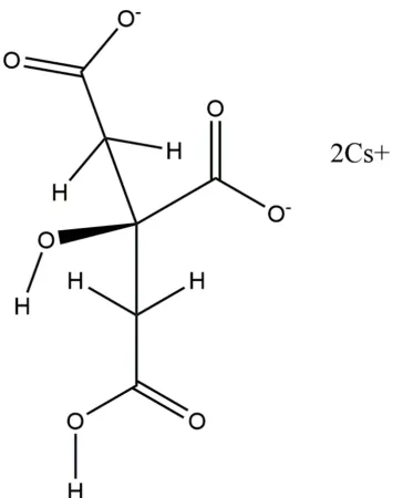
Figure 1
[image:2.610.82.264.109.334.2]The asymmetric unit of the title compound, with the atom numbering. The atoms are represented by 50% probability spheroids.
Figure 2
Comparison of the refined and optimized structures of dicesium hydrogen citrate. The refined structure is in red, and the DFT-optimized structure is in blue.
Table 1
Hydrogen-bond geometry (A˚ ,).
D—H A D—H H A D A D—H A
O12—H20 O12i 1.208 1.208 2.416 180.0 O17—H18 O16ii 0.999 1.634 2.632 178.2
Symmetry codes: (i)xþ1;yþ1;zþ1; (ii)x1 2;yþ
1 2;zþ
[image:2.610.46.296.469.714.2] [image:2.610.315.563.498.706.2]4. Database survey
Details of the comprehensive literature search for citrate structures are presented in Rammohan & Kaduk (2017a). A reduced cell search of the cell of dicesium hydrogen citrate in the Cambridge Structural Database (Groom et al., 2016) (increasing the default tolerance from 1.5 to 2.0%) yielded 100 hits, but combining the cell search with the elements C, H, Cs, and O only yielded no hits.
5. Synthesis and crystallization
Citric acid monohydrate, H3C6H5O7(H2O), (2.0796 g,
10.0 mmol) was dissolved in 20 ml deionized water. Cs2CO3
(3.2582 g, 10.0 mmol, Sigma–Aldrich) was added to the citric acid solution slowly with stirring. The resulting clear colourless solution was evaporated to dryness in a 333 K oven. Single crystals were isolated from the colourless solid.
6. Refinement
A single crystal was mounted in inert oil and transferred to the cold gas stream of a Bruker Kappa APEX CCD area detector system equipped with a Cu K sealed tube with MX optics. Despite suggestions from multiple programs that the space group was Pnma, all attempts to refine the structure in this space group yielded unreasonable disorder and non-positive-definite displacement coefficients. Presumably the poor crystal quality and/or twinning were the source of the problems. The best refinement using single crystal data was obtained using space groupP212121.
A portion of the sample was ground in a mortar and pestle, and blended with NIST SRM 640b silicon internal standard. The powder pattern indicated that the sample contained about 24 wt% CsHC6H5O7(Rammohan & Kaduk, 2017f), which was
included as phase 2 in the refinement. The Si internal standard was included as phase 3.
Initial Rietveld refinements used the single crystalP212121
model, but were unstable. The ADDSYM module of
PLATON (Spek, 2009) suggested the presence of an
addi-tional center of symmetry, and that the correct space group wasPnma(with a transformation of axes). Refinement in the higher-symmetry space group was uneventful. Pseudo-Voigt profile coefficients were as parameterized in Thompsonet al.
(1987) with profile coefficients for Simpson’s rule integration of the pseudo-Voigt function according to Howard (1982). The asymmetry correction of Fingeret al.(1994) was applied, and microstrain broadening by Stephens (1999). The structure was refined by the Rietveld method using GSAS/EXPGUI
(Larson & Von Dreele, 2004; Toby, 2001). All C—C and C—O bond lengths were restrained, as were all bond angles. The hydrogen atoms were included at fixed positions, which were recalculated during the course of the refinement using Mate-rials Studio(Dassault Systemes, 2014). The limited resolution of the powder data precluded refining displacement coeffi-cients, which were fixed at typical values for alkali metal citrates. Diffraction data are displayed in Fig. 4. Crystal data, data collection and structure refinement details are summar-ized in Table 2.
7. DFT calculations
After the Rietveld refinement, a density functional geometry optimization (fixed experimental unit cell) was carried out using CRYSTAL14 (Dovesiet al., 2014). The basis sets for the C, H, and O atoms were those of Peintingeret al.(2012), and the basis set for Cs was that of Sophia et al. (2014). The calculation was run on eight 2.1 GHz Xeon cores (each with 6 Gb RAM) of a 304-core Dell Linux cluster at IIT, used 8k -points and the B3LYP functional, and took about 13 h. The
Uisovalues from the Rietveld refinement were assigned to the
optimized fractional coordinates.
research communications
Acta Cryst.(2017). E73, 231–234 Rammohanet al. 2Cs+C
[image:3.610.313.567.69.256.2]6H6O72
233
Figure 3
Crystal structure of Cs2HC6H5O7, viewed down the a-axis. CsO9
[image:3.610.45.299.71.215.2]polyhedra are green.
Figure 4
Rietveld plot for the refinement of Cs2HC6H5O7. The red crosses
represent the observed data points, and the green line is the calculated pattern. The magenta curve is the difference pattern, plotted at the same scale as the other patterns. The row of black tick marks indicates the reflection positions, the row of red tick marks indicates the positions of the CsH2C6H5O7impurity peaks, and the blue tick marks indicate the Si
Acknowledgements
We thank Andrey Rogachev for the use of computing resources at IIT.
References
Bravais, A. (1866). In Etudes Cristallographiques. Paris: Gauthier Villars.
Bruker (2009).DIFFRAC. Measurement. Bruker AXS Inc., Madison Wisconsin, USA.
Crystal Impact (2015). DIAMOND. Crystal Impact GbR, Bonn, Germany.
Dassault Systemes (2014). Materials Studio. BIOVIA, San Diego, California, USA.
Donnay, J. D. H. & Harker, D. (1937).Am. Mineral.22, 446–467. Dovesi, R., Orlando, R., Erba, A., Zicovich-Wilson, C. M., Civalleri,
B., Casassa, S., Maschio, L., Ferrabone, M., De La Pierre, M., D’Arco, P., Noe¨l, Y., Causa`, M., Re´rat, M. & Kirtman, B. (2014).Int. J. Quantum Chem.114, 1287–1317.
Finger, L. W., Cox, D. E. & Jephcoat, A. P. (1994).J. Appl. Cryst.27, 892–900.
Friedel, G. (1907).Bull. Soc. Fr. Mineral.30, 326–455.
Groom, C. R., Bruno, I. J., Lightfoot, M. P. & Ward, S. C. (2016).Acta Cryst.B72, 171–179.
Howard, C. J. (1982).J. Appl. Cryst.15, 615–620.
Kaduk, J. A. & Stern, C. (2016a). CSD Communication 1446457– 1446458.
Kaduk, J. A. & Stern, C. (2016b). CSD Communication 1446460– 1446461.
Larson, A. C. & Von Dreele, R. B. (2004).General Structure Analysis System, (GSAS). Report LAUR, 86–784 Los Alamos National Laboratory, New Mexico, USA.
Macrae, C. F., Bruno, I. J., Chisholm, J. A., Edgington, P. R., McCabe, P., Pidcock, E., Rodriguez-Monge, L., Taylor, R., van de Streek, J. & Wood, P. A. (2008).J. Appl. Cryst.41, 466–470.
Peintinger, M. F., Vilela Oliveira, D. & Bredow, T. (2012). Comput. Chem., doi: 10.1002/jcc.23153.
Rammohan, A. & Kaduk, J. A. (2016a).Acta Cryst.E72, 170–173. Rammohan, A. & Kaduk, J. A. (2016b).Acta Cryst.E72, 403–406. Rammohan, A. & Kaduk, J. A. (2016c).Acta Cryst.E72, 793–796. Rammohan, A. & Kaduk, J. A. (2016d).Acta Cryst.E72, 854–857. Rammohan, A. & Kaduk, J. A. (2016e).Acta Cryst.E72, 1159–1162. Rammohan, A. & Kaduk, J. A. (2017a).Acta Cryst. B.Submitted. Rammohan, A. & Kaduk, J. A. (2017b).Acta Cryst.E73, 92–95. Rammohan, A. & Kaduk, J. A. (2017c).Acta Cryst.E73, 227–230. Rammohan, A. & Kaduk, J. A. (2017d).Acta Cryst.E73, 250–253. Rammohan, A. & Kaduk, J. A. (2017e).Acta Cryst.E73, 286–290. Rammohan, A. & Kaduk, J. A. (2017f).Acta Cryst.E73, 133–136. Rammohan, A., Sarjeant, A. A. & Kaduk, J. A. (2016).Acta Cryst.
E72, 943–946.
Sheldrick, G. M. (2015).Acta Cryst.A71, 3–8.
Sophia, G., Baranek, P., Sarrazin, M., Rerat, M. & Dovesi, R. (2014).
Systematic influence of atomic substitution on the phase diagram of ABO3ferroelectric perovskites.
Spek, A. L. (2009).Acta Cryst.D65, 148–155. Stephens, P. W. (1999).J. Appl. Cryst.32, 281–289.
Streek, J. van de & Neumann, M. A. (2014).Acta Cryst.B70, 1020– 1032.
Thompson, P., Cox, D. E. & Hastings, J. B. (1987).J. Appl. Cryst.20, 79–83.
Toby, B. H. (2001).J. Appl. Cryst.34, 210–213. Westrip, S. P. (2010).J. Appl. Cryst.43, 920–925.
234
Rammohanet al. 2Cs+C6H6O72 Acta Cryst.(2017). E73, 231–234
[image:4.610.45.559.93.324.2]research communications
Table 2
Experimental details.
Phase 1 Phase 2
Crystal data
Chemical formula 2Cs+C6H6O72
C6H7CsO7
Mr 455.92 324.02
Crystal system, space group Orthorhombic,Pnma Orthorhombic,Pna21
Temperature (K) 300 300
a,b,c(A˚ ) 9.8466 (3), 15.8872 (5), 6.5959 (2) 8.7362, 20.5351, 5.1682
V(A˚3) 1031.82 (6) 927.17
Z 4 4
Radiation type K1,K2,= 1.540629, 1.544451 A˚ K1,K2,= 1.540629, 1.544451 A˚
Specimen shape, size (mm) Flat sheet, 2424 Flat sheet, 2424
Data collection
Diffractometer Bruker D2 Phaser Bruker D2 Phaser
Specimen mounting Standard PMMA holder Standard PMMA holder
Data collection mode Reflection Reflection
Scan method Step Step
2values (
) 2min= 5.042 2max= 70.050 2step= 0.020 2min= 5.042 2max= 70.050 2step= 0.020
Refinement
Rfactors and goodness of fit Rp= 0.050,Rwp= 0.062,Rexp= 0.030, R(F2) = 0.081,2= 4.494
Rp= 0.050,Rwp= 0.062,Rexp= 0.030, R(F2) = 0.081,2= 4.494
No. of parameters 57 57
No. of restraints 18 18
supporting information
sup-1
Acta Cryst. (2017). E73, 231-234
supporting information
Acta Cryst. (2017). E73, 231-234 [https://doi.org/10.1107/S2056989017000792]
Crystal structure of dicesium hydrogen citrate from laboratory single-crystal
and powder X-ray diffraction data and DFT comparison
Alagappa Rammohan, Amy A. Sarjeant and James A. Kaduk
Computing details
Data collection: DIFFRAC.Measurement (Bruker, 2009) for RAMM016C_phase_2. Program(s) used to solve structure:
SHELXT (Sheldrick, 2015) for RAMM016C_phase_1, RAMM016C_phase_2. Molecular graphics: DIAMOND (Crystal
Impact, 2015) for RAMM016C_phase_2. Software used to prepare material for publication: publCIF (Westrip, 2010) for
RAMM016C_phase_2.
(RAMM016C_phase_1) Dicesium hydrogen citrate
Crystal data
2Cs+·C6H6O72− Mr = 455.92
Orthorhombic, Pnma Hall symbol: -P 2ac 2n a = 9.8466 (3) Å b = 15.8872 (5) Å
c = 6.5959 (2) Å V = 1031.82 (6) Å3 Z = 4
Dx = 2.935 Mg m−3 T = 300 K
Fractional atomic coordinates and isotropic or equivalent isotropic displacement parameters (Å2)
x y z Uiso*/Ueq
C1 0.485 (3) 0.4027 (17) 0.728 (4) 0.03*
C2 0.536 (3) 0.3251 (14) 0.837 (3) 0.03*
C3 0.503 (4) 0.25 0.697 (5) 0.03*
C6 0.595 (3) 0.25 0.507 (4) 0.03*
H7 0.4982 0.8239 0.0123 0.039*
H8 0.3388 0.8299 0.1638 0.039*
O11 0.389 (2) 0.4449 (13) 0.807 (4) 0.03*
O12 0.549 (3) 0.4350 (14) 0.580 (3) 0.03*
O15 0.529 (3) 0.25 0.340 (4) 0.03*
O16 0.724 (3) 0.25 0.524 (4) 0.03*
O17 0.367 (4) 0.25 0.627 (4) 0.03*
H18 0.3147 0.25 0.748 0.039*
Cs19 0.28419 (18) 0.39202 (15) 0.2547 (10) 0.02*
supporting information
sup-2
Acta Cryst. (2017). E73, 231-234
Geometric parameters (Å, º)
C1—C2 1.510 (2) O12—H20 1.25 (2)
C1—O11 1.270 (2) O15—C6 1.275 (6)
C1—O12 1.273 (6) O15—Cs19 3.35 (2)
C2—C1 1.510 (2) O15—Cs19ii 3.35 (2)
C2—C3 1.540 (2) O15—Cs19viii 3.43 (2)
C2—H7i 1.05 (3) O15—Cs19vii 3.43 (2)
C2—H8i 1.23 (3) O16—C6 1.273 (6)
C3—C2 1.540 (2) O16—Cs19viii 2.971 (18)
C3—C2ii 1.540 (2) O16—Cs19vii 2.971 (18)
C3—C6 1.550 (2) O17—C3 1.421 (6)
C3—O17 1.421 (6) O17—H18 0.95 (3)
C6—C3 1.550 (2) O17—Cs19 3.43 (2)
C6—O15 1.275 (6) O17—Cs19ii 3.43 (2)
C6—O16 1.273 (6) H18—O17 0.95 (3)
H7—C2iii 1.05 (3) Cs19—O11ix 3.24 (2)
H8—C2iii 1.23 (3) Cs19—O11 3.88 (2)
O11—C1 1.270 (2) Cs19—O11x 3.12 (2)
O11—Cs19 3.88 (2) Cs19—O12 3.44 (3)
O11—Cs19iv 3.24 (2) Cs19—O12vi 3.38 (3)
O11—Cs19v 3.12 (2) Cs19—O12xi 3.27 (2)
O12—C1 1.273 (6) Cs19—O15 3.35 (2)
O12—Cs19 3.44 (3) Cs19—O15xii 3.43 (2)
O12—Cs19vi 3.38 (3) Cs19—O16xii 2.971 (18)
O12—Cs19vii 3.27 (2) Cs19—O17 3.43 (2)
C2—C1—O11 119 (2) Cs19—O17—Cs19ii 82.2 (6)
C2—C1—O12 122 (2) O11ix—Cs19—O11x 93.4 (5)
O11—C1—O12 118 (3) O11ix—Cs19—O12 106.0 (6)
C1—C2—C3 106.4 (17) O11ix—Cs19—O12vi 85.9 (5)
C1—C2—H7iii 111 (2) O11ix—Cs19—O12xv 63.7 (6)
C1—C2—H8iii 106 (2) O11ix—Cs19—O15 95.7 (6)
C3—C2—H7iii 119 (3) O11ix—Cs19—O15xvi 103.7 (6)
C3—C2—H8iii 105 (2) O11ix—Cs19—O16xvi 72.3 (6)
H7iii—C2—H8iii 108.8 (18) O11ix—Cs19—O17 138.4 (6)
C2—C3—C2ii 102 (3) O11x—Cs19—O12 100.4 (6)
C2—C3—C6 111.1 (18) O11x—Cs19—O12vi 63.6 (6)
C2—C3—O17 113 (2) O11x—Cs19—O12xv 60.9 (6)
C2ii—C3—C6 111.1 (18) O11x—Cs19—O15 159.0 (5)
C2ii—C3—O17 113 (2) O11x—Cs19—O15xvi 99.6 (5)
C6—C3—O17 107 (2) O11x—Cs19—O16xvi 126.1 (6)
C3—C6—O15 114 (3) O11x—Cs19—O17 126.6 (6)
C3—C6—O16 121 (3) O12—Cs19—O12vi 43.1 (7)
C1—O11—Cs19iv 118.2 (17) O12—Cs19—O12xv 156.1 (7)
C1—O11—Cs19v 143.2 (19) O12—Cs19—O15 58.9 (5)
Cs19iv—O11—Cs19v 98.2 (7) O12—Cs19—O15xvi 142.9 (5)
supporting information
sup-3
Acta Cryst. (2017). E73, 231-234
C1—O12—Cs19vi 109 (2) O12—Cs19—O17 60.4 (5)
C1—O12—Cs19xiii 141.7 (17) O12vi—Cs19—O12xv 113.1 (5)
Cs19—O12—Cs19vi 136.9 (7) O12vi—Cs19—O15 98.2 (5)
Cs19—O12—Cs19xiii 94.3 (6) O12vi—Cs19—O15xvi 161.4 (5)
C6—O15—Cs19 120.8 (14) O12vi—Cs19—O16xvi 156.0 (7)
C6—O15—Cs19ii 120.8 (14) O12vi—Cs19—O17 100.8 (6)
C6—O15—Cs19xiv 77.5 (15) O12xv—Cs19—O15 139.8 (6)
C6—O15—Cs19xiii 77.5 (15) O12xv—Cs19—O15xvi 59.7 (6)
Cs19—O15—Cs19ii 84.7 (7) O12xv—Cs19—O16xvi 66.4 (6)
Cs19—O15—Cs19xiv 159.7 (9) O12xv—Cs19—O17 141.9 (7)
Cs19—O15—Cs19xiii 93.05 (18) O15—Cs19—O15xvi 96.59 (14)
Cs19ii—O15—Cs19xiv 93.05 (18) O15—Cs19—O16xvi 74.7 (5)
Cs19ii—O15—Cs19xiii 159.7 (9) O15—Cs19—O17 42.8 (7)
Cs19xiv—O15—Cs19xiii 82.1 (6) O15xvi—Cs19—O16xvi 40.7 (6)
C6—O16—Cs19xiv 98.2 (15) O15xvi—Cs19—O17 82.6 (7)
C6—O16—Cs19xiii 98.2 (15) O16xvi—Cs19—O17 89.5 (5)
Cs19xiv—O16—Cs19xiii 98.8 (8) O12—H20—O12vi 180.0
Symmetry codes: (i) −x+1, y−1/2, −z+1; (ii) x, −y+1/2, z; (iii) −x+1, y+1/2, −z+1; (iv) x, y, z+1; (v) −x+1/2, −y+1, z+1/2; (vi) −x+1, −y+1, −z+1; (vii)
x+1/2, y, −z+1/2; (viii) x+1/2, −y+1/2, −z+1/2; (ix) x, y, z−1; (x) −x+1/2, −y+1, z−1/2; (xi) x−1/2, y, −z+1/2; (xii) x−1/2, −y+1/2, −z+1/2; (xiii) x+3/2, y, −z+3/2; (xiv) x+3/2, −y+3/2, −z+3/2; (xv) x+1/2, y, −z+3/2; (xvi) x+1/2, −y+3/2, −z+3/2.
(RAMM016C_phase_2) cesium dihydrogen citrate
Crystal data
C6H7CsO7 Mr = 324.02
Orthorhombic, Pna21 Hall symbol: P 2c -2n a = 8.7362 Å
b = 20.5351 Å
c = 5.1682 Å V = 927.17 Å3 Z = 4
Dx = 2.321 Mg m−3 T = 300 K
Fractional atomic coordinates and isotropic or equivalent isotropic displacement parameters (Å2)
x y z Uiso*/Ueq
C1 0.1877 0.0459 0.281 0.065*
C2 0.3465 0.0446 0.166 0.009*
C3 0.4423 0.0965 0.304 0.009*
C4 0.6089 0.0896 0.212 0.009*
C5 0.7062 0.1464 0.317 0.065*
C6 0.3805 0.1664 0.241 0.065*
O7 0.1302 −0.0065 0.333 0.065*
O8 0.1066 0.0875 0.223 0.065*
O9 0.371 0.1861 0.009 0.065*
O10 0.351 0.2038 0.417 0.065*
O11 0.7164 0.1977 0.185 0.065*
O12 0.7293 0.1503 0.553 0.065*
O13 0.4359 0.0847 0.577 0.065*
H14 0.4007 −0.0062 0.1958 0.012*
H15 0.3339 0.0538 −0.0498 0.012*
supporting information
sup-4
Acta Cryst. (2017). E73, 231-234
H17 0.6502 0.0404 0.2549 0.012*
H18 0.6012 0.0942 −0.0229 0.012*
Cs19 0.04535 0.20017 0.7594 0.0503*
H20 0.0694 −0.0507 0.5686 0.039*
H21 0.67528 0.243 0.2524 0.039*
Geometric parameters (Å, º)
C1—C2 1.5095 O12—C5 1.2389
C1—O7 1.2176 O12—O11 2.1395
C1—O8 1.1496 O12—Cs19i 3.1321
C2—C1 1.5095 O12—Cs19v 3.6262
C2—C3 1.5313 O13—C3 1.4327
C2—H14 1.1559 O13—H16 1.1764
C2—H15 1.1365 H14—C2 1.1559
C3—C2 1.5313 H14—H15 1.8627
C3—C4 1.5377 H15—C2 1.1365
C3—C6 1.5678 H15—H14 1.8627
C3—O13 1.4327 H16—O9viii 1.9851
C4—C3 1.5377 H16—O13 1.1764
C4—C5 1.5419 H16—Cs19 3.6122
C4—H17 1.0955 H16—Cs19v 3.6143
C4—H18 1.2195 H17—C4 1.0955
C5—C4 1.5419 H17—H18 1.8615
C5—O11 1.2582 H18—C4 1.2195
C5—O12 1.2389 H18—H17 1.8615
C5—Cs19i 3.9020 Cs19—C5ix 3.9020
C5—H21 2.0297 Cs19—C6 4.0289
C6—C3 1.5678 Cs19—C6viii 3.9050
C6—O9 1.2682 Cs19—C6x 3.972
C6—O10 1.2181 Cs19—O8 3.6503
C6—Cs19ii 3.9050 Cs19—O8viii 3.3735
C6—Cs19 4.0289 Cs19—O9viii 3.1371
C6—Cs19iii 3.972 Cs19—O9x 3.0722
O7—C1 1.2176 Cs19—O10 3.2042
O7—O8 2.0228 Cs19—O10vi 3.1469
O7—H20 1.6089 Cs19—O11xi 3.6193
O8—C1 1.1496 Cs19—O11vi 3.9299
O8—O7 2.0228 Cs19—O11x 3.3867
O8—Cs19ii 3.3735 Cs19—O12ix 3.1321
O8—Cs19 3.6503 Cs19—O12vi 3.6262
O8—H20iv 1.8900 Cs19—H16 3.6122
O9—C6 1.2682 Cs19—H16vi 3.6143
O9—O10 2.1468 Cs19—Cs19ii 5.1682
O9—H16ii 1.9851 Cs19—Cs19viii 5.1682
O9—Cs19ii 3.1371 Cs19—Cs19vi 4.8238
O9—Cs19iii 3.0722 Cs19—Cs19v 4.8238
supporting information
sup-5
Acta Cryst. (2017). E73, 231-234
O10—O9 2.1468 Cs19—H21vi 3.0848
O10—Cs19 3.2042 Cs19—H21x 3.0236
O10—Cs19v 3.1469 H20—O7 1.6089
O10—H21vi 2.0673 H20—O8xii 1.8900
O11—C5 1.2582 H20—Cs19iv 3.6027
O11—O12 2.1395 H21—C5 2.0297
O11—Cs19vii 3.6193 H21—O10v 2.0673
O11—Cs19iii 3.3867 H21—O11 1.0563
O11—Cs19v 3.9299 H21—Cs19iii 3.0236
O11—H21 1.0563 H21—Cs19v 3.0848
C2—C1—O7 116.7731 O9x—Cs19—O10vi 59.4859
C2—C1—O8 118.4826 O9x—Cs19—O11xi 50.343
O7—C1—O8 117.3875 O9x—Cs19—O11x 58.3514
C1—C2—C3 107.8548 O9x—Cs19—O12ix 87.3923
C1—C2—H14 109.8752 O9x—Cs19—Cs19ii 114.8276
C1—C2—H15 107.1232 O9x—Cs19—Cs19viii 65.1724
C3—C2—H14 110.0087 O9x—Cs19—Cs19vi 39.5136
C3—C2—H15 113.2215 O9x—Cs19—Cs19v 97.2637
H14—C2—H15 108.6894 O9x—Cs19—H21vi 104.5664
C2—C3—C4 108.0085 O9x—Cs19—H21x 62.5411
C2—C3—C6 110.6267 O10—Cs19—O10vi 97.1512
C2—C3—O13 108.6407 O10—Cs19—O11xi 176.0673
C4—C3—C6 110.2492 O10—Cs19—O11x 88.658
C4—C3—O13 109.0185 O10—Cs19—O12ix 123.6515
C6—C3—O13 110.2395 O10—Cs19—Cs19ii 56.4773
C3—C4—C5 110.0762 O10—Cs19—Cs19viii 123.5227
C3—C4—H17 109.5203 O10—Cs19—Cs19vi 138.138
C3—C4—H18 104.3867 O10—Cs19—Cs19v 40.1379
C5—C4—H17 116.414 O10—Cs19—H21vi 38.3222
C5—C4—H18 108.7936 O10—Cs19—H21x 98.2538
H17—C4—H18 106.9223 O10vi—Cs19—O11xi 85.5337
C4—C5—O11 118.7882 O10vi—Cs19—O11x 102.4289
C4—C5—O12 119.0252 O10vi—Cs19—O12ix 62.4676
O11—C5—O12 117.9141 O10vi—Cs19—Cs19ii 55.783
C3—C6—O9 120.7284 O10vi—Cs19—Cs19viii 124.217
C3—C6—O10 119.6742 O10vi—Cs19—Cs19vi 41.0239
O9—C6—O10 119.4116 O10vi—Cs19—Cs19v 102.8686
C1—O7—H20 143.3052 O10vi—Cs19—H21vi 58.9237
C1—O8—Cs19ii 142.4291 O10vi—Cs19—H21x 115.7564
C6—O9—Cs19ii 118.5237 O11xi—Cs19—O11x 87.9548
C6—O9—Cs19iii 127.377 O11xi—Cs19—O12ix 60.1617
Cs19ii—O9—Cs19iii 101.9433 O11xi—Cs19—Cs19ii 127.4261
C6—O10—Cs19 125.0345 O11xi—Cs19—Cs19viii 52.5739
C6—O10—Cs19v 134.4984 O11xi—Cs19—Cs19vi 44.5151
Cs19—O10—Cs19v 98.8382 O11xi—Cs19—Cs19v 136.4653
C5—O11—Cs19vii 113.4209 O11xi—Cs19—H21vi 144.4574
supporting information
sup-6
Acta Cryst. (2017). E73, 231-234
C5—O11—H21 122.3074 O11x—Cs19—O12ix 144.3543
Cs19vii—O11—Cs19iii 86.9593 O11x—Cs19—Cs19ii 130.5024
Cs19vii—O11—H21 117.2654 O11x—Cs19—Cs19viii 49.4976
Cs19iii—O11—H21 61.2332 O11x—Cs19—Cs19vi 97.8643
C5—O12—Cs19i 120.0024 O11x—Cs19—Cs19v 48.5256
C3—O13—H16 114.4184 O11x—Cs19—H21vi 98.9194
O8viii—Cs19—O9viii 60.0614 O11x—Cs19—H21x 17.8322
O8viii—Cs19—O9x 107.5655 O12ix—Cs19—Cs19ii 70.0882
O8viii—Cs19—O10 106.0226 O12ix—Cs19—Cs19viii 109.9118
O8viii—Cs19—O10vi 156.1712 O12ix—Cs19—Cs19vi 48.7345
O8viii—Cs19—O11xi 71.6205 O12ix—Cs19—Cs19v 159.551
O8viii—Cs19—O11x 83.8832 O12ix—Cs19—H21vi 99.1359
O8viii—Cs19—O12ix 99.0601 O12ix—Cs19—H21x 138.084
O8viii—Cs19—Cs19ii 135.2548 Cs19ii—Cs19—Cs19viii 180.0
O8viii—Cs19—Cs19viii 44.7452 Cs19ii—Cs19—Cs19vi 90.0
O8viii—Cs19—Cs19vi 115.7608 Cs19ii—Cs19—Cs19v 90.0
O8viii—Cs19—Cs19v 98.4731 Cs19ii—Cs19—H21vi 31.8529
O8viii—Cs19—H21vi 143.5507 Cs19ii—Cs19—H21x 147.4235
O8viii—Cs19—H21x 66.8377 Cs19viii—Cs19—Cs19vi 90.0
O9viii—Cs19—O9x 110.3022 Cs19viii—Cs19—Cs19v 90.0
O9viii—Cs19—O10 58.2306 Cs19viii—Cs19—H21vi 148.1471
O9viii—Cs19—O10vi 141.1001 Cs19viii—Cs19—H21x 32.5765
O9viii—Cs19—O11xi 117.9614 Cs19vi—Cs19—Cs19v 129.7922
O9viii—Cs19—O11x 52.3974 Cs19vi—Cs19—H21vi 99.9449
O9viii—Cs19—O12ix 155.4194 Cs19vi—Cs19—H21x 100.1484
O9viii—Cs19—Cs19ii 114.2806 Cs19v—Cs19—H21vi 60.4158
O9viii—Cs19—Cs19viii 65.7194 Cs19v—Cs19—H21x 59.755
O9viii—Cs19—Cs19vi 149.3491 H21vi—Cs19—H21x 115.5707
O9viii—Cs19—Cs19v 38.5431 O11—H21—Cs19iii 100.9346
O9viii—Cs19—H21vi 92.8906 O11—H21—Cs19v 137.5968
O9viii—Cs19—H21x 49.3517 Cs19iii—H21—Cs19v 115.5707
O9x—Cs19—O10 128.8561
Symmetry codes: (i) x+1, y, z; (ii) x, y, z−1; (iii) x+1/2, −y+1/2, z−1; (iv) −x, −y, z−1/2; (v) x+1/2, −y+1/2, z; (vi) x−1/2, −y+1/2, z; (vii) x+1, y, z−1; (viii)
x, y, z+1; (ix) x−1, y, z; (x) x−1/2, −y+1/2, z+1; (xi) x−1, y, z+1; (xii) −x, −y, z+1/2.
(RAMM016C_phase_3) silicon
Crystal data
Si
Mr = 28.09
Cubic, Fd3m
Hall symbol: -F 4vw 2vw a = 5.43105 Å
V = 160.20 Å3 Z = 8
Dx = 2.329 Mg m−3 T = 300 K
Fractional atomic coordinates and isotropic or equivalent isotropic displacement parameters (Å2)
x y z Uiso*/Ueq
supporting information
sup-7
Acta Cryst. (2017). E73, 231-234
Geometric parameters (Å, º)
Si1—Si1i 2.3517 Si1—Si1iii 2.3517
Si1—Si1ii 2.3517 Si1—Si1iv 2.3517
Si1i—Si1—Si1ii 109.4712 Si1ii—Si1—Si1iii 109.4712
Si1i—Si1—Si1iii 109.4712 Si1ii—Si1—Si1iv 109.4712
Si1i—Si1—Si1iv 109.4712 Si1iii—Si1—Si1iv 109.4712
Symmetry codes: (i) x+1/4, y+1/4, −z; (ii) −z, x+1/4, y+1/4; (iii) y+1/4, −z, x+1/4; (iv) −x, −y, −z.
(ramm016c_DFT)
Crystal data
C6H6Cs2O7 Mr = 455.92
Orthorhombic, Pnma Hall symbol: -P 2ac 2n a = 9.8466 Å
b = 15.8872 Å c = 6.5959 Å V = 1031.82 Å3 Z = 4
T = 300 K
Data collection
h = → k = →
l = →
Fractional atomic coordinates and isotropic or equivalent isotropic displacement parameters (Å2)
x y z Uiso*/Ueq
C1 0.48590 0.40995 0.73839 0.03000*
C2 0.53924 0.32807 0.82202 0.03000*
H7 0.49922 0.81932 0.02513 0.03900*
H8 0.35061 0.83203 0.17117 0.03900*
O11 0.39754 0.45079 0.82841 0.03000*
O12 0.54129 0.43424 0.56822 0.03000*
Cs19 0.28894 0.39193 0.26540 0.02000*
C3 0.50141 0.25000 0.69412 0.03000*
C6 0.58468 0.25000 0.49542 0.03000*
O15 0.52651 0.25000 0.32675 0.03000*
O16 0.71279 0.25000 0.52034 0.03000*
O17 0.36003 0.25000 0.64658 0.03000*
H18 0.30582 0.25000 0.77454 0.03900*
H20 0.50000 0.50000 0.50000 0.03900*
Bond lengths (Å)
C1—C2 1.507 O12—H20 1.208
C1—O11 1.237 C3—C2iii 1.546
C1—O12 1.306 C3—C6 1.546
C2—C3 1.546 C3—O17 1.427
C2—H7i 1.086 C6—O15 1.251
supporting information
sup-8
Acta Cryst. (2017). E73, 231-234
H7—C2ii 1.086 O17—H18 0.999
H8—C2ii 1.087 H20—O12iv 1.208
Symmetry codes: (i) −x+1, y−1/2, −z+1; (ii) −x+1, y+1/2, −z+1; (iii) x, −y+1/2, z; (iv) −x+1, −y+1, −z+1.
Hydrogen-bond geometry (Å, º)
D—H···A D—H H···A D···A D—H···A
O12—H20···O12iv 1.208 1.208 2.416 180.0
O17—H18···O16v 0.999 1.634 2.632 178.2