Crystal structure of
1-methoxy-5-methyl-N
-phenyl-1,2,3-triazole-4-carboxamide
Inna S. Khazhieva,a* Tatiana V. Glukhareva,aPavel A. Slepukhinband Yury Yu. Morzherina
aUral Federal University, Mira 19 Ekaterinburg 620002, Russian Federation, and bI. Postovsky Institute of Organic Synthesis, Kovalevskoy 22 Ekaterinburg 620090,
Russian Federation. *Correspondence e-mail: i.s.khazhieva@urfu.ru
Received 17 September 2015; accepted 22 September 2015
Edited by W. T. A. Harrison, University of Aberdeen, Scotland
The title compound, C11H12N4O2,was prepared via the
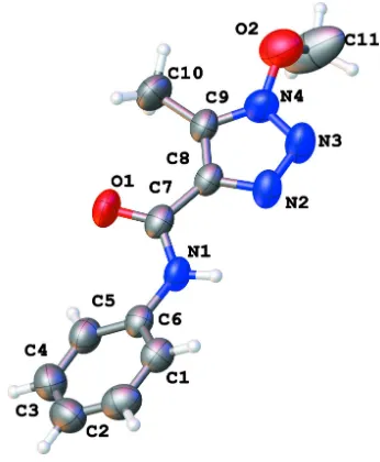
transformation of sodium 4-acetyl-1-phenyl-1H -[1.2.3]triazo-late under the action of methoxyamine hydrochloride. The dihedral angle between the triazole and phenyl rings is 25.12 (16) and the C atom of the methoxy group deviates
from the triazole plane by 0.894 (4)A˚ . The conformation of the CONHR-group is consolodated by an intramolecular N— H N hydrogen bond to an N-atom of the triazole ring, which closes anS(5) ring. In the crystal, weak N—H N hydrogen bonds link the molecules intoC(6) [010] chains.
Keywords:crystal structure; 1,2,3-triazole; rearrangements; hydrogen bonding.
CCDC reference:1426448
1. Related literature
For biological activities of 1.2.3-triazoles, see: Sathish Kumar & Kavitha (2013); Khazhievaet al.(2015a). For the synthesis, see: Khazhievaet al.(2015b).
2. Experimental
2.1. Crystal data
C11H12N4O2 Mr= 232.25 Monoclinic,P21=c a= 11.4637 (8) A˚
b= 6.4345 (13) A˚
c= 15.822 (3) A˚
= 100.367 (12)
V= 1148.0 (3) A˚3
Z= 4
MoKradiation
= 0.10 mm1 T= 295 K
0.210.160.09 mm
2.2. Data collection
Agilent Xcalibur S CCD diffractometer 7259 measured reflections
2302 independent reflections 1077 reflections withI> 2(I)
Rint= 0.040
2.3. Refinement
R[F2> 2(F2)] = 0.055 wR(F2) = 0.147
S= 1.00 2302 reflections 160 parameters
H atoms treated by a mixture of independent and constrained refinement
max= 0.43 e A˚ 3
min=0.22 e A˚ 3
Table 1
Hydrogen-bond geometry (A˚ ,).
D—H A D—H H A D A D—H A
N1—H1 N2 0.86 (2) 2.33 (3) 2.780 (4) 113 (2)
N1—H1 N3i
0.86 (2) 2.41 (2) 3.184 (3) 150 (2)
Symmetry code: (i)xþ2;y1 2;zþ
1 2.
Data collection: CrysAlis PRO (Agilent, 2006); cell refinement: CrysAlis PRO; data reduction: CrysAlis PRO; program(s) used to solve structure: SHELXS97(Sheldrick, 2008); program(s) used to refine structure:SHELXS97(Sheldrick, 2008); molecular graphics: publCIF (Westrip, 2010); software used to prepare material for publication:publCIF(Westrip, 2010).
Acknowledgements
We thank the Russian Foundation for Basic Research (grant 13–03-00137), State task Ministry of Education and Science of the Russian Federation No. 4.560.2014-K and the Project Enhance Competitiveness of the Ural Federal University (Project 5–100-2020)
Supporting information for this paper is available from the IUCr electronic archives (Reference: HB7511).
References
Agilent (2006). CrysAlis PRO. Agilent Technologies UK Ltd, Yarnton, England.
Khazhieva, I. S., Glukhareva, T. V., El’tsov, O. S., Morzherin, Yu. Yu., Minin, A. A., Pozdina, V. A. & Ulitko, M. V. (2015b).Khim. Farm. Zh.49, 12–15. Khazhieva, I. S., Glukhareva, T. V. & Morzherin, Yu. Yu. (2015a).Chim. Tech.
Acta,2, 52–58.
Sathish Kumar, S. & Kavitha, H. P. (2013).Mini-Rev. Org. Chem.10, 40–65.
supporting information
Acta Cryst. (2015). E71, o798 [doi:10.1107/S2056989015017776]
Crystal structure of 1-methoxy-5-methyl-
N
-phenyl-1,2,3-triazole-4-carboxamide
Inna S. Khazhieva, Tatiana V. Glukhareva, Pavel A. Slepukhin and Yury Yu. Morzherin
S1. Synthesis and crystallization
The titled compound was prepared as previously reported (Khazhieva et al., 2015b). Crystals were obtained by slow
Figure 1
The molecular structure of (I), with 50% probability displacement ellipsoids for non-H atoms.
1-Methoxy-5-methyl-N-phenyl-1,2,3-triazole-4-carboxamide
Crystal data
C11H12N4O2
Mr = 232.25 Monoclinic, P21/c
a = 11.4637 (8) Å
b = 6.4345 (13) Å
c = 15.822 (3) Å
β = 100.367 (12)°
V = 1148.0 (3) Å3
Z = 4
F(000) = 488
Dx = 1.344 Mg m−3 Melting point: 310 K
Mo Kα radiation, λ = 0.71073 Å Cell parameters from 1077 reflections
θ = 2.9–26.4°
µ = 0.10 mm−1
Data collection
Agilent Xcalibur S CCD diffractometer
Radiation source: fine-focus sealed tube Graphite monochromator
ω scans
7259 measured reflections 2302 independent reflections
1077 reflections with I > 2σ(I)
Rint = 0.040
θmax = 26.4°, θmin = 2.9°
h = −7→14
k = −5→8
l = −19→19
Refinement
Refinement on F2 Least-squares matrix: full
R[F2 > 2σ(F2)] = 0.055
wR(F2) = 0.147
S = 1.00 2302 reflections 160 parameters 0 restraints
Primary atom site location: structure-invariant direct methods
Secondary atom site location: difference Fourier map
Hydrogen site location: inferred from neighbouring sites
H atoms treated by a mixture of independent and constrained refinement
w = 1/[σ2(F
o2) + (0.0682P)2] where P = (Fo2 + 2Fc2)/3 (Δ/σ)max < 0.001
Δρmax = 0.43 e Å−3 Δρmin = −0.22 e Å−3
Special details
Geometry. All e.s.d.'s (except the e.s.d. in the dihedral angle between two l.s. planes) are estimated using the full covariance matrix. The cell e.s.d.'s are taken into account individually in the estimation of e.s.d.'s in distances, angles and torsion angles; correlations between e.s.d.'s in cell parameters are only used when they are defined by crystal symmetry. An approximate (isotropic) treatment of cell e.s.d.'s is used for estimating e.s.d.'s involving l.s. planes.
Refinement. Refinement of F2 against ALL reflections. The weighted R-factor wR and goodness of fit S are based on F2, conventional R-factors R are based on F, with F set to zero for negative F2. The threshold expression of F2 > σ(F2) is used only for calculating R-factors(gt) etc. and is not relevant to the choice of reflections for refinement. R-factors based on F2 are statistically about twice as large as those based on F, and R-factors based on ALL data will be even larger.
Fractional atomic coordinates and isotropic or equivalent isotropic displacement parameters (Å2)
x y z Uiso*/Ueq
O1 0.61315 (16) 0.0563 (3) 0.15704 (14) 0.0777 (7)
C8 0.7887 (2) 0.1712 (4) 0.24262 (18) 0.0475 (7)
C6 0.7399 (2) −0.2566 (4) 0.08075 (18) 0.0496 (7)
C7 0.7204 (2) 0.0365 (4) 0.17728 (19) 0.0533 (7)
N2 0.90562 (18) 0.1400 (4) 0.27423 (17) 0.0605 (7)
N4 0.8463 (2) 0.3915 (4) 0.33811 (19) 0.0708 (8)
C9 0.7489 (2) 0.3375 (4) 0.28291 (19) 0.0553 (8)
N1 0.7844 (2) −0.1083 (4) 0.14353 (16) 0.0530 (6)
N3 0.9416 (2) 0.2771 (4) 0.3343 (2) 0.0759 (8)
O2 0.8515 (2) 0.5302 (4) 0.40535 (18) 0.0956 (8)
C1 0.7975 (2) −0.4450 (5) 0.0824 (2) 0.0589 (8)
H1A 0.8634 −0.4721 0.1246 0.071*
C5 0.6434 (3) −0.2172 (5) 0.0169 (2) 0.0641 (8)
H5A 0.6049 −0.0896 0.0148 0.077*
C3 0.6605 (3) −0.5520 (6) −0.0411 (2) 0.0809 (10)
C2 0.7571 (3) −0.5924 (5) 0.0214 (2) 0.0739 (9)
H2A 0.7955 −0.7200 0.0225 0.089*
C4 0.6048 (3) −0.3651 (6) −0.0430 (2) 0.0769 (10)
H4A 0.5395 −0.3381 −0.0857 0.092*
C11 0.9070 (4) 0.7045 (6) 0.3901 (3) 0.137 (2)
H11A 0.8970 0.8073 0.4321 0.205*
H11B 0.8741 0.7551 0.3337 0.205*
H11C 0.9900 0.6765 0.3933 0.205*
C10 0.6343 (3) 0.4512 (5) 0.2740 (2) 0.0818 (10)
H10A 0.6298 0.5214 0.3268 0.123*
H10B 0.5699 0.3543 0.2609 0.123*
H10C 0.6292 0.5511 0.2284 0.123*
H1 0.858 (2) −0.109 (4) 0.1666 (17) 0.048 (8)*
Atomic displacement parameters (Å2)
U11 U22 U33 U12 U13 U23
O1 0.0365 (12) 0.1021 (17) 0.0930 (17) 0.0057 (10) 0.0076 (11) −0.0186 (13)
C8 0.0393 (15) 0.0458 (16) 0.0589 (18) 0.0007 (12) 0.0134 (13) 0.0056 (14)
C6 0.0414 (15) 0.0538 (18) 0.0549 (19) −0.0035 (14) 0.0117 (14) 0.0045 (16)
C7 0.0418 (16) 0.0582 (18) 0.062 (2) −0.0016 (14) 0.0146 (15) 0.0066 (16)
N2 0.0444 (14) 0.0516 (15) 0.0836 (19) −0.0032 (11) 0.0062 (13) −0.0019 (14)
N4 0.0607 (17) 0.0658 (17) 0.089 (2) −0.0011 (13) 0.0206 (15) −0.0268 (16)
C9 0.0400 (16) 0.065 (2) 0.0624 (19) −0.0028 (14) 0.0117 (15) −0.0013 (16)
N1 0.0354 (13) 0.0558 (15) 0.0661 (17) 0.0045 (11) 0.0041 (12) −0.0011 (13)
N3 0.0465 (15) 0.0710 (17) 0.107 (2) 0.0004 (13) 0.0059 (14) −0.0188 (17)
O2 0.0967 (18) 0.0961 (18) 0.102 (2) −0.0125 (14) 0.0395 (15) −0.0152 (16)
C1 0.0573 (17) 0.0563 (19) 0.064 (2) 0.0021 (15) 0.0140 (15) 0.0062 (17)
C5 0.0512 (18) 0.073 (2) 0.067 (2) 0.0069 (15) 0.0089 (16) 0.0035 (19)
C3 0.077 (2) 0.093 (3) 0.075 (3) −0.018 (2) 0.018 (2) −0.024 (2)
C2 0.081 (2) 0.061 (2) 0.085 (3) −0.0020 (18) 0.030 (2) −0.004 (2)
C4 0.061 (2) 0.098 (3) 0.070 (2) −0.005 (2) 0.0055 (17) −0.010 (2)
C11 0.171 (4) 0.055 (2) 0.221 (5) −0.019 (2) 0.133 (4) −0.005 (3)
C10 0.0566 (19) 0.098 (2) 0.092 (3) 0.0195 (17) 0.0172 (17) −0.016 (2)
Geometric parameters (Å, º)
O1—C7 1.220 (3) C1—C2 1.372 (4)
C8—N2 1.359 (3) C1—H1A 0.9300
C8—C9 1.365 (3) C5—C4 1.359 (4)
C8—C7 1.463 (4) C5—H5A 0.9300
C6—C1 1.379 (4) C3—C4 1.360 (5)
C6—C5 1.381 (4) C3—C2 1.370 (5)
C6—N1 1.405 (3) C3—H3A 0.9300
C7—N1 1.354 (3) C2—H2A 0.9300
N2—N3 1.308 (3) C4—H4A 0.9300
N4—N3 1.327 (3) C11—H11A 0.9600
N4—O2 1.382 (3) C11—H11C 0.9600
C9—C10 1.488 (4) C10—H10A 0.9600
N1—H1 0.85 (3) C10—H10B 0.9600
O2—C11 1.333 (4) C10—H10C 0.9600
N2—C8—C9 109.5 (2) C4—C5—C6 120.0 (3)
N2—C8—C7 122.6 (2) C4—C5—H5A 120.0
C9—C8—C7 127.8 (2) C6—C5—H5A 120.0
C1—C6—C5 119.6 (3) C4—C3—C2 120.0 (3)
C1—C6—N1 118.1 (3) C4—C3—H3A 120.0
C5—C6—N1 122.3 (3) C2—C3—H3A 120.0
O1—C7—N1 124.1 (3) C3—C2—C1 120.2 (3)
O1—C7—C8 120.6 (2) C3—C2—H2A 119.9
N1—C7—C8 115.3 (2) C1—C2—H2A 119.9
N3—N2—C8 109.2 (2) C5—C4—C3 120.7 (3)
N3—N4—C9 115.2 (2) C5—C4—H4A 119.6
N3—N4—O2 118.1 (3) C3—C4—H4A 119.6
C9—N4—O2 125.9 (2) O2—C11—H11A 109.5
N4—C9—C8 101.5 (2) O2—C11—H11B 109.5
N4—C9—C10 123.7 (3) H11A—C11—H11B 109.5
C8—C9—C10 134.8 (3) O2—C11—H11C 109.5
C7—N1—C6 126.3 (3) H11A—C11—H11C 109.5
C7—N1—H1 113.4 (17) H11B—C11—H11C 109.5
C6—N1—H1 120.2 (17) C9—C10—H10A 109.5
N2—N3—N4 104.6 (2) C9—C10—H10B 109.5
C11—O2—N4 111.1 (3) H10A—C10—H10B 109.5
C2—C1—C6 119.6 (3) C9—C10—H10C 109.5
C2—C1—H1A 120.2 H10A—C10—H10C 109.5
C6—C1—H1A 120.2 H10B—C10—H10C 109.5
Hydrogen-bond geometry (Å, º)
D—H···A D—H H···A D···A D—H···A
N1—H1···N2 0.86 (2) 2.33 (3) 2.780 (4) 113 (2)
N1—H1···N3i 0.86 (2) 2.41 (2) 3.184 (3) 150 (2)