Received 3 June 2019 Accepted 8 July 2019
Edited by L. Van Meervelt, Katholieke Universi-teit Leuven, Belgium
Keywords:crystal structure; dihydroindole-dione; hydrogen bond; micelle;-stacking.
CCDC reference:1938997
Supporting information:this article has supporting information at journals.iucr.org/e
Crystal structure, DFT study and Hirshfeld surface
analysis of 1-nonyl-2,3-dihydro-1
H
-indole-2,3-dione
Ibtissam Rayni,aYouness El Bakri,b* Chin-Hung Lai,c,dJihad Sebhaoui,aEl Mokhtar Essassiaand Joel T. Maguee
aLaboratoire de Chimie Organique He´te´rocyclique, Centre de Recherche des Sciences des Me´dicaments, URAC 21, Poˆle
de Compe´tence Pharmacochimie, Av Ibn Battouta, BP 1014, Faculte´ des Sciences, Universite´ Mohammed V, Rabat, Morocco,bOrganic Chemistry Department, Faculty of Science, RUDN University, Miklukho-Maklaya St. 6, 117198
Moscow, Russian Federation,cDepartment of Medical Applied Chemistry, Chung Shan Medical University, Taichung
40241, Taiwan,dDepartment of Medical Education, Chung Shan Medical University Hospital, 402 Taichung, Taiwan,
andeDepartment of Chemistry, Tulane University, New Orleans, LA 70118, USA. *Correspondence e-mail:
In the title molecule, C17H23NO2, the dihydroindole portion is planar (r.m.s.
deviation = 0.0157 A˚ ) and the nonyl substituent is in an ‘extended’ conformation. In the crystal, the nonyl chains intercalate and the dihydro-indoledione units are associated through C—H O hydrogen bonds to form micellar blocks. Based on the Hirshfeld surface analysis, the most important intermolecular interaction is the H H interaction.
1. Chemical context
Indoline-2,3-dione or indole-1H-2,3-dione, commonly known as isatin, is a well-known natural product found in plants of genusIsatisand inCouropita guianancis aubl(Da Silvaet al., 2001). It has also been isolated as a metabolic derivative of adrenaline in humans (Almeida et al., 2010). It was first obtained as an oxidation product of indigo in the early 19th century, and its current structure was proposed by Kekule´ (1869). Isatin is a core constituent of many alkaloids (Trostet al., 2009) and drugs (Aboul-Fadlet al., 2010) as well as dyes (Dome´nech et al., 2009), pesticides and analytical reagents. Isatin derivatives possess diverse activities such as anti-bacterial (Kassab et al., 2010), antiviral (Jarrahpour et al., 2007), anti-HIV (Sriramet al., 2006), anticancer (Gu¨rsoyet al., 2003) and anti-inflammatory (Sridharet al., 2001) activities. As a continuation of our research work devoted to the develop-ment of isatin derivatives (Ben-Yahiaet al., 2018; Rayniet al., 2019), we report in this work the synthesis and the Hirshfeld surface analysis of a new indoline-2,3-dione derivative obtained by the action of nonyl bromide on isatin under phase-transfer catalysis conditions.
2. Structural commentary
The molecular structure of the title compound is shown in Fig. 1. The dihydroindole skeleton is planar to within 0.0286 (8) A˚ (r.m.s. deviation of the fitted atoms = 0.0157 A˚) with Cl being the furthest from the mean plane. The nonyl chain is in an ‘extended’ conformation and is well out of the mean plane of the dihydroindole unit, as indicated by the C1— N1—C9—C10 torsion angle of69.94 (12).
3. Supramolecular features
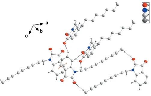
In the crystal, the molecules pack in a typical micellar manner with the dihydroindoldione head groups associated through C2—H2 O2i, C3—H3 O1iiand C9—H9B O1ihydrogen bonds (Table 1) and the nonyl ‘tails’ intercalating and aided by paired C17—H17B O2iii hydrogen bonds (Table 1 and Fig. 2). The micellar blocks are associated through-stacking interactions between inversion-related C1–C6 rings [centroid– centroid distance = 3.6470 (7) A˚ ; Figs. 2 and 3].
4. Database survey
A search of the Cambridge Crystallographic Database (Version 5.40 updated to April 2019; Groom et al., 2016) provided structures of 11 derivatives of the dihydroindole-2,3-dione skeleton having a saturated carbon chain of at least three atoms bound to nitrogen. Thus, in place of then-nonyl
chain (R) in the title compound, there are ones with R= 3-bromopropyl (AKOBIN; Qachchachiet al., 2016a),n-propyl (AKOCOU; Qachchachi et al., 2016b), n-octyl (CIQDOX; Qachchachiet al., 2013), 2,3-dibenzoylethane (FUBLIZ; Zˇ ari
et al., 2015),n-dodecyl (GITTEK; Qachchachi et al., 2014a), cyclopentyl (JOWSOF; Mironova et al., 2015), 3-carboxy-methylpropane (JOWSUL; Mironova et al., 2015), 2-cyano-ethane (LIVSIU; Qachchachi et al., 2014b), n-tetradecyl (TUPSIH; Mamari et al., 2010a) and n-decyl (TUPSON; Mamariet al., 2010b). In addition, there is one structure with two dihydroindole-2,3-dione moieties connected by a –(CH2)6– linkage (OJIGOF; Qachchachiet al., 2016c). In all of
[image:2.610.316.567.69.229.2] [image:2.610.46.296.92.152.2]these compounds, the dihydroindole-2,3-dione skeleton is planar and the first two carbon atoms from the nitrogen are rotated so that the N–C–C plane is nearly perpendicular to the plane of the dihydroindole-2,3-dione. Additionally, the C—C distances corresponding to the C7—C8 distance in the title structure [1.5554 (15) A˚ ] are in the range 1.543 (4)– 1.563 (6) A˚ . Generally, the carbon chains are in an ‘extended’ conformation.
Table 1
Hydrogen-bond geometry (A˚ ,).
D—H A D—H H A D A D—H A
C2—H2 O2i 0.992 (13) 2.412 (13) 3.3737 (13) 163.3 (10)
C3—H3 O1ii 0.997 (14) 2.454 (15) 3.2734 (14) 139.0 (11)
C9—H9B O1i 0.994 (13) 2.546 (13) 3.5012 (13) 161.0 (10)
C17—H17B O2iii 0.98 (2) 2.49 (2) 3.3941 (17) 153.3 (15)
Symmetry codes: (i)x;yþ3 2;z
1
2; (ii)x;yþ 1 2;z
1
2; (iii)xþ1;yþ2;zþ1.
Figure 2
Detail of the intermolecular interactions. C—H O hydrogen bonds and
[image:2.610.46.298.531.717.2]-stacking interactions are shown, respectively, by black and orange dashed lines. H atoms not involved in hydrogen bonds are omitted for clarity.
Figure 3
Packing viewed along the b-axis direction with intermolecular inter-actions depicted as in Fig. 2. H atoms not involved in hydrogen bonds are omitted for clarity.
Figure 1
[image:2.610.313.567.551.705.2]5. Calculation of the electronic structure
The structure in the gas phase of the title compound was optimized by means of density functional theory. The DFT calculation was performed using the hybrid B3LYP method, which is based on the idea of Becke and considers a mixture of the exact (HF) and DFT exchange utilizing the B3 functional, together with the LYP correlation functional (Becke, 1993; Leeet al., 1988; Miehlichet al., 1989). The B3LYP calculation was performed in conjunction with the def2-SVP basis set (Weigend & Ahlrichs, 2005). After obtaining the converged geometry, the harmonic vibrational frequencies were calcu-lated on the same theoretical level to confirm that the number of imaginary frequencies is zero for the stationary point. Both the geometry optimization and the harmonic vibrational frequency analysis of the title compound were performed using theGaussian 16program (Frischet al., 2016). The result of the B3LYP geometry optimization for the title compound (shown in Fig. 4) was compared to that of the crystallographic study with selected geometric parameters for the gas-phase and solid-phase structures summarized in Table 2. This shows that there is a clear discrepancy between the B3LYP-opti-mized geometry and the X-ray geometry. To quantify this, the
openBabelprogram was then used to convert the experimental CIF file to aGaussian.gjf input file (O’Boyleet al., 2011). The structure compared built in theChemCraftprogram (graphical
software for visualization of quantum chemistry computations; https://www.chemcraftprog.com) was finally used to obtain a weighted r.m.s. deviation of 0.5808 A˚ with r.m.s.d. values of of 0.6297, 0.5213, 0.2231, and 0.5977 A˚ , respectively, for the H, C, N and O atoms.
6. Hirshfeld surface analysis
Both the definition of a molecule in a condensed phase and the recognition of distinct entities in molecular liquids and crystals are fundamental concepts in chemistry. Based on Hirshfeld’s partitioning scheme, Spackman et al. (1997) proposed a method to divide the electron distribution in a crystalline phase into molecular fragments (Spackman & Byrom, 1997; McKinnon et al., 2004; Spackman & Jayatilaka, 2009). Their proposed method partitioned the crystal into regions where the electron distribution of a sum of spherical atoms for the molecule dominates over the corresponding sum of the crystal. In this study, the Hirshfeld surface analysis of the title compound was performed utilizing the CrystalExplorer
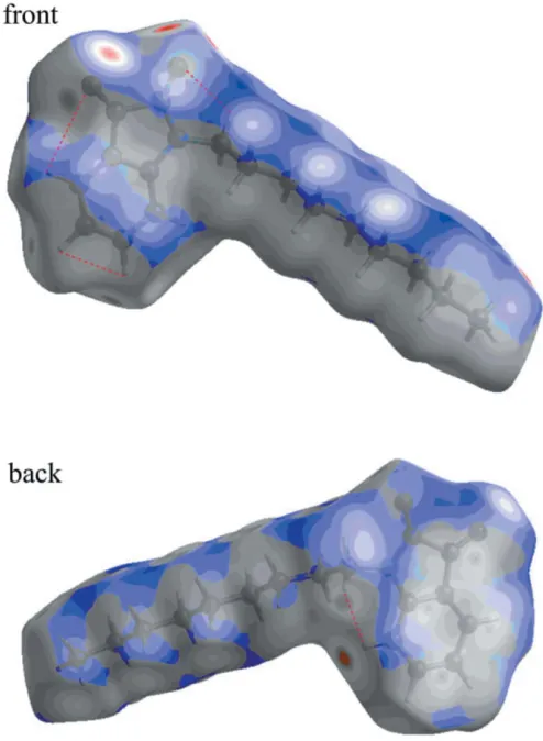
program (Turneret al., 2017). The standard resolution mol-ecular Hirshfeld surface (dnorm) of the title compound is
depicted in Fig. 5. This surface can be used to identify very close intermolecular interactions. The value ofdnorm is
nega-Figure 5
[image:3.610.44.297.102.250.2]ThednormHirshfeld surface of the title compound (red: negative, white: zero, blue: positive; scale:0.2101 to 1.3375 a.u.).
Table 2
The B3LYP-optimized and X-ray structural parameters (A˚ ,) for the title compound.
B3LYP X-ray
C1—C2 1.394 1.3806 (13)
C2—C3 1.404 1.3899 (16)
C3—C4 1.402 1.3868 (16)
C4—C5 1.400 1.3871 (16)
C5—C6 1.393 1.3862 (15)
C6—C7 1.473 1.4599 (13)
C6—C1 1.413 1.4009 (13)
C7—C8 1.568 1.5554 (15)
C8—N1 1.390 1.3603 (13)
N1—C1 1.404 1.4127 (13)
C7—O1 1.206 1.2126 (12)
C8—O2 1.206 1.2106 (13)
N1—C9 1.454 1.4606 (13)
[image:3.610.314.561.371.708.2]N1—C8—C7 105.9 106.20 (8)
Figure 4
[image:3.610.45.292.603.704.2]tive (positive) when intermolecular contacts are shorter (longer) than the van der Waals radii. The dnorm value is
mapped onto the Hirshfeld surface using red, white or blue colours. The red regions represent closer contacts with a negative dnormvalue while the blue regions represent longer
contacts with a positive dnorm value. The white regions
represent contacts equal to the van der Waals separation and have a dnorm value of zero. As depicted in Fig. 5, important
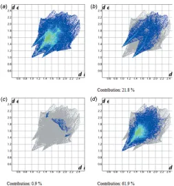
interactions in the title compound are H O and H N hydrogen bonds. The two-dimensional fingerprint plots (Fig. 6) highlight particular atom-pair contacts and enable the separation of contributions from different interaction types that overlap in the full fingerprint. The most important interactions involving the hydrogen atoms in the title compound are the H H contactso. The H H, H O/ O H and H N/N H contacts make contribututions of 61.9, 21.8 and 0.9%, respectively, to the Hirshfeld surface.
7. Synthesis and crystallization
To a solution of isatin (0.5 g, 3.4 mmol) dissolved in 25 ml of
N,N-dimethylformamide, 1-bromooctane (0.7 ml, 3.4 mmol), potassium carbonate (0.61 g, 4.4 mmol) and a catalytic amount of tetra-n-butylammonium bromide (0.1 g, 0.4 mmol) were added. The mixture was stirred for 48 h and the reaction monitored by thin layer chromatography. The mixture was filtered and the solvent removed under vacuum. The solid obtained was recrystallized from ethanol to afford the title compound as orange–red crystals.
8. Refinement
Crystal data, data collection and structure refinement details are summarized in Table 3.
Acknowledgements
We thank the National Center for High-performance Computing (Taiwan) for providing computing time.
Funding information
This publication was prepared with the support of the RUDN University Program 5–100. The support of NSF–MRI grant No. 1228232 for the purchase of the diffractometer and Tulane University for support of the Tulane Crystallography Laboratory are gratefully acknowledged.
References
Aboul-Fadl, T., Bin-Jubair, F. A. S. & Aboul-Wafa, O. (2010).Eur. J. Med. Chem.45, 4578–4586.
Almeida, M. R., Leita˜o, G. G., Silva, B. V., Barbosa, J. P. & Pinto, A. C. J. (2010).J. Braz. Chem. Soc.21, 764–769.
Becke, A. D. (1993).J. Chem. Phys.98, 5648–5652.
Ben-Yahia, A., El Bakri, Y. E., Lai, C.-H., Essassi, E. M. & Mague, J. T. (2018).Acta Cryst.E74, 1857–1861.
Brandenburg, K. & Putz, H. (2012). DIAMOND, Crystal Impact GbR, Bonn, Germany.
[image:4.610.45.298.66.336.2]Bruker (2016). APEX3, SAINT and SADABS, Bruker AXS Inc., Madison, Wisconsin, USA.
Figure 6
Fingerprint plots for the title compound: (a) full and delineated into (b) H O/O H, (c) H N/N H and (d) H H contacts.
Table 3
Experimental details.
Crystal data
Chemical formula C17H23NO2
Mr 273.36
Crystal system, space group Monoclinic,P21/c
Temperature (K) 150
a,b,c(A˚ ) 16.2512 (4), 7.6859 (2), 13.0989 (3)
() 106.640 (1)
V(A˚3) 1567.60 (7)
Z 4
Radiation type CuK
(mm1) 0.59
Crystal size (mm) 0.240.200.14
Data collection
Diffractometer Bruker D8 VENTURE PHOTON
100 CMOS
Absorption correction Multi-scan (SADABS; Krauseet
al., 2015)
Tmin,Tmax 0.82, 0.92
No. of measured, independent and observed [I> 2(I)] reflections
11594, 3128, 2879
Rint 0.029
(sin/)max(A˚
1
) 0.625
Refinement
R[F2> 2(F2)],wR(F2),S 0.035, 0.096, 1.05
No. of reflections 3128
No. of parameters 274
H-atom treatment All H-atom parameters refined
max, min(e A˚
3
) 0.22,0.14
[image:4.610.312.567.90.366.2]Dome´nech, A., Dome´nech-Carbo´, M. T., Sa´nchez del Rı´o, M., Va´zquez de Agredos Pascual, M. L. & Lima, E. (2009). New J. Chem.33, 2371–2379.
Frisch, M. J., Trucks, G. W., Schlegel, H. B., Scuseria, G. E., Robb, M. A., Cheeseman, J. R., Scalmani, G., Barone, V., Petersson, G. A., Nakatsuji, H., Li, X., Caricato, M., Marenich, A. V., Bloino, J., Janesko, B. G., Gomperts, R., Mennucci, B., Hratchian, H. P., Ortiz, J. V., Izmaylov, A. F., Sonnenberg, J. L., Williams-Young, D., Ding, F., Lipparini, F., Egidi, F., Goings, J., Peng, B., Petrone, A., Henderson, T., Ranasinghe, D., Zakrzewski, V. G., Gao, J., Rega, N., Zheng, G., Liang, W., Hada, M., Ehara, M., Toyota, K., Fukuda, R., Hasegawa, J., Ishida, M., Nakajima, T., Honda, Y., Kitao, O., Nakai, H., Vreven, T., Throssell, K., Montgomery, J. A., Peralta, J. E. Jr, Ogliaro, F., Bearpark, M. J., Heyd, J. J., Brothers, E. N., Kudin, K. N., Staroverov, V. N., Keith, T. A., Kobayashi, R., Normand, J., Raghavachari, K., Rendell, A. P., Burant, J. C., Iyengar, S. S., Tomasi, J., Cossi, M., Millam, J. M., Klene, M., Adamo, C., Cammi, R., Ochterski, J. W., Martin, R. L., Morokuma, K., Farkas, O., Foresman, J. B. & Fox, D. J. (2016). Gaussian 16, Revision A. 03. Gaussian, Inc., Wallingford CT.
Groom, C. R., Bruno, I. J., Lightfoot, M. P. & Ward, S. C. (2016).Acta Cryst.B72, 171–179.
Gu¨rsoy, A. & Karalı´, N. (2003).Eur. J. Med. Chem.38, 633–643. Jarrahpour, A., Khalili, D., De Clercq, E., Salmi, C. & Brunel, J. M.
(2007).Molecules,12, 1720–1730.
Kassab, S., Hegazy, G., Eid, N., Amin, K. & El-Gendy, A. (2010). Nucleosides Nucleotides Nucleic Acids,29, 72–80.
Kekule´, A. (1869).Ber. Dtsch. Chem. Ges.2, 748–749.
Krause, L., Herbst-Irmer, R., Sheldrick, G. M. & Stalke, D. (2015).J. Appl. Cryst.48, 3–10.
Lee, C., Yang, W. & Parr, R. G. (1988).Phys. Rev. B,37, 785–789. Mamari, K., Zouihri, H., Essassi, E. M. & Ng, S. W. (2010a). Acta
Cryst. E66, o1410.
Mamari, K., Zouihri, H., Essassi, E. M. & Ng, S. W. (2010b).Acta Cryst.E66, o1411.
McKinnon, J. J., Spackman, M. A. & Mitchell, A. S. (2004). Acta Cryst.B60, 627–668.
Miehlich, B., Savin, A., Stoll, H. & Preuss, H. (1989).Chem. Phys. Lett.157, 200–206.
Mironova, E. V., Bogdanov, A. V., Krivolapov, D. B., Musin, L. I., Litvinov, I. A. & Mironov, V. F. (2015).J. Mol. Struct.1079, 87– 93.
O’Boyle, N. M., Banck, M., James, C. A., Morley, C., Vandermeersch, T. & Hutchison, G. R. (2011).J. Cheminform,3, 33.
Qachchachi, F.-Z., Kandri Rodi, Y., Essassi, E. M., Bodensteiner, M. & El Ammari, L. (2014b).Acta Cryst.E70, o361–o362.
Qachchachi, F.-Z., Kandri Rodi, Y., Essassi, E. M., Kunz, W. & El Ammari, L. (2013).Acta Cryst.E69, o1801.
Qachchachi, F. Z., Kandri Rodi, Y., Haoudi, A., Essassi, E. M., Capet, F. & Zouihri, H. (2016a).IUCrData,1, x160593.
Qachchachi, F. Z., Kandri Rodi, Y., Haoudi, A., Essassi, E. M., Capet, F. & Zouihri, H. (2016b).IUCrData,1, x160609.
Qachchachi, F. Z., Kandri Rodi, Y., Haoudi, A., Essassi, E. M., Capet, F. & Zouihri, H. (2016c).IUCrData,1, x160542.
Qachchachi, F.-Z., Ouazzani Chahdi, F., Misbahi, H., Bodensteiner, M. & El Ammari, L. (2014a).Acta Cryst.E70, o229.
Rayni, I., El Bakri, Y., Lai, C.-H., El Ghayati, L., Essassi, E. M. & Mague, J. T. (2019).Acta Cryst.E75, 21–25.
Sheldrick, G. M. (2015a).Acta Cryst.A71, 3–8. Sheldrick, G. M. (2015b).Acta Cryst.C71, 3–8.
Silva, J. F. M. da, Garden, S. J. & Pinto, A. C. J. (2001).J. Braz. Chem. Soc.12, 273–324.
Spackman, M. A. & Byrom, P. G. (1997).Chem. Phys. Lett.267, 215– 220.
Spackman, M. A. & Jayatilaka, D. (2009).CrystEngComm,11, 19– 32.
Sridhar, S. K. & Ramesh, A. (2001). Biol. Pharm. Bull. 24, 1149– 1152.
Sriram, D., Yogeeswari, P., Myneedu, N. S. & Saraswat, V. (2006). Bioorg. Med. Chem. Lett.16, 2127–2129.
Trost, B. & Brennan, M. (2009).Synthesis, pp. 3003–3025.
Turner, M. J., McKinnon, J. J., Wolff, S. K., Grimwood, D. J., Spackman, P. R., Jayatilaka, D. & Spackman, M. A. (2017). CrystalExplorer17.University of Western Australia.
Weigend, F. & Ahlrichs, R. (2005).Phys. Chem. Chem. Phys.7, 3297– 3305.
sup-1
Acta Cryst. (2019). E75, 1140-1144
supporting information
Acta Cryst. (2019). E75, 1140-1144 [https://doi.org/10.1107/S2056989019009691]
Crystal structure, DFT study and Hirshfeld surface analysis of
1-nonyl-2,3-di-hydro-1
H
-indole-2,3-dione
Ibtissam Rayni, Youness El Bakri, Chin-Hung Lai, Jihad Sebhaoui, El Mokhtar Essassi and Joel T.
Mague
Computing details
Data collection: APEX3 (Bruker, 2016); cell refinement: SAINT (Bruker, 2016); data reduction: SAINT (Bruker, 2016); program(s) used to solve structure: SHELXT (Sheldrick, 2015a); program(s) used to refine structure: SHELXL2018
(Sheldrick, 2015b); molecular graphics: DIAMOND (Brandenburg & Putz, 2012); software used to prepare material for publication: SHELXTL (Sheldrick, 2008).
1-Nonyl-2,3-dihydro-1H-indole-2,3-dione
Crystal data
C17H23NO2 Mr = 273.36
Monoclinic, P21/c a = 16.2512 (4) Å
b = 7.6859 (2) Å
c = 13.0989 (3) Å
β = 106.640 (1)°
V = 1567.60 (7) Å3
Z = 4
F(000) = 592
Dx = 1.158 Mg m−3
Cu Kα radiation, λ = 1.54178 Å Cell parameters from 9962 reflections
θ = 5.1–74.4°
µ = 0.59 mm−1 T = 150 K Block, orange-red 0.24 × 0.20 × 0.14 mm
Data collection
Bruker D8 VENTURE PHOTON 100 CMOS diffractometer
Radiation source: INCOATEC IµS micro–focus source
Mirror monochromator
Detector resolution: 10.4167 pixels mm-1 ω scans
Absorption correction: multi-scan
(SADABS; Krause et al., 2015)
Tmin = 0.82, Tmax = 0.92
11594 measured reflections 3128 independent reflections 2879 reflections with I > 2σ(I)
Rint = 0.029
θmax = 74.4°, θmin = 6.4° h = −18→19
k = −9→8
l = −16→15
Refinement
Refinement on F2
Least-squares matrix: full
R[F2 > 2σ(F2)] = 0.035 wR(F2) = 0.096 S = 1.05 3128 reflections 274 parameters 0 restraints
Primary atom site location: structure-invariant direct methods
Secondary atom site location: difference Fourier map
Hydrogen site location: difference Fourier map All H-atom parameters refined
w = 1/[σ2(F
o2) + (0.0474P)2 + 0.2775P]
sup-2
Acta Cryst. (2019). E75, 1140-1144
(Δ/σ)max = 0.001
Δρmax = 0.22 e Å−3
Δρmin = −0.14 e Å−3
Extinction correction: SHELXL2018 (Sheldrick, 2015b), Fc*=kFc[1+0.001xFc2λ3/sin(2θ)]-1/4
Extinction coefficient: 0.0114 (9)
Special details
Geometry. All esds (except the esd in the dihedral angle between two l.s. planes) are estimated using the full covariance matrix. The cell esds are taken into account individually in the estimation of esds in distances, angles and torsion angles; correlations between esds in cell parameters are only used when they are defined by crystal symmetry. An approximate (isotropic) treatment of cell esds is used for estimating esds involving l.s. planes.
Refinement. Refinement of F2 against ALL reflections. The weighted R-factor wR and goodness of fit S are based on F2,
conventional R-factors R are based on F, with F set to zero for negative F2. The threshold expression of F2 > 2sigma(F2) is
used only for calculating R-factors(gt) etc. and is not relevant to the choice of reflections for refinement. R-factors based on F2 are statistically about twice as large as those based on F, and R- factors based on ALL data will be even larger.
Fractional atomic coordinates and isotropic or equivalent isotropic displacement parameters (Å2)
x y z Uiso*/Ueq
sup-3
Acta Cryst. (2019). E75, 1140-1144
H14B 0.5448 (9) 0.595 (2) 0.3575 (12) 0.057 (4)* C15 0.59615 (7) 0.79507 (16) 0.28902 (9) 0.0403 (3) H15A 0.6056 (9) 0.921 (2) 0.3018 (11) 0.059 (4)* H15B 0.5542 (9) 0.7861 (17) 0.2165 (11) 0.048 (3)* C16 0.67845 (8) 0.70487 (17) 0.28662 (10) 0.0448 (3) H16A 0.7217 (10) 0.725 (2) 0.3567 (13) 0.064 (4)* H16B 0.6688 (10) 0.575 (2) 0.2808 (13) 0.068 (4)* C17 0.71335 (9) 0.76729 (19) 0.19762 (11) 0.0494 (3) H17A 0.6695 (11) 0.753 (2) 0.1291 (14) 0.070 (5)* H17B 0.7272 (12) 0.892 (3) 0.2051 (14) 0.081 (5)* H17C 0.7687 (12) 0.699 (2) 0.1952 (14) 0.076 (5)*
Atomic displacement parameters (Å2)
U11 U22 U33 U12 U13 U23
O1 0.0578 (5) 0.0554 (5) 0.0250 (4) 0.0034 (4) 0.0169 (3) 0.0028 (3) O2 0.0739 (6) 0.0483 (5) 0.0427 (5) −0.0124 (4) 0.0191 (4) −0.0180 (4) N1 0.0383 (5) 0.0331 (4) 0.0272 (4) −0.0027 (3) 0.0110 (3) −0.0016 (3) C1 0.0299 (5) 0.0327 (5) 0.0254 (4) 0.0014 (4) 0.0072 (3) −0.0003 (3) C2 0.0360 (5) 0.0430 (6) 0.0286 (5) −0.0006 (4) 0.0129 (4) −0.0041 (4) C3 0.0421 (6) 0.0468 (6) 0.0384 (6) −0.0016 (5) 0.0148 (4) −0.0141 (5) C4 0.0477 (6) 0.0365 (6) 0.0510 (7) −0.0068 (5) 0.0178 (5) −0.0096 (5) C5 0.0418 (6) 0.0376 (6) 0.0394 (6) −0.0019 (4) 0.0149 (4) 0.0029 (4) C6 0.0338 (5) 0.0353 (5) 0.0251 (4) 0.0021 (4) 0.0086 (3) 0.0016 (4) C7 0.0385 (5) 0.0408 (5) 0.0222 (4) 0.0039 (4) 0.0076 (4) 0.0020 (4) C8 0.0426 (6) 0.0390 (5) 0.0270 (5) 0.0006 (4) 0.0083 (4) −0.0035 (4) C9 0.0394 (6) 0.0341 (5) 0.0360 (5) −0.0014 (4) 0.0113 (4) 0.0058 (4) C10 0.0365 (5) 0.0390 (6) 0.0357 (5) −0.0010 (4) 0.0092 (4) 0.0072 (4) C11 0.0375 (5) 0.0393 (6) 0.0381 (5) −0.0022 (4) 0.0095 (4) 0.0080 (4) C12 0.0367 (6) 0.0422 (6) 0.0387 (6) −0.0016 (4) 0.0094 (4) 0.0068 (4) C13 0.0371 (6) 0.0461 (6) 0.0370 (6) −0.0011 (4) 0.0086 (4) 0.0070 (5) C14 0.0368 (6) 0.0457 (6) 0.0378 (6) −0.0014 (4) 0.0088 (4) 0.0058 (4) C15 0.0384 (6) 0.0450 (6) 0.0364 (5) −0.0016 (4) 0.0090 (4) 0.0052 (4) C16 0.0388 (6) 0.0523 (7) 0.0435 (6) −0.0006 (5) 0.0118 (5) 0.0066 (5) C17 0.0496 (7) 0.0541 (7) 0.0486 (7) −0.0067 (6) 0.0205 (6) −0.0001 (6)
Geometric parameters (Å, º)
sup-4
Acta Cryst. (2019). E75, 1140-1144
C3—H3 0.997 (14) C14—C15 1.5163 (15) C4—C5 1.3871 (16) C14—H14A 1.007 (15) C4—H4 0.971 (16) C14—H14B 1.002 (16) C5—C6 1.3862 (15) C15—C16 1.5147 (17) C5—H5 0.980 (14) C15—H15A 0.986 (17) C6—C7 1.4599 (13) C15—H15B 1.000 (14) C7—C8 1.5554 (15) C16—C17 1.5132 (16) C9—C10 1.5233 (15) C16—H16A 0.995 (17) C9—H9A 0.989 (14) C16—H16B 1.011 (18) C9—H9B 0.994 (13) C17—H17A 0.979 (18) C10—C11 1.5204 (14) C17—H17B 0.98 (2) C10—H10A 1.006 (15) C17—H17C 1.050 (18)
sup-5
Acta Cryst. (2019). E75, 1140-1144
C9—C10—H10A 109.3 (8) C16—C17—H17B 111.0 (10) C11—C10—H10B 109.4 (7) H17A—C17—H17B 106.5 (15) C9—C10—H10B 109.0 (7) C16—C17—H17C 112.2 (9) H10A—C10—H10B 104.9 (11) H17A—C17—H17C 108.7 (14) C12—C11—C10 113.04 (9) H17B—C17—H17C 108.6 (14) C12—C11—H11A 110.9 (8)
C8—N1—C1—C2 178.81 (10) C1—C6—C7—C8 −1.98 (10) C9—N1—C1—C2 −6.62 (15) C1—N1—C8—O2 177.89 (11) C8—N1—C1—C6 −0.19 (11) C9—N1—C8—O2 3.47 (18) C9—N1—C1—C6 174.37 (9) C1—N1—C8—C7 −1.07 (11) C6—C1—C2—C3 −2.13 (15) C9—N1—C8—C7 −175.49 (9) N1—C1—C2—C3 178.96 (9) O1—C7—C8—O2 2.55 (17) C1—C2—C3—C4 0.59 (16) C6—C7—C8—O2 −177.09 (11) C2—C3—C4—C5 1.19 (18) O1—C7—C8—N1 −178.47 (10) C3—C4—C5—C6 −1.41 (17) C6—C7—C8—N1 1.89 (11) C4—C5—C6—C1 −0.11 (15) C8—N1—C9—C10 103.80 (12) C4—C5—C6—C7 179.33 (11) C1—N1—C9—C10 −69.94 (12) C2—C1—C6—C5 1.95 (15) N1—C9—C10—C11 167.45 (9) N1—C1—C6—C5 −178.98 (9) C9—C10—C11—C12 −173.60 (9) C2—C1—C6—C7 −177.62 (9) C10—C11—C12—C13 175.57 (9) N1—C1—C6—C7 1.45 (11) C11—C12—C13—C14 −178.95 (10) C5—C6—C7—O1 −1.08 (19) C12—C13—C14—C15 177.47 (10) C1—C6—C7—O1 178.42 (11) C13—C14—C15—C16 −179.94 (10) C5—C6—C7—C8 178.52 (11) C14—C15—C16—C17 174.71 (10)
Hydrogen-bond geometry (Å, º)
D—H···A D—H H···A D···A D—H···A
C2—H2···O2i 0.992 (13) 2.412 (13) 3.3737 (13) 163.3 (10)
C3—H3···O1ii 0.997 (14) 2.454 (15) 3.2734 (14) 139.0 (11)
C9—H9B···O1i 0.994 (13) 2.546 (13) 3.5012 (13) 161.0 (10)
C17—H17B···O2iii 0.98 (2) 2.49 (2) 3.3941 (17) 153.3 (15)