research communications
Acta Cryst.(2020). E76, 443–445 https://doi.org/10.1107/S2056989020002479
443
Received 22 January 2020Accepted 20 February 2020
Edited by A. J. Lough, University of Toronto, Canada
‡ Additional correspondence author, email: [email protected].
Keywords:crystal structure; pyrazolopyridine; biological activity.
Supporting information:this article has supporting information at journals.iucr.org/e
Crystal structure analysis of ethyl
3-(4-chlorophen-yl)-1,6-dimethyl-4-methylsulfanyl-1
H
-pyrazolo-[3,4-
b
]pyridine-5-carboxylate
H. Surya Prakash Rao,a,b* Ramalingam Gunasundariband Jayaraman Muthukumaranc‡
aDepartment of Chemistry and Biochemistry, School of Basic Sciences and Research, Sharda University, Greater Noida 201306, India,bDepartment of Chemistry, Pondicherry University, Puducherry 605014, India, andcDepartment of Biotechnology, School of Engineering and Technology, Sharda University, Greater Noida 201306, India. *Correspon-dence e-mail: [email protected]
In the title compound, C18H18ClN3O2S, the dihedral angle between the fused
pyrazole and pyridine rings is 3.81 (9). The benzene ring forms dihedral angles of 35.08 (10) and 36.26 (9)with the pyrazole and pyridine rings, respectively. In the crystal, weak C—H O hydrogen bonds connect molecules along [100].
1. Chemical context
The nitrogen-containing heterocyclic motif is a component in many medicinally important drugs. Molecules built around the pyrazolopyridine core structure exhibit diverse medicinal properties that include anti-microbial, anti-viral, anti-fungal, anti-hypertensive, analgesic, anti-cancer, anti-inflammatory, anti-Alzheimer’s, anti-diabetic, anti-nociceptive, anti-tuber-culosis, and anti-leishmanial activities (Hardy, 1984; Hawaset al.2019; de Melloet al.2004; Panchalet al.2019; El-Goharyet al.2019). In addition, some pyrazolopyridines have found uses for the treatment of hemorrhagic stress, infertility, and drug addiction (Parmar et al. 1974). Specifically, they act as inhi-bitors of enzymes such as glycogen synthase kinase-3 (With-eringtonet al.2003) and as inhibitors for adenosine receptors (Timo´teoet al.2008). Furthermore, they have been identified as promising inhibitors of cycline dependent kinase, xanthine oxidase, interleukin-6 (IL-6), tumor necrosis factor alpha (TNF-), phosphodiesterase-4, NAD(P)H oxidases and cholesterol formation (Go¨khan-Kelekc¸iet al.2007; Panchalet al. 2019; Fathy et al. 2015). Considering the aforementioned importance of derivatives of pyrazolopyridine, we have carried out a single-crystal X-ray diffraction study on the title compound and have analyzed the structure in terms of geometrical parameters, conformation, and intermolecular hydrogen-bonding interactions.
2. Structural commentary
The title compound has pyrazole[3,4-b]pyridine motif that is decorated by several substituents shown in Fig. 1. The chlorophenyl (C6H4Cl) group attached to the
pyrazolo-pyridine moiety exhibits an ( )anticlinal conformation [N3— C7—C6—C3 = 141.96 (19)], as does the methylthio (SCH3)
group attached to the pyrazolopyridine unit [C11—S1—C12— C13 = 128.93 (15)] while the –COOC
2H5group attached to
the pyrazolopyridine moiety has an (+)anti-periplanar conformation [N1—C14—C13—C16 = 177.00 (15), as do the methyl group attached to the pyridine sub-structure [C9—
N1—C14—C15 = 176.20 (16)] and the methyl group
attached to the pyrazole ring (NCH3) [C10—N2—C9—C8:
178.42 (19)]. The fused pyrazole and pyridine rings are not exactly planar, subtending a dihedral angle of 3.81 (9). The dihedral angle between the planes of the benzene and pyra-zole rings is 35.08 (10) and that between the benzene and pyridine rings is 36.26 (9).
3. Supramolecular features
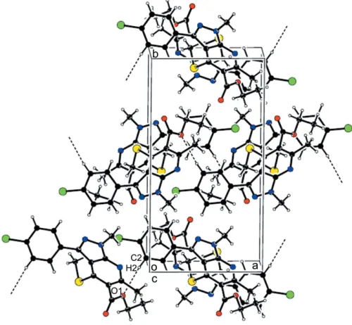
In the crystal, weak C—H O hydrogen bonds link molecules into chains along [100] (Table 1 and Fig. 2).
4. Database survey
A search for the pyrazolopyridine scaffold in the Cambridge Structural Database (CSD, Version 5.40; Groomet al., 2016) gave 236 hits. Of these, the structures most closely related to the title compound are FIZLEI (ethyl 2,7-diamino-3,4-di-cyano-5-phenylpyrazolo[1,5-a]pyridine-6-carboxylate; Naiket
al. 2019), ALAFID (Wu et al. 2016), DAWKAQ
{[2-(4-chlorophenyl)pyrazolo[1,5-a]pyridin-3-yl(phenyl)methanone; Ravi et al. 2017}, NADPIU [3-(4-chlorophenyl)pyrazolo[1,5-a]pyridine; Wuet al.2016] and ZOJWAW (Barrettet al.1996). The geometrical parameters of the –COOCH2CH3substituent
in the title compound are comparable with those reported for
FIZLEI. Similarly, the geometrical parameters of the –C6H4Cl
unit in the title compound are comparable with those for in
DAWKAQ and NADPIU. The bond lengths of the
pyrazolo[3,4-b]pyridine scaffold of the title compound are closer to those in NADPIU. The pyrazolopyridine moiety (N1–N3/C7–C9/C12–C14) of the title compound is approxi-mately plan, as is also observed for FIZLEI, ALAFID, DAWKAQ, NADPIU and ZOJWAW. Apart from the CSD database, two other important databases, namely Drug Bank (database for FDA-approved drugs, drugs under investigation or in clinical trials,etc; Lawet al.2013) and ZINC (database for commercially available compounds; Irwinet al.2005) were also surveyed. The former database is used for drug repur-posing or drug re-profiling studies, and latter for high-throughput virtual screening against the binding site of drug target proteins to identify promising and putative inhibitors. In the Drug Bank database, there were 31 hits, based on a 0.5 similarity threshold, whereas the ZINC search gave only three
hits (ZINCIDs: ZINC45166781, ZINC3852638 and
ZINC39053824). Out of 31 molecules identified in the Drug Bank database, two molecules were in the approved drug category namely riciguat (accession No: DB08931, similarity score: 0.55) and teletristat ethyl (accession No: DB12095, similarity score: 0.511). The remaining 29 molecules belong to the experimental, investigational or other categories.
444
Raoet al. C18H18ClN3O2S Acta Cryst.(2020). E76, 443–445research communications
Figure 1
[image:2.610.45.297.76.237.2]The molecular structure of the title compound with the atom-numbering scheme and displacement ellipsoids drawn at the 50% probability level
Figure 2
[image:2.610.311.568.88.124.2]The crystal packing of title compound, viewed along thecaxis, showing the weak intermolecular C—H O hydrogen bonds as dotted lines
Table 1
Hydrogen-bond geometry (A˚ ,).
D—H A D—H H A D A D—H A
C2—H2 O1i 0.93 2.59 3.513 (2) 170
[image:2.610.314.564.480.711.2]5. Synthesis and crystallization:
To a solution of 3-(4-chlorophenyl)-1-methyl-1H -pyrazol-5-amine (125 mg, 0.65 mmol) and ethyl 2-(bis(methylthio)-methylene)-3-oxobutanoate (145 mg, 0.65 mmol) in toluene (5 ml) under a blanket of dry N2, a catalytic amount of
tri-fluoroacetic acid (TFA; 30 mol%) was added. The resulting mixture was refluxed for 12 h, while monitoring progress by TLC (hexane:ethyl acetate, 99:1). After completion of the reaction, the resulting mixture was subjected to purification by column chromatography to furnish 182 mg of the title compound in 75% yield as a colourless solid, m.p. 415.85 K,Rf
= 0.3 (hexane:ethyl acetate 99:01). A sample suitable for single-crystal X-ray analysis was obtained by recrystallization from dry methanol.
6. Refinement
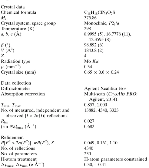
Crystal data, data collection and structure refinement details are summarized in Table 2. Hydrogen atoms were placed in calculated positions, with C—H = 0.93–0.97 A˚ and refined using a riding model withUiso(H) = 1.2Ueq(C) or 1.5Ueq
(C-methyl).
Acknowledgements
The authors thank the DST–FIST Single Crystal XRD facility at the Department of Chemistry, Pondicherry University, for the diffraction data and Dr Clara Gomes (FCT–UNL, Portugal) for the CSD database survey. RG thanks the Department of Chemistry for facilities, and the UGC and CSIR for a fellowship. JM thanks Dr Amit Kumar Singh (Sharda University, India) for support.
References
Agilent (2014).CrysAlis PRO. Agilent Technologies Ltd, Yarnton, England.
Barrett, D., Sasaki, H., Kinoshita, T. & Sakane, K. (1996).Chem. Commun.pp. 61–62.
El-Gohary, N. S., Gabr, M. T. & Shaaban, M. I. (2019).Bioorg. Chem.
89, 1–13.
Farrugia, L. J. (2012).J. Appl. Cryst.45, 849–854.
Fathy, U., Younis, A. & Awad, H. M. (2015).J. Chem. Pharm. Res.7, 4–12.
Go¨khan-Kelekc¸i, N., Yabanog˘lu, S., Ku¨peli, E., Salgin, U., Ozgen, O., Uc¸ar, G., Yes¸ilada, E., Kendi, E., Yes¸ilada, A. & Bilgin, A. A. A. (2007).Bioorg. Med. Chem.15, 5775–5786.
Groom, C. R., Bruno, I. J., Lightfoot, M. P. & Ward, S. C. (2016).Acta Cryst.B72, 171–179.
Hardy, C. R. (1984). InAdvances in Heterocyclic Chemistry, Vol. 36, pp. 343–409. New York: Academic Press.
Hawas, S. S., Gohary, N. S., Gabr, M. T., Shaaban, M. I. & El-Ashmawy, M. B. (2019).Synth. Commun.49, 2466–2487.
Irwin, J. J. & Shoichet, B. K. (2005).J. Chem. Inf. Model.45, 177–182. Law, V., Knox, C., Djoumbou, Y., Jewison, T., Guo, A. C., Liu, Y., Maciejewski, A., Arndt, D., Wilson, M., Neveu, V., Tang, A., Gabriel, G., Ly, C., Adamjee, S., Dame, Z. T., Han, B., Zhou, Y. & Wishart, D. S. (2013).Nucleic Acids Res.42, D1091–D1097. Macrae, C. F., Sovago, I., Cottrell, S. J., Galek, P. T. A., McCabe, P.,
Pidcock, E., Platings, M., Shields, G. P., Stevens, J. S., Towler, M. & Wood, P. A. (2020).J. Appl. Cryst.53, 226–235.
Mello, H. de, Echevarria, A., Bernardino, A. M., Canto-Cavalheiro, M. & Leon, L. L. (2004).J. Med. Chem.47, 5427–5432.
Naik, N. S., Shastri, L. A., Shastri, S. L., Chougala, B. M., Shaikh, F., Madar, J. M., Kulkarni, R. C., Dodamani, S., Jalalpure, S., Joshi, S. D. & Sunagar, V. (2019).ChemistrySelect,4, 285–297.
Panchal, V., Variya, H. H. & Patel, G. R. (2019).Int. J. Appl. Eng. Res, 14, 43–50.
Parmar, S. S., Pandey, B. R., Dwivedi, C. & Ali, B. (1974).J. Med. Chem.17, 1031–1033.
Ravi, C., Samanta, S., Mohan, D. C., Reddy, N. N. K. & Adimurthy, S. (2017).Synthesis,49, 2513–2522.
Sheldrick, G. M. (2015a).Acta Cryst.A71, 3–8. Sheldrick, G. M. (2015b).Acta Cryst.C71, 3–8. Spek, A. L. (2020).Acta Cryst.E76, 1–11.
Timo´teo, M. A., Oliveira, L., Campesatto-Mella, E., Barroso, A., Silva, C., Magalha˜es-Cardoso, M. T., Alves-do-Prado, W. & Correia-de-Sa´, P. (2008).Neurochem. Int.52, 834–845.
Witherington, J., Bordas, V., Garland, S. L., Hickey, D. M., Ife, R. J., Liddle, J., Saunders, M., Smith, D. G. & Ward, R. W. (2003).Bioorg. Med. Chem. Lett.13, 1577–1580.
Wu, H.-C., Chu, J.-H., Li, C.-W., Hwang, L.-C. & Wu, M.-J. (2016).
Organometallics,35, 288–300.
research communications
[image:3.610.44.290.89.366.2]Acta Cryst.(2020). E76, 443–445 Raoet al. C18H18ClN3O2S
445
Table 2
Experimental details.
Crystal data
Chemical formula C18H18ClN3O2S
Mr 375.86
Crystal system, space group Monoclinic,P21/a
Temperature (K) 298
a,b,c(A˚ ) 8.9995 (5), 16.7778 (11),
12.3595 (8) (
) 98.892 (6)
V(A˚3) 1843.8 (2)
Z 4
Radiation type MoK
(mm 1) 0.34
Crystal size (mm) 0.650.60.24
Data collection
Diffractometer Agilent Xcalibur Eos
Absorption correction Multi-scan (CrysAlis PRO; Agilent, 2014)
Tmin,Tmax 0.857, 1.000
No. of measured, independent and observed [I> 2(I)] reflections
13882, 4340, 3323
Rint 0.027
(sin/)max(A˚ 1) 0.682
Refinement
R[F2> 2(F2)],wR(F2),S 0.049, 0.161, 1.10
No. of reflections 4340
No. of parameters 230
H-atom treatment H-atom parameters constrained
max,min(e A˚ 3) 0.30, 0.41
Computer programs: CrysAlis PRO (Agilent, 2014), SHELXT(Sheldrick, 2015a),
SHELXL2018(Sheldrick, 2015b),ORTEP-3 for Windows(Farrugia, 2012),PLATON
supporting information
sup-1 Acta Cryst. (2020). E76, 443-445
supporting information
Acta Cryst. (2020). E76, 443-445 [https://doi.org/10.1107/S2056989020002479]
Crystal structure analysis of ethyl
3-(4-chlorophenyl)-1,6-dimethyl-4-methyl-sulfanyl-1
H
-pyrazolo[3,4-
b
]pyridine-5-carboxylate
H. Surya Prakash Rao, Ramalingam Gunasundari and Jayaraman Muthukumaran
Computing details
Data collection: CrysAlis PRO (Agilent, 2014); cell refinement: CrysAlis PRO (Agilent, 2014); data reduction: CrysAlis
PRO (Agilent, 2014); program(s) used to solve structure: SHELXT (Sheldrick, 2015a); program(s) used to refine
structure: SHELXL2018 (Sheldrick, 2015b); molecular graphics: ORTEP-3 for Windows (Farrugia, 2012), PLATON
(Spek, 2020) and Mercury (Macrae et al., 2020); software used to prepare material for publication: ORTEP-3 for
Windows (Farrugia, 2012), PLATON (Spek, 2020) and Mercury (Macrae et al., 2020).
Ethyl 3-(4-chlorophenyl)-1,6-dimethyl-4-methylsulfanyl-1H-pyrazolo[3,4-b]pyridine-5-carboxylate
Crystal data
C18H18ClN3O2S
Mr = 375.86
Monoclinic, P21/a
a = 8.9995 (5) Å
b = 16.7778 (11) Å
c = 12.3595 (8) Å
β = 98.892 (6)°
V = 1843.8 (2) Å3
Z = 4
F(000) = 784
Dx = 1.354 Mg m−3
Mo Kα radiation, λ = 0.71073 Å
Cell parameters from 4472 reflections
θ = 3.9–29.0°
µ = 0.34 mm−1
T = 298 K
Block, colourless 0.65 × 0.6 × 0.24 mm
Data collection
Agilent Xcalibur Eos diffractometer
Radiation source: Enhance (Mo) X-ray Source
Detector resolution: 15.9821 pixels mm-1
ω scans
Absorption correction: multi-scan (CrysAlis PRO; Agilent, 2014) Tmin = 0.857, Tmax = 1.000
13882 measured reflections 4340 independent reflections 3323 reflections with I > 2σ(I) Rint = 0.027
θmax = 29.0°, θmin = 3.9°
h = −11→12
k = −22→22
l = −15→16
Refinement
Refinement on F2
Least-squares matrix: full R[F2 > 2σ(F2)] = 0.049
wR(F2) = 0.161
S = 1.10
4340 reflections 230 parameters 0 restraints
Primary atom site location: structure-invariant direct methods
Secondary atom site location: difference Fourier map
Hydrogen site location: inferred from neighbouring sites
supporting information
sup-2 Acta Cryst. (2020). E76, 443-445
w = 1/[σ2(F
o2) + (0.1P)2] where P = (Fo2 + 2Fc2)/3
(Δ/σ)max = 0.006
Δρmax = 0.30 e Å−3 Δρmin = −0.41 e Å−3
Special details
Geometry. All esds (except the esd in the dihedral angle between two l.s. planes) are estimated using the full covariance matrix. The cell esds are taken into account individually in the estimation of esds in distances, angles and torsion angles; correlations between esds in cell parameters are only used when they are defined by crystal symmetry. An approximate (isotropic) treatment of cell esds is used for estimating esds involving l.s. planes.
Fractional atomic coordinates and isotropic or equivalent isotropic displacement parameters (Å2)
x y z Uiso*/Ueq
S1 0.38706 (5) −0.06089 (3) 0.65888 (4) 0.04572 (19)
Cl1 −0.26383 (7) 0.13979 (4) 0.63284 (7) 0.0751 (3)
O1 0.77428 (16) −0.12709 (8) 0.67467 (12) 0.0460 (4)
N1 0.73909 (16) 0.02099 (9) 0.95211 (12) 0.0363 (4)
N2 0.57052 (18) 0.12776 (9) 0.97183 (14) 0.0404 (4)
O2 0.62392 (19) −0.20707 (9) 0.75585 (14) 0.0599 (4)
N3 0.42863 (18) 0.15124 (10) 0.92994 (14) 0.0409 (4)
C9 0.6093 (2) 0.06010 (10) 0.92252 (14) 0.0335 (4)
C7 0.37469 (19) 0.09887 (11) 0.85219 (15) 0.0347 (4)
C14 0.75435 (19) −0.04529 (11) 0.89531 (15) 0.0340 (4)
C3 0.1239 (2) 0.04255 (12) 0.77078 (17) 0.0421 (5)
H3 0.161730 −0.008533 0.785668 0.051*
C8 0.48642 (19) 0.03902 (11) 0.84112 (14) 0.0323 (4)
C15 0.8936 (2) −0.09402 (12) 0.93020 (17) 0.0430 (5)
H15A 0.968939 −0.061386 0.972304 0.064*
H15B 0.930807 −0.113697 0.866557 0.064*
H15C 0.869892 −0.138103 0.974026 0.064*
C6 0.21737 (19) 0.10825 (11) 0.79948 (15) 0.0345 (4)
C12 0.51150 (19) −0.02733 (11) 0.77555 (14) 0.0329 (4)
C2 −0.0231 (2) 0.05183 (12) 0.72086 (17) 0.0435 (5)
H2 −0.083292 0.007504 0.701057 0.052*
C5 0.0072 (2) 0.19448 (12) 0.73163 (18) 0.0463 (5)
H5 −0.032853 0.245383 0.719599 0.056*
C13 0.64421 (19) −0.06941 (10) 0.80566 (14) 0.0322 (4)
C4 0.1552 (2) 0.18420 (12) 0.78090 (17) 0.0410 (4)
H4 0.214097 0.228738 0.801982 0.049*
C10 0.6609 (3) 0.17282 (13) 1.05805 (19) 0.0525 (5)
H10A 0.723443 0.137017 1.105641 0.079*
H10B 0.596079 0.201357 1.099350 0.079*
H10C 0.722908 0.209945 1.026288 0.079*
C16 0.6758 (2) −0.14269 (11) 0.74373 (15) 0.0372 (4)
C1 −0.0800 (2) 0.12802 (13) 0.70063 (17) 0.0439 (5)
C11 0.3529 (3) 0.02945 (16) 0.57990 (18) 0.0586 (6)
H11A 0.303494 0.067547 0.620089 0.088*
H11B 0.290191 0.017846 0.511611 0.088*
supporting information
sup-3 Acta Cryst. (2020). E76, 443-445
C17 0.8220 (3) −0.19386 (15) 0.6139 (2) 0.0620 (7)
H17A 0.741429 −0.209688 0.556464 0.074*
H17B 0.847277 −0.238963 0.662333 0.074*
C18 0.9549 (3) −0.16898 (19) 0.5657 (2) 0.0784 (9)
H18A 0.928634 −0.124659 0.517341 0.118*
H18B 0.988342 −0.212571 0.525243 0.118*
H18C 1.034070 −0.153518 0.623018 0.118*
Atomic displacement parameters (Å2)
U11 U22 U33 U12 U13 U23
S1 0.0408 (3) 0.0500 (3) 0.0424 (3) 0.0014 (2) −0.0061 (2) −0.0113 (2)
Cl1 0.0410 (3) 0.0734 (5) 0.1034 (6) 0.0093 (3) −0.0123 (3) −0.0214 (4)
O1 0.0491 (8) 0.0400 (8) 0.0522 (8) 0.0015 (6) 0.0179 (7) −0.0078 (6)
N1 0.0350 (8) 0.0336 (8) 0.0387 (8) −0.0032 (6) 0.0005 (6) −0.0019 (7)
N2 0.0426 (9) 0.0337 (9) 0.0426 (9) −0.0007 (6) −0.0009 (7) −0.0074 (7)
O2 0.0696 (10) 0.0392 (9) 0.0749 (11) −0.0140 (7) 0.0236 (8) −0.0115 (8)
N3 0.0411 (9) 0.0371 (9) 0.0436 (9) 0.0035 (7) 0.0040 (7) −0.0016 (7)
C9 0.0356 (9) 0.0319 (9) 0.0327 (9) −0.0041 (7) 0.0042 (7) −0.0007 (7)
C7 0.0375 (9) 0.0305 (9) 0.0368 (9) 0.0014 (7) 0.0084 (7) 0.0017 (7)
C14 0.0308 (8) 0.0332 (9) 0.0370 (9) −0.0045 (7) 0.0027 (7) 0.0027 (8)
C3 0.0426 (10) 0.0332 (10) 0.0522 (12) 0.0002 (8) 0.0122 (9) 0.0019 (9)
C8 0.0312 (8) 0.0317 (9) 0.0339 (9) −0.0015 (7) 0.0048 (7) 0.0008 (7)
C15 0.0360 (9) 0.0422 (11) 0.0476 (11) 0.0032 (8) −0.0030 (8) −0.0006 (9)
C6 0.0323 (8) 0.0373 (10) 0.0357 (9) 0.0010 (7) 0.0105 (7) 0.0015 (8)
C12 0.0306 (8) 0.0361 (9) 0.0319 (9) −0.0044 (7) 0.0049 (7) 0.0006 (7)
C2 0.0375 (10) 0.0416 (11) 0.0531 (12) −0.0053 (8) 0.0122 (8) −0.0061 (9)
C5 0.0408 (10) 0.0391 (11) 0.0590 (13) 0.0088 (8) 0.0075 (9) −0.0040 (10)
C13 0.0320 (8) 0.0312 (9) 0.0334 (9) −0.0030 (7) 0.0050 (7) −0.0004 (7)
C4 0.0376 (9) 0.0355 (10) 0.0511 (11) −0.0009 (8) 0.0101 (8) −0.0049 (9)
C10 0.0624 (13) 0.0420 (12) 0.0490 (12) −0.0044 (10) −0.0044 (10) −0.0149 (10)
C16 0.0333 (9) 0.0364 (10) 0.0405 (10) 0.0003 (7) 0.0015 (7) −0.0031 (8)
C1 0.0349 (10) 0.0494 (12) 0.0487 (11) 0.0037 (8) 0.0098 (8) −0.0067 (9)
C11 0.0595 (13) 0.0778 (17) 0.0367 (10) 0.0189 (12) 0.0022 (9) 0.0032 (11)
C17 0.0615 (14) 0.0577 (15) 0.0692 (15) 0.0088 (11) 0.0179 (12) −0.0218 (12)
C18 0.0790 (19) 0.092 (2) 0.0719 (17) 0.0399 (16) 0.0366 (15) 0.0135 (16)
Geometric parameters (Å, º)
S1—C12 1.7752 (18) C15—H15C 0.9600
S1—C11 1.803 (3) C6—C4 1.396 (3)
Cl1—C1 1.746 (2) C12—C13 1.388 (2)
O1—C16 1.347 (2) C2—C1 1.385 (3)
O1—C17 1.450 (2) C2—H2 0.9300
N1—C14 1.333 (2) C5—C1 1.383 (3)
N1—C9 1.340 (2) C5—C4 1.387 (3)
N2—C9 1.359 (2) C5—H5 0.9300
supporting information
sup-4 Acta Cryst. (2020). E76, 443-445
N2—C10 1.449 (2) C4—H4 0.9300
O2—C16 1.195 (2) C10—H10A 0.9600
N3—C7 1.337 (2) C10—H10B 0.9600
C9—C8 1.420 (2) C10—H10C 0.9600
C7—C8 1.442 (2) C11—H11A 0.9600
C7—C6 1.473 (2) C11—H11B 0.9600
C14—C13 1.426 (2) C11—H11C 0.9600
C14—C15 1.502 (3) C17—C18 1.476 (3)
C3—C2 1.380 (3) C17—H17A 0.9700
C3—C6 1.399 (3) C17—H17B 0.9700
C3—H3 0.9300 C18—H18A 0.9600
C8—C12 1.415 (2) C18—H18B 0.9600
C15—H15A 0.9600 C18—H18C 0.9600
C15—H15B 0.9600
C12—S1—C11 101.91 (10) C1—C5—H5 120.4
C16—O1—C17 117.03 (16) C4—C5—H5 120.4
C14—N1—C9 114.90 (15) C12—C13—C14 122.00 (17)
C9—N2—N3 111.25 (15) C12—C13—C16 120.27 (16)
C9—N2—C10 127.70 (17) C14—C13—C16 117.74 (16)
N3—N2—C10 121.05 (16) C5—C4—C6 121.27 (18)
C7—N3—N2 107.38 (15) C5—C4—H4 119.4
N1—C9—N2 124.05 (17) C6—C4—H4 119.4
N1—C9—C8 128.45 (16) N2—C10—H10A 109.5
N2—C9—C8 107.43 (16) N2—C10—H10B 109.5
N3—C7—C8 110.17 (16) H10A—C10—H10B 109.5
N3—C7—C6 117.73 (15) N2—C10—H10C 109.5
C8—C7—C6 131.99 (16) H10A—C10—H10C 109.5
N1—C14—C13 121.99 (16) H10B—C10—H10C 109.5
N1—C14—C15 116.87 (16) O2—C16—O1 124.26 (18)
C13—C14—C15 121.14 (17) O2—C16—C13 124.64 (17)
C2—C3—C6 121.48 (19) O1—C16—C13 111.07 (15)
C2—C3—H3 119.3 C5—C1—C2 121.04 (19)
C6—C3—H3 119.3 C5—C1—Cl1 119.79 (16)
C12—C8—C9 115.20 (15) C2—C1—Cl1 119.16 (16)
C12—C8—C7 140.99 (16) S1—C11—H11A 109.5
C9—C8—C7 103.74 (15) S1—C11—H11B 109.5
C14—C15—H15A 109.5 H11A—C11—H11B 109.5
C14—C15—H15B 109.5 S1—C11—H11C 109.5
H15A—C15—H15B 109.5 H11A—C11—H11C 109.5
C14—C15—H15C 109.5 H11B—C11—H11C 109.5
H15A—C15—H15C 109.5 O1—C17—C18 108.3 (2)
H15B—C15—H15C 109.5 O1—C17—H17A 110.0
C4—C6—C3 117.87 (17) C18—C17—H17A 110.0
C4—C6—C7 120.25 (17) O1—C17—H17B 110.0
C3—C6—C7 121.83 (17) C18—C17—H17B 110.0
C13—C12—C8 116.96 (16) H17A—C17—H17B 108.4
supporting information
sup-5 Acta Cryst. (2020). E76, 443-445
C8—C12—S1 125.32 (13) C17—C18—H18B 109.5
C3—C2—C1 119.14 (19) H18A—C18—H18B 109.5
C3—C2—H2 120.4 C17—C18—H18C 109.5
C1—C2—H2 120.4 H18A—C18—H18C 109.5
C1—C5—C4 119.13 (19) H18B—C18—H18C 109.5
C9—N2—N3—C7 −0.4 (2) C9—C8—C12—S1 −173.01 (13)
C10—N2—N3—C7 179.58 (18) C7—C8—C12—S1 3.2 (3)
C14—N1—C9—N2 178.40 (16) C11—S1—C12—C13 −128.93 (15)
C14—N1—C9—C8 1.6 (3) C11—S1—C12—C8 51.29 (17)
N3—N2—C9—N1 −175.78 (16) C6—C3—C2—C1 −1.2 (3)
C10—N2—C9—N1 4.2 (3) C8—C12—C13—C14 −2.9 (3)
N3—N2—C9—C8 1.6 (2) S1—C12—C13—C14 177.35 (13)
C10—N2—C9—C8 −178.42 (19) C8—C12—C13—C16 177.08 (15)
N2—N3—C7—C8 −0.9 (2) S1—C12—C13—C16 −2.7 (2)
N2—N3—C7—C6 175.72 (15) N1—C14—C13—C12 −3.1 (3)
C9—N1—C14—C13 3.7 (2) C15—C14—C13—C12 176.81 (16)
C9—N1—C14—C15 −176.20 (16) N1—C14—C13—C16 177.00 (15)
N1—C9—C8—C12 −7.2 (3) C15—C14—C13—C16 −3.1 (3)
N2—C9—C8—C12 175.55 (15) C1—C5—C4—C6 0.1 (3)
N1—C9—C8—C7 175.22 (17) C3—C6—C4—C5 −2.5 (3)
N2—C9—C8—C7 −2.00 (19) C7—C6—C4—C5 −179.85 (17)
N3—C7—C8—C12 −174.7 (2) C17—O1—C16—O2 −1.7 (3)
C6—C7—C8—C12 9.4 (4) C17—O1—C16—C13 176.52 (18)
N3—C7—C8—C9 1.81 (19) C12—C13—C16—O2 −79.4 (3)
C6—C7—C8—C9 −174.18 (18) C14—C13—C16—O2 100.5 (2)
C2—C3—C6—C4 3.1 (3) C12—C13—C16—O1 102.39 (19)
C2—C3—C6—C7 −179.64 (17) C14—C13—C16—O1 −77.7 (2)
N3—C7—C6—C4 35.3 (2) C4—C5—C1—C2 1.8 (3)
C8—C7—C6—C4 −148.98 (19) C4—C5—C1—Cl1 −177.07 (15)
N3—C7—C6—C3 −141.96 (19) C3—C2—C1—C5 −1.3 (3)
C8—C7—C6—C3 33.8 (3) C3—C2—C1—Cl1 177.61 (15)
C9—C8—C12—C13 7.2 (2) C16—O1—C17—C18 −166.05 (19)
C7—C8—C12—C13 −176.6 (2)
Hydrogen-bond geometry (Å, º)
D—H···A D—H H···A D···A D—H···A
C2—H2···O1i 0.93 2.59 3.513 (2) 170