Received 8 October 2019 Accepted 19 November 2019
Edited by C. Rizzoli, Universita degli Studi di Parma, Italy
Keywords:crystal structure; chromanone deri-vative; flavanone derideri-vative; lipophilicity index; Hirshfeld surface analysis.
CCDC references:1966749; 1966750
Supporting information:this article has supporting information at journals.iucr.org/e
Crystal structures of (
E
)-3-(4-hydroxybenzylidene)-chroman-4-one and (
E
)-3-(3-hydroxybenzylidene)-2-phenylchroman-4-one
Kamil Suchojad,aAnna Dołe˛ga,b Angelika Adamus-Grabicka,cElz.bieta Budziszc and Magdalena Małeckaa*
aDepartment of Physical Chemistry, Faculty of Chemistry, University of Lodz, Pomorska 163/165, 90-236 Ło´dz´, Poland, bDepartment of Inorganic Chemistry, Gdan´sk University of Technology, G. Narutowicza 11/12., 80-233 Gdan´sk, Poland, andcDepartment of Cosmetic Raw Materials Chemistry, Faculty of Pharmacy, Medical University of Lodz, Muszynskiego 1, 90-151 Ło´dz´, Poland. *Correspondence e-mail: magdalena.malecka@chemia.uni.lodz.pl
The synthesis and crystal structures of (E )-3-(4-hydroxybenzylidene)chroman-4-one, C16H12O3,I, and (E)-3-(3-hydroxybenzylidene)-2-phenylchroman-4-one, C22H16O3, II, are reported. These compounds are of interest with respect to biological activity. Both structures display intermolecular C—H O and O— H O hydrogen bonding, forming layers in the crystal lattice. The crystal structure of compound I is consolidated by–interactions. The lipophilicity (logP) was determined as it is one of the parameters qualifying compounds as potential drugs. The logP value for compound I is associated with a larger contribution of C H interaction in the Hirshfeld surface.
1. Chemical context
Chromanone (chroman-4-one) and flavanone (2-phenyl-chroman-4-one) belong to the class of heterocyclic compounds and are composed of a benzene ring fused to a 2,3-dihydro- -pyranone ring (Emami & Ghanbarimasir, 2015). 3-Aryl-idenechromanones/flavanones and their derivatives are natu-rally occurring homoisoflavones, and can be obtained by condensing the corresponding aryl aldehydes with chroma-none/flavanone. These compounds were synthesized for the first time by Robinson in the early 1920s by the condensation reaction of chromanone or flavanone with the appropriate aryl aldehyde using a catalyst (alcohol potassium hydroxide) (Perkinet al.,1926). In 1979, Levai and Schag synthesizedE -3-arylidenechroman-4-one using piperidine as a catalyst (Levai & Schag, 1979). Several years later, in 1993, Pijewska and coworkers (Pijewska et al., 1993) obtained the series of 3-arylideneflavanones derivatives substituted by various groups using flavanones with aromatic aldehydes in the presence of piperidine. Flavonoid compounds belong to one of the largest and most interesting groups of chemical compounds. They are of interest to many scientists because they show biological properties (Nijveldtet al., 2001; Williams et al., 2004). Natural and synthetic flavonoids have a wide range of antioxidant, allergic, inflammatory, anti-microbial, anti-coagulant, anti-cholesterol or anti-cancer activities (Czaplin´skaet al., 2012).
2. Structural commentary
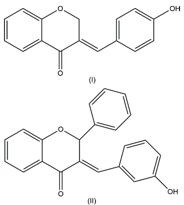
The molecular structures ofIandIIare shown in Fig. 1. The main chroman skeleton of each molecule consists of a benzene ring fused with a pyran ring. In position 3 of the chroman moiety, a para-hydroxybenzylidene (I) or a meta -hydroxy-benzylidene (II) substituent is connected to give theE-isomer, similar to the previously mentioned structure (Kupcewicz, et al., 2013). Moreover in compoundII, the chroman moiety is subsituted at position 2 by a phenyl ring. The pyran rings adopt an envelope conformation with puckering parameters QT= 0.371 (2) A˚ ,’2= 233.8 (4),2= 120.0 (3)forI, andQT = 0.423 (3) A˚ ,’2= 65.9 (5),2= 58.5 (4)forII. The dihedral angles between the hydroxybenzylidene rings and the main chroman skeleton are 47.54 (8) and 69.46 (12), respectively, forIandII(Fig. 2).
3. Supramolecular features
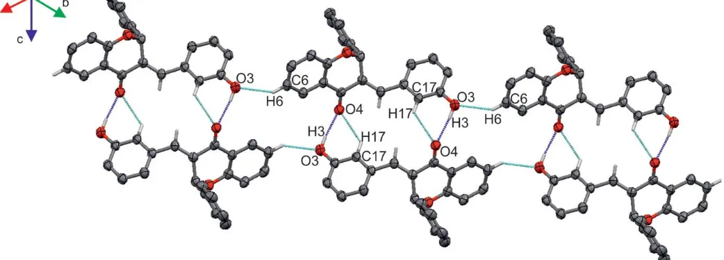
In the crystal packing ofI, molecules are connected into layers parallel to the bc plane via C—H O and O—H O hydrogen bonds (Table 1, Fig. 3). The stability of the layers is further enhanced by – stacking interactions occurring between the benzene rings fused with the pyran rings and the
aromatic rings of adjacent hydroxybenzylidene groups (Table 2). In the crystal packing of II, molecules are also linked by O—H O and C—H O hydrogen bonds (Table 3, Fig. 4) into layers parallel to theabplane.
4. Database survey
A search of the Cambridge Structural Database (CSD version 5.40, last update November 2018; Groomet al., 2016) using the scheme presented in Fig. 5 found 41 chromanone (Ishikawaet al., 2013a,b; Zimmermanet al., 2015; Marx, Sureshet al., 2007; Katrusiak et al., 1987; Brien et al., 2012; Sureshet al., 2007; Boonsriet al., 2005; Birunthaet al., 2018; Talhiet al., 2016; Wu, Liuet al., 2011; Marxet al., 2008; Chenget al., 2011; Valkonen et al., 2012; Lepitreet al., 2017; Gopaul, Shaikh, Koorbanallyet al., 2012; Gopaul, Koorbanallyet al., 2012; Marx, Manivannan et al., 2007; Sureshet al., 2007; Marxet al., 2008; Hassaineet al., 2016; Chantraprommaet al., 2006; Zhanget al., 2012; Augus-tine et al., 2008; Gopaul, Shaikh, Ramjugernathet al., 2012; Gopaul & Koorbanally, 2012; Zhang et al., 2013) and four flavanone structures (Zhonget al., 2013; Kupcewiczet al., 2013; Wu, Zenget al., 2011; Monserratet al., 2013). In the flavanone structures, the phenyl substituent at the C2position is always nearly perpendicular to the chroman moiety, with the
1908
Suchojadet al. C16H12O3and C22H16O3 Acta Cryst.(2019). E75, 1907–1913
[image:2.610.79.262.80.284.2]research communications
Table 1
Hydrogen-bond geometry (A˚ ,) forI.
D—H A D—H H A D A D—H A
C11—H11 O3i 0.95 2.55 3.264 (2) 132
C11—H11 O3ii 0.95 2.52 3.194 (2) 129
O3—H3 O4iii 0.84 1.85 2.6852 (19) 172
C2—H2A O1iv 0.99 2.53 3.397 (3) 147
C11—H11 O4 0.95 2.45 2.818 (2) 103
Symmetry codes: (i)x1;yþ1 2;z
1
2; (ii)x;yþ 1 2;z
1
2; (iii)xþ1;yþ 1 2;zþ
1 2;
[image:2.610.312.565.531.725.2](iv)x1;y;z.
Figure 1
The molecular structures of compounds I and II with displacement ellipsoids drawn at the 50% probability level.
Figure 2
[image:2.610.47.294.613.720.2]Table 2
Geometrical parameters (A˚ ,) for the–stacking interactions for compoundI.
Cg(1) andCg(2) are the centroids of the C5–C10 and C12–C17 rings, respectively;refers to the dihedral angle between planes (I) and (J);refers to the angle between theCg(I))–Cg(J) vector and normal to plane (I);refers to the angle between theCg(I))–Cg(J) vector and normal to plane (J).
Cg(I) Cg(J) Cg(I)_Perp Cg(J)_Perp
Cg(1) Cg(1)i 3.8508 (13) 3.5260 (9) 3.5259 (9) 0.03 (10) 23.7 23.7
Cg(1) Cg(1)ii 3.8512 (13) 3.5260 (9) 3.5262 (9) 0.03 (10) 23.7 23.7
Cg(2) Cg(2)i 3.8510 (13) 3.3739 (8) 3.3738 (8) 0.03 (10) 28.8 28.8
Cg(2) Cg(2)ii 3.8510 (13) 3.3740 (8) 3.3738 (8) 0.03 (10) 28.8 28.8
[image:3.610.45.569.119.523.2]Symmetry codes: (i)1 +x,y,z; (ii) 1 +x,y,z.
Figure 3
Partial packing of compoundIshowing the O—H O (blue dotted lines) and C—H O (cyan dotted lines) hydrogen-bonding network.
Figure 4
[image:3.610.44.296.236.295.2]Partial packing of compoundIIshowing the O—H O (blue dotted lines) and C—H O (cyan dotted lines) hydrogen-bonding network. Table 3
Hydrogen-bond geometry (A˚ ,) forII.
D—H A D—H H A D A D—H A
O3—H3 O4i 0.84 1.89 2.728 (3) 172
C17—H17 O4i 0.95 2.49 3.184 (4) 130
C6—H6 O3ii 0.95 2.45 3.265 (4) 143
C11—H11 O4 0.95 2.43 2.807 (3) 103
Symmetry codes: (i)xþ1;yþ1;zþ1; (ii)xþ1;y1;z.
Figure 5
[image:3.610.46.567.545.732.2]C(phen)—C2—C3—C4 torsion angle in the range 82.44– 107.90. In both chromanone and flavanone structures, the pyran ring adopts a slightly distorted envelope conformation. In the 41 chromanone derivatives, the bond distances and angles within the chroman moiety are in good agreement with those found in compoundI.
5. Experimental and theoretical lipophilicity of compounds I and II
Lipophilicity is one of the descriptors that is currently used in the design of new drugs and in assessing the activity of medicinal substances (Jo´z´wiak et al., 2001). Most often, the increase in lipophilicity increases the biological activity of compounds as a result of the affinity of substances with biological membranes and better permeability (Dołowy, 2009). However, a further increase in lipophilicity results in greater affinity for lipids and hinders the transport of compound molecules through the aqueous phase. That is why it is important to choose substances with optimal hydrophobic and hydrophilic properties and partition coefficient logP (Dołowy, 2009).
The experimental lipophilicity (logP) of compoundsIandII was determined using the RP–TLC method. The values of logPobtained are 2.95 and 3.98, respectively forIandII, the difference being due to the different, bulky substituent at the C2 position of the pyran ring. The theoretical values of lipo-philicity (miLogP) also show the same trend, the value for compoundIis lower (miLogP =3.14) than that for compound II (miLogP = 4.70). This is in agreement with the values
previously reported for similar arylidenochromanone/flava-none derivatives (Adamus-Grabickaet al., 2018). The theo-retical values of lipophilicity were calculated using the online Molinspiration Cheminformatics software (http://www.molin-spiration.com). According to the ‘rule of five’ proposed by Lipinskiet al. (2001), compoundsI and IImay be potential anti-cancer drugs, the most important parameters according to Lipinski being the logP value (logP < 5) and molar mass (< 500 Da).
6. Hirshfeld surface analysis and lipophilicity index versusC H contact
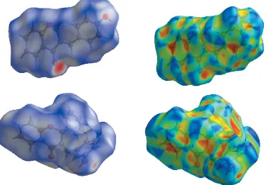
As the Hirshfeld surface (HS) analysis may provide useful descriptors for QSAR study (Kupcewicz,et al., 2016) and the lipophilicity parameter in biologically active compounds is associated with the contribution of intermolecular interactions to the Hirshfeld surface (Małecka & Budzisz, 2014), we generated the Hirshfeld surfaces (Hirshfeld, 1977; Spackman & Jayatilaka, 2009) using the CrystalExplorer program (Turneret al., 2017) for chromone and flavanone derivatives for which the lipophilicity parameters are available, i.e. compound I, II, 3-(4-chlorobenzylidene)-2-phenyl-2,3-di-hydro-4H-chromen-4-one (III; Kupcewiczet al., 2013), (E )-3-(4-N,N-diethylaminobenzylidene)chroman-4-one (IV; Adam-us-Grabicka et al., 2018) and (E)-3-(4-N,N -diethylamino-benzylidene)-2-phenylchroman-4-one (V; Adamus-Grabicka et al., 2018).
The Hirshfeld surfaces were mapped over dnorm (Fig. 6). The red, white and blue regions visible on thednormsurfaces
1910
Suchojadet al. C16H12O3and C22H16O3 Acta Cryst.(2019). E75, 1907–1913
[image:4.610.120.494.77.338.2]research communications
Figure 6
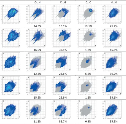
indicate contacts with distances shorter, longer and equal to the van der Waals radii. The decomposition of the HS into 2D fingerprint plots for particular contacts is presented in Fig. 7, together with the relative percentage of contributions of different contacts. The dominant interaction in all derivatives is the H H interaction. The contribution to the Hirshfeld surface is in the range 39.2– 55.5% forIIIandV. Comparing
the C C contacts, we can observe a large spread of percen-tage contribution ranging from 0.3% for V to 13.1% for compound I. This is also reflected in the presence of –
[image:5.610.49.564.71.577.2]stacking interactions observed in compoundI(Table 2). As in our previous studies (Małeckaet al., 2014; Kupcewicz et al., 2103), we found a relationship between the logP value and the fraction of the Hirshfeld surface covered by different Figure 7
intermolecular interactions. The increase of logPcorresponds in fact to increasing the C H contribution in the Hirshfeld surface. Furthermore, for compoundsI–V, the contribution of the O H interaction in the Hirshfeld surface is inversely proportional to the value of logP.
7. Synthesis and crystallization
The synthesis of compounds I and II is based on the condensation of chromanone or flavanone with an aryl alde-hyde in the presence of piperidine (Fig. 8). CompoundIwas prepared according to a slightly modified procedure with respect to that described in the literature (Levai & Schag, 1979). A mechanically stirred mixture of chroman-4-one (0.01 mol),p-methoxybenzaldehyde (0.01 mol) and five drops of piperidine was heated at 413 K in an oil bath for four h. The progress of the synthesis was controlled by thin layer chro-matography (TLC) using toluene/methanol (9:1v/v) as eluent. After cooling the reaction mixture was left for 24 h at room temperature. The solidified product was filtered and
crystal-lized from methanol. CompoundI was obtained as a yellow powder. The isolated solid was further recrystallized by slow evaporation at room temperature of an acetone solution. Yield: 64%, M.p.: 501–502.5 K. MS (ESI+): m/z 253.3 C16H12O3 [M+H]+. IR (KBr): 3126 (O—H), 2809 (C— Haromat), 1652 (C O), 1608, 1578 (C C), 1164 (C–O—C), 751 ( C—H).1HNMR (600 MHz, DMSO-d6)(ppm): 5.42 (1H,s, CH), 6.86–7.86 (8H,m, C—Haromat), 7.87 (2H,d,JAB = 18 Hz C2—H), 10.12 (1H, s, OH). Analysis calculated for C16H12O3(M= 252.23 g mol1) % C: 76.18; % H: 4.81; % O: 19.01. Found % C: 75.3; % H: 5.01; % O: 19.69.
CompoundIIwas synthesized according to the procedure described by Pijewskaet al., (1993). A mixture of 2-phenyl-chroman-4-one (0.01 mol), 3-hydroxybenzaldehyde (0.01 mol) and five drops of piperidine was heated under reflux in an oil bath with mechanical stirring. The reaction proceeded at 413 K for 5 h. The progress of the reaction was controlled by TLC (eluent: toluene/methanol, 9:1 v/v). After cooling at room temperature, the mixture was dissolved in methanol. After 24 h compound II precipitated as a light-cream fine crystalline powder and was purified by crystallization from methanol. Crystal suitable for X-ray analysis were obtained by slow evaporation of an ethanol solution at room temperature. Yield: 52.4%. M.p.: 482–483 K. MS (ESI+): m/z 329.2 C22H16O3 [M+H]+. IR (KBr): 3297 (O—H), 3054 (C— Haromat), 2351 (C—Haliph), 1663 (C O), 1608, 1590, 1504 (C C), 1141 (C—O—C), 757 ( C—H).1HNMR (600 MHz, DMSO-d6)(ppm): 6.57 (1H,s, C2—H), 5.69 (1H,s, CH), 6.89–7.91 (14H, m, CHaromat), 8.12 (1H, s, OH). Analysis calculated For C22H16O3 (M = 328.19 g mol1) %C: 80.51;
1912
Suchojadet al. C16H12O3and C22H16O3 Acta Cryst.(2019). E75, 1907–1913
[image:6.610.45.563.89.359.2]research communications
Table 4
Experimental details.
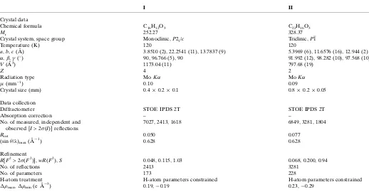
I II
Crystal data
Chemical formula C16H12O3 C22H16O3
Mr 252.27 328.37
Crystal system, space group Monoclinic,P21/c Triclinic,P1
Temperature (K) 120 120
a,b,c(A˚ ) 3.8510 (2), 22.2541 (11), 13.7837 (9) 5.3969 (6), 11.6576 (16), 12.944 (2)
,,(
) 90, 96.766 (5), 90 91.992 (12), 98.282 (10), 97.568 (10)
V(A˚3) 1173.04 (11) 797.68 (19)
Z 4 2
Radiation type MoK MoK
(mm1) 0.10 0.09
Crystal size (mm) 0.40.20.1 0.80.20.05
Data collection
Diffractometer STOE IPDS 2T STOE IPDS 2T
Absorption correction – –
No. of measured, independent and observed [I> 2 (I)] reflections
7027, 2413, 1618 6849, 3281, 1804
Rint 0.050 0.077
(sin/)max(A˚
1) 0.628 0.628
Refinement
R[F2> 2 (F2)],wR(F2),S 0.048, 0.115, 1.03 0.068, 0.200, 0.94
No. of reflections 2413 3281
No. of parameters 173 228
H-atom treatment H-atom parameters constrained H-atom parameters constrained
max,min(e A˚
3) 0.19,0.19 0.23,0.29
Computer programs:X-AREAandX-RED32(Stoe & Cie, 2002),SHELXT(Sheldrick, 2015a),SHELXL2014/7(Sheldrick, 2015b) andpublCIF(Westrip, 2010).
Figure 8
[image:6.610.43.297.649.719.2]%H: 4.87; % O: 14.62. Found %C: 79.99; %H: 5.11; % O: 14.90.
8. Refinement
Crystal data, data collection and structure refinement details are summarized in Table 4. All hydrogen atoms were fixed geometrically at calculated positions (O—H = 0.84 A˚ , C—H = 0.95–0.99 A˚ ) and refined as riding model with Uiso(H) = 1.5Ueq(O) or 1.2Ueq(C). A rotating model was used for the hydroxy groups.
Funding information
Funding for this research was provided by: Uniwesytet Ło´dzki, Uniwersytet Medyczny w Łodzi (grant No. SGB_148_Sucho-jad_Kamil to K. Suchojad; grant No. 502-03/3-066-02/502-34-118 to A. Adamus-Grabicka, E. Budzisz).
References
Adamus-Grabicka, A., Markowicz-Piasecka, M., Ponczek, M. B., Kusz, J., Małecka, M., Krajewska, U. & Budzisz, E. (2018).
Molecules,23, 3172–3188.
Augustine, T., Vithiya, S. M., Ramkumar, V. & Kanakam, C. C. (2008).
Acta Cryst.E64, o2080.
Biruntha, K., Reuben Jonathan, D., Mohamooda Sumaya, U., Dravida Thendral, E. R. A. & Usha, G. (2018). IUCrData, 3, x181273.
Boonsri, S., Chantrapromma, S., Fun, H.-K., Karalai, C., Kanjana-opas, A. & Anjum, S. (2005).Acta Cryst.E61, o3930–o3932. Brien, K. A., Bandi, R. K., Behera, A. K., Mishra, B. K., Majumdar, P.,
Satam, V., Savagian, M., Tzou, S., Lee, M., Zeller, M., Robles, A. J., Mooberry, S., Pati, H. & Lee, M. (2012).Arch. Pharm. Pharm. Med. Chem.345, 341–348.
Chantrapromma, S., Boonsri, S., Fun, H.-K., Anjum, S. & Kanjana-opas, A. (2006).Acta Cryst.E62, o1254–o1256.
Cheng, X.-M., Huang, Z.-T. & Zheng, Q.-Y. (2011).Tetrahedron,67, 9093–9098.
Czaplin´ska, M., Czepas, J. & Gwoz´dzin´ski, K. (2012).Post. Bioch.58, 235–242.
Dołowy, M. (2009).Farm. Pol.65(10), 689–693.
Emami, S. & Ghanbarimasir, Z. (2015).Eur. J. Med. Chem.93, 539– 563.
Gopaul, K. & Koorbanally, N. A. (2012). Private Communication (refcode ????). CCDC, Cambridge, England.
Gopaul, K., Koorbanally, N. A., Shaikh, M. M., Su, H. & Ramjugernath, D. (2012).Acta Cryst.E68, o3062.
Gopaul, K., Shaikh, M. M., Koorbanally, N. A., Ramjugernath, D. & Omondi, B. (2012).Acta Cryst.E68, o1972.
Gopaul, K., Shaikh, M., Ramjugernath, D., Koorbanally, N. A. & Omondi, B. (2012).Acta Cryst.E68, o1006.
Groom, C. R., Bruno, I. J., Lightfoot, M. P. & Ward, S. C. (2016).Acta Cryst.B72, 171–179.
Hassaine, R., Talhi, O., Taibi, N., Almeida Paz, F., Bensaid, O., Bachari, K. & Silva, A. M. S. (2016).Synlett,27, 465–470. Hirshfeld, H. L. (1977).Theor. Chim. Acta,44, 129–138. Ishikawa, Y. & Motohashi, Y. (2013a).Acta Cryst.E69, o1225. Ishikawa, Y. & Motohashi, Y. (2013b).Acta Cryst.E69, o1226.
Jo´z´wiak, K., Szumiło, H. & Soczewin´ski, E. (2001).Wiad. Chem.55, 1047–1073.
Katrusiak, A., Ratajczak-Sitarz, M., Kałuski, Z. & Orlov, V. D. (1987).
Acta Cryst.C43, 103–105.
Kupcewicz, B., Balcerowska-Czerniak, G., Małecka, M., Paneth, P., Krajewska, U. & Rozalski, M. (2013).Bioorg. Med. Chem. Lett.23, 4102–4106.
Kupcewicz, B., Małecka, M., Zapadka, M., Krajewska, U., Rozalski, M. & Budzisz, E. (2016).Bioorg. Med. Chem. Lett.26, 3336–3341. Lepitre, T., Denhez, C., Moncol, J., Othman, M., Lawson, A. M. &
Daı¨ch, A. (2017).J. Org. Chem.82, 12188–12201. Levai, A. & Schag, J. B. (1979).Pharmazie,34, 748–749.
Lipinski, C. A., Lombardo, F., Dominy, B. W. & Feeney, P. J. (2001).
Adv. Drug Deliv. Rev.46, 3–26.
Małecka, M. & Budzisz, E. (2014).CrystEngComm16, 6654–6663. Marx, A., Manivannan, V., Suresh, R., Kanagam, C. C. &
Bu¨yu¨kgu¨ngo¨r, O. (2007).Acta Cryst.E63, o4383.
Marx, A., Suresh, R., Kanagam, C. C., Manivannan, V. & Bu¨yu¨kgu¨ngo¨r, O. (2007).Acta Cryst.E63, o4530.
Marx, A., Suresh, R., Kanakam, C. C., Manivannan, V. & Vasam, C. S. (2008).Acta Cryst.E64, o27.
Monserrat, J.-P., Tiwari, K. N., Quentin, L., Pigeon, P., Jaouen, G., Vessie`res, A., Chabot, G. G. & Hillard, E. A. (2013).J. Organomet. Chem.734, 78–85.
Nijveldt, R. J., van Nood, E., van Hoorn, D., Boelens, P. G., van Norren, K. & van Leeuwen, P. (2001).Am. J. Clin. Nutr.74, 418– 425.
Perkin, W. H., Raˆy, J. N. & Robinson, R. (1926).J. Chem. Soc.129, 941–953.
Pijewska, L., Kamecki, J. & Perka-Karolczak, W. (1993).Pharmazie, 48, 254–257.
Sheldrick, G. M. (2015a).Acta Cryst.A71, 3–8. Sheldrick, G. M. (2015b).Acta Cryst.C71, 3–8.
Spackman, M. A. & Jayatilaka, D. (2009).CrystEngComm,11, 19–32. Stoe & Cie (2002).X-AREAandX-RED32. Stoe & Cie, Darmstadt,
Germany.
Suresh, R., Kanagam, C. C., Umarani, P. R., Manivannan, V. & Bu¨yu¨kgu¨ngo¨r, O. (2007).Acta Cryst.E63, o4248.
Talhi, O., Brodziak-Jarosz, L., Panning, J., Orlikova, B., Zwergel, C., Tzanova, T., Philippot, S., Pinto, D. C. G. A., Paz, F. A. A., Gerha¨user, C., Dick, T. P., Jacob, C., Diederich, M., Bagrel, D., Kirsch, G. & Silva, A. M. S. (2016).Eur. J. Org. Chem.pp. 965–975. Turner, M. J., McKinnon, J. J., Wolff, S. K., Grimwood, D. J., Spackman, P. R., Jayatilaka, D. & Spackman, M. A. (2017).
CrystalExplorer17. The University of Western Australia.
Valkonen, A., Laihia, K., Kolehmainen, E., Kauppinen, R. & Perje´si, P. (2012).Struct. Chem.23, 209–217.
Westrip, S. P. (2010).J. Appl. Cryst.43, 920–925.
Williams, R. J., Spencer, J. P. & Rice-Evans, C. (2004).Free Radical Biol. Med.36, 838–849.
Wu, Ch., Liu, Y., Zeng, H., Liu, L., Wang, D. & Chen, Y. (2011).Org. Biomol. Chem.9, 253–256.
Wu, Ch., Zeng, H., Liu, L., Wang, D. & Chen, Y. (2011).Tetrahedron, 67, 1231–1237.
Zhang, H.-J., Becker, P., Huang, H., Pirwerdjan, R., Pan, F.-F. & Bolm, C. (2012).Adv. Synth. Catal.354, 2157–2161.
Zhang, Y., Lv, Z., Zhang, M. & Li, K. (2013).Tetrahedron,69, 8839– 8846.
Zhong, N.-J., Liu, L., Wang, D. & Chen, Y.-J. (2013).Chem. Commun.
49, 3697–3699.
supporting information
sup-1
Acta Cryst. (2019). E75, 1907-1913
supporting information
Acta Cryst. (2019). E75, 1907-1913 [https://doi.org/10.1107/S2056989019015639]
Crystal structures of (
E
)-3-(4-hydroxybenzylidene)chroman-4-one and (
E
)-3-(3-hydroxybenzylidene)-2-phenylchroman-4-one
Kamil Suchojad, Anna Do
łę
ga, Angelika Adamus-Grabicka, El
ż
bieta Budzisz and Magdalena
Ma
ł
ecka
Computing details
For both structures, data collection: X-AREA (Stoe & Cie, 2002); cell refinement: X-AREA (Stoe & Cie, 2002); data
reduction: X-AREA (Stoe & Cie, 2002), X-RED32 (Stoe & Cie, 2002); program(s) used to solve structure: SHELXT
(Sheldrick, 2015a); program(s) used to refine structure: SHELXL2014/7 (Sheldrick, 2015b); software used to prepare
material for publication: SHELXL2014/7 (Sheldrick, 2015b), publCIF (Westrip, 2010).
(E)-3-(4-Hydroxybenzylidene)chroman-4-one (I)
Crystal data
C16H12O3
Mr = 252.27
Monoclinic, P21/c
a = 3.8510 (2) Å b = 22.2541 (11) Å c = 13.7837 (9) Å β = 96.766 (5)° V = 1173.04 (11) Å3
Z = 4 F(000) = 528
Dx = 1.428 Mg m−3
Melting point: 220 K
Mo Kα radiation, λ = 0.71073 Å Cell parameters from 219 reflections θ = 4.1–28.9°
µ = 0.10 mm−1
T = 120 K
Needle, light-yellow 0.4 × 0.2 × 0.1 mm
Data collection
STOE IPDS 2T diffractometer
Radiation source: GeniX Mo, 0.05 x 0.05 mm2 microfocus
Detector resolution: 6.67 pixels mm-1
rotation method, ω scans 7027 measured reflections
2413 independent reflections 1618 reflections with I > 2σ(I) Rint = 0.050
θmax = 26.5°, θmin = 3.7°
h = −4→4 k = −27→27 l = −16→17
Refinement
Refinement on F2
Least-squares matrix: full R[F2 > 2σ(F2)] = 0.048
wR(F2) = 0.115
S = 1.02 2413 reflections 173 parameters 0 restraints
Primary atom site location: difference Fourier map
Secondary atom site location: difference Fourier map
Hydrogen site location: inferred from neighbouring sites
sup-2
Acta Cryst. (2019). E75, 1907-1913
w = 1/[σ2(F
o2) + (0.0546P)2 + 0.2287P]
where P = (Fo2 + 2Fc2)/3
(Δ/σ)max < 0.001
Δρmax = 0.19 e Å−3
Δρmin = −0.19 e Å−3
Special details
Geometry. All esds (except the esd in the dihedral angle between two l.s. planes) are estimated using the full covariance matrix. The cell esds are taken into account individually in the estimation of esds in distances, angles and torsion angles; correlations between esds in cell parameters are only used when they are defined by crystal symmetry. An approximate (isotropic) treatment of cell esds is used for estimating esds involving l.s. planes.
Fractional atomic coordinates and isotropic or equivalent isotropic displacement parameters (Å2)
x y z Uiso*/Ueq
O3 0.5127 (4) 0.21588 (6) 0.68339 (10) 0.0282 (4)
H3 0.5952 0.1823 0.6701 0.042*
O4 −0.1988 (4) 0.39218 (6) 0.15977 (10) 0.0304 (4) O1 0.2599 (4) 0.50810 (6) 0.36926 (10) 0.0252 (4) C9 0.1347 (5) 0.52849 (8) 0.27851 (14) 0.0217 (4) C17 0.3626 (5) 0.25884 (8) 0.42639 (15) 0.0218 (4)
H17 0.4012 0.2441 0.3639 0.026*
C3 0.0393 (5) 0.40641 (8) 0.32620 (14) 0.0207 (4) C14 0.2335 (5) 0.29994 (8) 0.60754 (15) 0.0216 (4)
H14 0.1846 0.3135 0.6698 0.026*
C11 0.0949 (5) 0.34735 (8) 0.34238 (15) 0.0219 (4)
H11 0.0614 0.3229 0.2856 0.026*
C10 −0.0381 (5) 0.49118 (8) 0.20685 (15) 0.0217 (4) C12 0.1982 (5) 0.31486 (8) 0.43307 (14) 0.0200 (4) C13 0.1298 (5) 0.33398 (8) 0.52539 (15) 0.0219 (4)
H13 0.0104 0.3709 0.5318 0.026*
C8 0.2011 (6) 0.58826 (9) 0.25768 (15) 0.0256 (5)
H8 0.3229 0.6133 0.3061 0.031*
C15 0.4089 (5) 0.24590 (8) 0.59954 (15) 0.0208 (4) C7 0.0889 (6) 0.61086 (9) 0.16621 (16) 0.0277 (5)
H7 0.1330 0.6518 0.1523 0.033*
C4 −0.0782 (5) 0.42671 (9) 0.22612 (14) 0.0216 (4) C2 0.0920 (6) 0.45481 (8) 0.40230 (15) 0.0227 (4)
H2A −0.1381 0.4662 0.4220 0.027*
H2B 0.2363 0.4387 0.4607 0.027*
C6 −0.0882 (6) 0.57486 (9) 0.09380 (16) 0.0283 (5)
H6 −0.1649 0.5909 0.0311 0.034*
C5 −0.1499 (6) 0.51545 (9) 0.11493 (15) 0.0249 (5)
H5 −0.2708 0.4906 0.0661 0.030*
C16 0.4693 (5) 0.22483 (8) 0.50797 (15) 0.0217 (4)
H16 0.5830 0.1874 0.5018 0.026*
Atomic displacement parameters (Å2)
U11 U22 U33 U12 U13 U23
supporting information
sup-3
Acta Cryst. (2019). E75, 1907-1913
O4 0.0459 (10) 0.0235 (7) 0.0198 (8) −0.0049 (7) −0.0044 (7) −0.0004 (6) O1 0.0305 (8) 0.0209 (7) 0.0231 (8) −0.0046 (6) −0.0020 (6) 0.0006 (6) C9 0.0221 (11) 0.0231 (10) 0.0200 (11) 0.0031 (8) 0.0023 (8) 0.0014 (8) C17 0.0266 (11) 0.0229 (10) 0.0160 (10) −0.0013 (8) 0.0025 (8) −0.0018 (8) C3 0.0204 (10) 0.0223 (9) 0.0190 (11) −0.0003 (8) 0.0015 (8) 0.0008 (8) C14 0.0257 (11) 0.0199 (9) 0.0192 (11) −0.0036 (8) 0.0030 (8) −0.0029 (7) C11 0.0229 (11) 0.0242 (10) 0.0179 (11) −0.0002 (8) −0.0003 (8) −0.0023 (8) C10 0.0240 (11) 0.0200 (9) 0.0212 (11) 0.0025 (8) 0.0036 (9) 0.0006 (8) C12 0.0205 (10) 0.0215 (9) 0.0178 (10) −0.0019 (8) 0.0009 (8) −0.0008 (8) C13 0.0218 (10) 0.0192 (10) 0.0245 (11) −0.0005 (8) 0.0015 (8) −0.0001 (8) C8 0.0271 (12) 0.0228 (10) 0.0276 (12) −0.0012 (8) 0.0052 (9) −0.0029 (8) C15 0.0228 (10) 0.0198 (9) 0.0190 (10) −0.0023 (8) −0.0010 (8) 0.0035 (8) C7 0.0345 (13) 0.0200 (10) 0.0303 (13) 0.0021 (9) 0.0111 (10) 0.0028 (8) C4 0.0235 (11) 0.0234 (10) 0.0169 (11) 0.0013 (8) −0.0014 (8) −0.0001 (8) C2 0.0280 (11) 0.0203 (9) 0.0193 (11) −0.0016 (8) 0.0000 (9) 0.0005 (8) C6 0.0326 (12) 0.0275 (11) 0.0256 (12) 0.0066 (9) 0.0070 (9) 0.0055 (9) C5 0.0285 (12) 0.0250 (10) 0.0211 (11) 0.0024 (8) 0.0024 (9) −0.0008 (8) C16 0.0249 (11) 0.0169 (9) 0.0231 (11) 0.0006 (8) 0.0024 (9) −0.0005 (8)
Geometric parameters (Å, º)
O3—C15 1.354 (2) C11—H11 0.9500
O3—H3 0.8400 C10—C5 1.398 (3)
O4—C4 1.242 (2) C10—C4 1.471 (3)
O1—C9 1.364 (2) C12—C13 1.396 (3)
O1—C2 1.450 (2) C13—H13 0.9500
C9—C8 1.391 (3) C8—C7 1.379 (3)
C9—C10 1.397 (3) C8—H8 0.9500
C17—C16 1.378 (3) C15—C16 1.392 (3)
C17—C12 1.406 (3) C7—C6 1.394 (3)
C17—H17 0.9500 C7—H7 0.9500
C3—C11 1.346 (3) C2—H2A 0.9900
C3—C4 1.471 (3) C2—H2B 0.9900
C3—C2 1.501 (3) C6—C5 1.381 (3)
C14—C13 1.382 (3) C6—H6 0.9500
C14—C15 1.390 (3) C5—H5 0.9500
C14—H14 0.9500 C16—H16 0.9500
C11—C12 1.458 (3)
C15—O3—H3 109.5 C7—C8—H8 120.3
C9—O1—C2 115.95 (15) C9—C8—H8 120.3
O1—C9—C8 116.97 (18) O3—C15—C14 117.16 (18)
O1—C9—C10 122.55 (17) O3—C15—C16 122.95 (18)
C8—C9—C10 120.40 (18) C14—C15—C16 119.89 (18)
C16—C17—C12 121.80 (19) C8—C7—C6 121.30 (19)
C16—C17—H17 119.1 C8—C7—H7 119.4
C12—C17—H17 119.1 C6—C7—H7 119.4
sup-4
Acta Cryst. (2019). E75, 1907-1913
C11—C3—C2 125.41 (18) O4—C4—C3 123.19 (18)
C4—C3—C2 115.86 (16) C10—C4—C3 116.22 (17)
C13—C14—C15 120.37 (19) O1—C2—C3 113.35 (16)
C13—C14—H14 119.8 O1—C2—H2A 108.9
C15—C14—H14 119.8 C3—C2—H2A 108.9
C3—C11—C12 130.40 (19) O1—C2—H2B 108.9
C3—C11—H11 114.8 C3—C2—H2B 108.9
C12—C11—H11 114.8 H2A—C2—H2B 107.7
C5—C10—C9 118.78 (18) C5—C6—C7 118.8 (2)
C5—C10—C4 120.86 (18) C5—C6—H6 120.6
C9—C10—C4 120.22 (18) C7—C6—H6 120.6
C13—C12—C17 117.70 (18) C6—C5—C10 121.3 (2)
C13—C12—C11 124.65 (18) C6—C5—H5 119.4
C17—C12—C11 117.57 (17) C10—C5—H5 119.4
C14—C13—C12 120.83 (18) C17—C16—C15 119.31 (18)
C14—C13—H13 119.6 C17—C16—H16 120.3
C12—C13—H13 119.6 C15—C16—H16 120.3
C7—C8—C9 119.48 (19)
Hydrogen-bond geometry (Å, º)
D—H···A D—H H···A D···A D—H···A
C11—H11···O3i 0.95 2.55 3.264 (2) 132
C11—H11···O3ii 0.95 2.52 3.194 (2) 129
O3—H3···O4iii 0.84 1.85 2.6852 (19) 172
C2—H2A···O1iv 0.99 2.53 3.397 (3) 147
C11—H11···O4 0.95 2.45 2.818 (2) 103
Symmetry codes: (i) x−1, −y+1/2, z−1/2; (ii) x, −y+1/2, z−1/2; (iii) x+1, −y+1/2, z+1/2; (iv) x−1, y, z.
(E)-3-(3-Hydroxybenzylidene)-2-phenylchroman-4-one (II)
Crystal data
C22H16O3
Mr = 328.37
Triclinic, P1 a = 5.3969 (6) Å b = 11.6576 (16) Å c = 12.944 (2) Å α = 91.992 (12)° β = 98.282 (10)° γ = 97.568 (10)° V = 797.68 (19) Å3
Z = 2
F(000) = 344 Dx = 1.367 Mg m−3
Melting point: 210 K
Mo Kα radiation, λ = 0.71073 Å Cell parameters from 3650 reflections θ = 3.5–29.5°
µ = 0.09 mm−1
T = 120 K Plate, colourless 0.8 × 0.2 × 0.05 mm
Data collection
STOE IPDS 2T diffractometer
Radiation source: GeniX Mo, 0.05 x 0.05 mm2 microfocus
Detector resolution: 6.67 pixels mm-1
supporting information
sup-5
Acta Cryst. (2019). E75, 1907-1913
θmax = 26.5°, θmin = 3.5°
h = −6→6
k = −13→14 l = −16→16
Refinement
Refinement on F2
Least-squares matrix: full R[F2 > 2σ(F2)] = 0.068
wR(F2) = 0.200
S = 0.94 3281 reflections 228 parameters 0 restraints
Primary atom site location: difference Fourier map
Secondary atom site location: difference Fourier map
Hydrogen site location: inferred from neighbouring sites
H-atom parameters constrained w = 1/[σ2(F
o2) + (0.1159P)2]
where P = (Fo2 + 2Fc2)/3
(Δ/σ)max < 0.001
Δρmax = 0.23 e Å−3
Δρmin = −0.29 e Å−3
Special details
Geometry. All esds (except the esd in the dihedral angle between two l.s. planes) are estimated using the full covariance matrix. The cell esds are taken into account individually in the estimation of esds in distances, angles and torsion angles; correlations between esds in cell parameters are only used when they are defined by crystal symmetry. An approximate (isotropic) treatment of cell esds is used for estimating esds involving l.s. planes.
Fractional atomic coordinates and isotropic or equivalent isotropic displacement parameters (Å2)
x y z Uiso*/Ueq
O1 0.1107 (4) 0.13471 (17) 0.24184 (16) 0.0423 (6) O4 0.7889 (4) 0.30522 (17) 0.39197 (16) 0.0432 (6) O3 −0.1593 (4) 0.6939 (2) 0.44060 (17) 0.0503 (6)
H3 −0.0545 0.6964 0.4956 0.075*
C10 0.5289 (5) 0.1266 (2) 0.3391 (2) 0.0365 (7) C17 0.1145 (5) 0.5660 (2) 0.3820 (2) 0.0390 (7)
H17 0.1940 0.5611 0.4518 0.047*
C9 0.2888 (5) 0.0736 (3) 0.2913 (2) 0.0386 (7) C3 0.3849 (5) 0.3163 (3) 0.2945 (2) 0.0378 (7) C7 0.3920 (6) −0.1113 (3) 0.3446 (2) 0.0453 (8)
H7 0.3455 −0.1925 0.3474 0.054*
C5 0.6977 (6) 0.0575 (3) 0.3885 (2) 0.0405 (7)
H5 0.8608 0.0925 0.4207 0.049*
C4 0.5884 (5) 0.2528 (3) 0.3452 (2) 0.0370 (7) C15 −0.1803 (6) 0.6487 (3) 0.2598 (2) 0.0431 (8)
H15 −0.3077 0.6973 0.2454 0.052*
C16 −0.0743 (5) 0.6358 (3) 0.3619 (2) 0.0401 (7) C11 0.3763 (5) 0.4240 (3) 0.3311 (2) 0.0385 (7)
H11 0.5114 0.4544 0.3843 0.046*
C12 0.1891 (5) 0.5030 (2) 0.3014 (2) 0.0378 (7) C8 0.2210 (6) −0.0459 (3) 0.2943 (2) 0.0431 (7)
H8 0.0587 −0.0818 0.2620 0.052*
C13 0.0823 (6) 0.5165 (3) 0.1989 (3) 0.0442 (8)
H13 0.1332 0.4755 0.1427 0.053*
C14 −0.0998 (6) 0.5905 (3) 0.1791 (2) 0.0440 (8)
sup-6
Acta Cryst. (2019). E75, 1907-1913
C2 0.2058 (5) 0.2476 (2) 0.2083 (2) 0.0376 (7)
H2 0.0587 0.2908 0.1899 0.045*
C21 0.3266 (5) 0.2321 (3) 0.1100 (2) 0.0387 (7) C22 0.2451 (7) 0.1373 (3) 0.0408 (2) 0.0517 (9)
H22 0.1084 0.0819 0.0531 0.062*
C6 0.6322 (6) −0.0602 (3) 0.3915 (2) 0.0440 (8)
H6 0.7491 −0.1062 0.4251 0.053*
C26 0.5228 (6) 0.3134 (3) 0.0894 (2) 0.0493 (8)
H26 0.5783 0.3797 0.1357 0.059*
C23 0.3612 (8) 0.1222 (3) −0.0467 (3) 0.0632 (11)
H23 0.3042 0.0566 −0.0938 0.076*
C24 0.5596 (7) 0.2024 (4) −0.0654 (3) 0.0593 (10)
H24 0.6417 0.1913 −0.1244 0.071*
C25 0.6382 (6) 0.2991 (3) 0.0022 (3) 0.0558 (9)
H25 0.7716 0.3556 −0.0114 0.067*
Atomic displacement parameters (Å2)
U11 U22 U33 U12 U13 U23
supporting information
sup-7
Acta Cryst. (2019). E75, 1907-1913
Geometric parameters (Å, º)
O1—C9 1.369 (3) C15—H15 0.9500
O1—C2 1.453 (3) C11—C12 1.475 (4)
O4—C4 1.234 (3) C11—H11 0.9500
O3—C16 1.370 (4) C12—C13 1.391 (4)
O3—H3 0.8400 C8—H8 0.9500
C10—C5 1.397 (4) C13—C14 1.393 (4)
C10—C9 1.405 (4) C13—H13 0.9500
C10—C4 1.460 (4) C14—H14 0.9500
C17—C12 1.392 (4) C2—C21 1.526 (4)
C17—C16 1.388 (4) C2—H2 1.0000
C17—H17 0.9500 C21—C22 1.381 (4)
C9—C8 1.396 (4) C21—C26 1.388 (4)
C3—C11 1.335 (4) C22—C23 1.387 (5)
C3—C4 1.492 (4) C22—H22 0.9500
C3—C2 1.499 (4) C6—H6 0.9500
C7—C8 1.380 (4) C26—C25 1.381 (4)
C7—C6 1.396 (4) C26—H26 0.9500
C7—H7 0.9500 C23—C24 1.381 (5)
C5—C6 1.374 (4) C23—H23 0.9500
C5—H5 0.9500 C24—C25 1.383 (5)
C15—C14 1.378 (4) C24—H24 0.9500
C15—C16 1.383 (4) C25—H25 0.9500
C9—O1—C2 115.9 (2) C7—C8—H8 120.3
C16—O3—H3 109.5 C9—C8—H8 120.3
C5—C10—C9 118.8 (3) C12—C13—C14 119.7 (3)
C5—C10—C4 121.1 (3) C12—C13—H13 120.1
C9—C10—C4 119.8 (2) C14—C13—H13 120.1
C12—C17—C16 120.9 (3) C15—C14—C13 121.0 (3)
C12—C17—H17 119.5 C15—C14—H14 119.5
C16—C17—H17 119.5 C13—C14—H14 119.5
O1—C9—C8 117.0 (3) O1—C2—C3 111.0 (2)
O1—C9—C10 122.8 (3) O1—C2—C21 109.4 (2)
C8—C9—C10 120.2 (3) C3—C2—C21 112.3 (2)
C11—C3—C4 118.3 (3) O1—C2—H2 108.0
C11—C3—C2 127.3 (3) C3—C2—H2 108.0
C4—C3—C2 114.4 (2) C21—C2—H2 108.0
C8—C7—C6 121.1 (3) C22—C21—C26 118.9 (3)
C8—C7—H7 119.5 C22—C21—C2 121.1 (3)
C6—C7—H7 119.5 C26—C21—C2 120.1 (3)
C6—C5—C10 121.2 (3) C21—C22—C23 120.6 (3)
C6—C5—H5 119.4 C21—C22—H22 119.7
C10—C5—H5 119.4 C23—C22—H22 119.7
O4—C4—C10 123.2 (3) C5—C6—C7 119.3 (3)
O4—C4—C3 121.2 (3) C5—C6—H6 120.3
sup-8
Acta Cryst. (2019). E75, 1907-1913
C14—C15—C16 119.6 (3) C25—C26—C21 120.7 (3)
C14—C15—H15 120.2 C25—C26—H26 119.6
C16—C15—H15 120.2 C21—C26—H26 119.6
O3—C16—C15 118.4 (2) C24—C23—C22 120.1 (3)
O3—C16—C17 121.8 (3) C24—C23—H23 120.0
C15—C16—C17 119.8 (3) C22—C23—H23 120.0
C3—C11—C12 129.9 (3) C23—C24—C25 119.7 (3)
C3—C11—H11 115.1 C23—C24—H24 120.2
C12—C11—H11 115.1 C25—C24—H24 120.2
C17—C12—C13 118.9 (2) C26—C25—C24 120.0 (4)
C17—C12—C11 117.1 (3) C26—C25—H25 120.0
C13—C12—C11 124.1 (3) C24—C25—H25 120.0
C7—C8—C9 119.4 (3)
Hydrogen-bond geometry (Å, º)
D—H···A D—H H···A D···A D—H···A
O3—H3···O4i 0.84 1.89 2.728 (3) 172
C17—H17···O4i 0.95 2.49 3.184 (4) 130
C6—H6···O3ii 0.95 2.45 3.265 (4) 143
C11—H11···O4 0.95 2.43 2.807 (3) 103