A triclinic polymorph of dicadmium
divanadate(V)
Ahmed Ould Saleck,* Abderrazzak Assani, Mohamed Saadi and Lahcen El Ammari
Laboratoire de Chimie du Solide Applique´e, Faculte´ des Sciences, Universite´ Mohammed V-Agdal, Avenue Ibn Battouta, BP 1014, Rabat, Morocco Correspondence e-mail: a_ouldsaleck@yahoo.fr
Received 8 October 2013; accepted 21 October 2013
Key indicators: single-crystal X-ray study;T= 296 K; mean(V–O) = 0.001 A˚; Rfactor = 0.016;wRfactor = 0.037; data-to-parameter ratio = 21.3.
The title compound, Cd2V2O7, was obtained under
hydro-thermal conditions. Different from the known monoclinic
form, the new polymorph of Cd2V2O7has triclinic symmetry
and is isotypic with Ca2V2O7. The building units of the crystal
structure are two Cd2+cations, with coordination numbers of
six and seven, and two V atoms with a tetrahedral and a significantly distorted trigonal–pyramidal coordination
envir-onment, respectively. Two VO5 pyramids share an edge and
each pyramid is connected to one VO4 tetrahedron via a
corner atom, forming an isolated V4O14 8
anion. These anions are arranged in sheets parallel to (211) and are linked through
the Cd2+ cations into a three-dimensional framework
struc-ture.
Related literature
For Ca2V2O7, isotypic with the title compound, see: Trunovet
al. (1983). For the structure of the monoclinic polymorph of
Cd2V2O7, see: Au & Calvo (1967). For the thermal stability of
the monoclinic polymorph, see: Krasnenko & Rotermel
(2010). For applications of vanadates, see: Jin et al. (2013);
Valverde et al.(2012). For bond-valence analysis, see: Brown
& Altermatt (1985).
Crystal data
Cd2V2O7
Mr= 438.68
Triclinic,P1 a= 6.5974 (2) A˚ b= 6.8994 (2) A˚ c= 6.9961 (2) A˚ = 83.325 (1)
= 63.898 (1)
= 80.145 (1) V= 281.45 (1) A˚3
Z= 2
MoKradiation = 10.65 mm 1
T= 296 K
0.290.170.12 mm
Data collection
Bruker X8 APEX diffractometer Absorption correction: multi-scan
(SADABS; Bruker, 2009) Tmin= 0.164,Tmax= 0.376
10113 measured reflections 2134 independent reflections 2077 reflections withI> 2(I) Rint= 0.025
Refinement
R[F2> 2(F2)] = 0.016
wR(F2) = 0.037
S= 1.21 2134 reflections
100 parameters
max= 0.70 e A˚ 3 min= 1.53 e A˚ 3
Data collection:APEX2(Bruker, 2009); cell refinement:SAINT
(Bruker, 2009); data reduction:SAINT; program(s) used to solve structure:SHELXS97(Sheldrick, 2008); program(s) used to refine structure: SHELXL97 (Sheldrick, 2008); molecular graphics:
ORTEP-3 for Windows (Farrugia, 2012) and DIAMOND (Bran-denburg, 2006); software used to prepare material for publication:
publCIF(Westrip, 2010).
The authors thank the Unit of Support for Technical and
Scientific Research (UATRS, CNRST) for the X-ray
measurements.
Supplementary data and figures for this paper are available from the IUCr electronic archives (Reference: WM2776).
References
Au, P. K. L. & Calvo, C. (1967).Can. J. Chem.45, 2297–2302.
Brandenburg, K. (2006).DIAMOND. Crystal Impact GbR, Bonn, Germany. Brown, I. D. & Altermatt, D. (1985).Acta Cryst.B41, 244–247.
Bruker (2009).APEX2,SAINTandSADABS. Bruker AXS Inc., Madison, Wisconsin, USA.
Farrugia, L. J. (2012).J. Appl. Cryst.45, 849–854.
Jin, M., Lu, P., Yu, G. X., Cheng, Z. M., Chen, L. F. & Wang, J. A. (2013).Catal. Today,212, 142–148.
Krasnenko, T. I. & Rotermel, M. V. (2010).Russ. J. Inorg. Chem.55, 430–433. Sheldrick, G. M. (2008).Acta Cryst.A64, 112–122.
Trunov, V. K., Velikodnyi, Y. A., Murasheva, E. V. & Zhuravlev, V. D. (1983). Dokl. Akad. Nauk SSSR,270, 886–887.
Valverde, J. A., Echavarrı´a, A., Ribeiro, M. F., Palacio, L. A. & Eon, J. G. (2012).Catal. Today,192, 36–43.
Westrip, S. P. (2010).J. Appl. Cryst.43, 920–925. Structure Reports
Online
supporting information
Acta Cryst. (2013). E69, i79 [doi:10.1107/S1600536813028869]
A triclinic polymorph of dicadmium divanadate(V)
Ahmed Ould Saleck, Abderrazzak Assani, Mohamed Saadi and Lahcen El Ammari
S1. Comment
Vanadate-based compounds have received a great extent of interest and still remain promising functional materials. Their
structural diversity, mainly associated with the ability of vanadium to form different anions like (VO4)3-, (V2O7)4-,
(V4O12)4-, (V10O28)6-, is strongly required for catalysis applications (Jin et al., 2013; Valverde et al., 2012).
A bibliographic analysis revealed that pyrovanadates with the (V2O7)4- anion can adopt different symmetries. For
example, Cu2V2O7, Mn2V2O7 and Mg2V2O7 are polymorphic and exhibit both monoclinic and triclinic varieties, the
Cu-member additionally an orthorhombic form. In the case of Co2V2O7, Ni2V2O7, and Cd2V2O7 only one monoclinic form is
yet known, for Zn2V2O7 two monoclinic forms are reported. Ca2V2O7 and Ba2V2O7 crystallize in the triclinic system,
Sr2V2O7 is likewise polymorphic, with triclinic and tetragonal varieties. We report here on the crystal structure
determination of a new form of Cd2V2O7 that was hydrothermally synthesized. In contrast to the known monoclinic
polymorph (Au & Calvo, 1967) that is stable from ambient temperature to 1173 K (Krasnenko et al., 2010), this new
form has triclinic symmetry and is isotypic with Ca2V2O7 (Trunov et al., 1983).
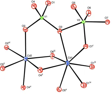
The structure of the title compound is built up from two types of vanadium sites and two types of cadmium sites, each
with a different oxygen coordination as shown in Fig. 1. The coordination environment of V1 is tetrahedral with V1–O
distances in the range 1.6882 (13) Å - 1.7708 (13) Å. V2 is surrounded by five oxygen atoms with four V2—O distances
ranging from 1.6612 (14) Å to 1.8535 (13) Å and the fifth O atom at a longer distance V2–O1 = 2.0348 (13) Å, forming a
distorted trigonal V2O55- pyramid. The bond valence sum calculation (Brown & Altermatt, 1985) for V1 and V2 are as
expected, viz. 4.99 and 5.11 valence units, respectively. This result confirms the involvement of O1 in the V2
environment. The V2O55- pyramids build a dimeric unit (V2)2O86- by sharing an edge. A corner atom (O1) of each of the
pyramids is also part of a V1O4 tetrahedron. This linkeage leads to the formation of a centrosymmetric (V4O14)8- anion.
This type of anion is rarely encountered in the crystal chemistry of pyrovanadates. Like in the monoclinic Cd2V2O7
variety (Au and Calvo, 1967), (V2O7)4- groups made up from two corner-sharing VO4 tetrahedra are more commonly
observed.
The two independent cadmium sites Cd1 and Cd2 are surrounded by six and seven oxygen atoms, respectively, in the
form of distorted polyhedra. The Cd—O distance range from 2.2401 (13) Å to 2.5300 (13) Å for Cd1O7 and from
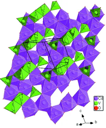
2.2449 (14) Å to 2.4562 (14) Å for Cd2O6. As shown in Fig. 2, edge-sharing CdO6 and CdO7 polyhedra built up sheets
parallel to (211) with 8-membered open rings. Two adjacent layers are linked by the (V4O14)8- groups into a
three-dimensional framework (Fig. 3). In the monoclinic Cd2V2O7 polymorph, the CdO6 octahedra share edges and form sheets
with six-membered rings that are linked by (V2O7)4- groups.
S2. Experimental
Crystals of the title compound were isolated from the hydrothermal treatment of cadmium oxide, ammonium
filled to 50% with distilled water and under autogeneous pressure at 493 K for four days. After being filtered off, washed
with deionized water and air dried, the reaction product consists of colourless sheet-shaped crystals corresponding to the
title compound.
S3. Refinement
The highest peak and the deepest hole in the final Fourier map are at 1.69 Å and 0.08 Å, from O2 and Cd2, respectively.
Reflections (200) and (020) were omitted from the refinement due to large differences between observed and calculated
[image:3.610.129.488.204.506.2]intensities.
Figure 1
The coordination environment of the Cd and V sites in the crystal structure of the title compound, triclinic Cd2V2O7.
Displacement ellipsoids are drawn at the 70% probability level. [Symmetry codes: (i) -x, -y + 1, -z + 1; (ii) -x + 1, -y, -z +
Figure 2
Figure 3
A three dimensional view of the crystal structure of triclinic Cd2V2O7 showing the stacking of the cadium and vanadium
layers that extend parallel to (211).
Dicadmium divanadate(V)
Crystal data
Cd2V2O7
Mr = 438.68 Triclinic, P1 Hall symbol: -P 1
a = 6.5974 (2) Å
b = 6.8994 (2) Å
c = 6.9961 (2) Å
α = 83.325 (1)°
β = 63.898 (1)°
γ = 80.145 (1)°
V = 281.45 (1) Å3
Z = 2
F(000) = 396
Dx = 5.176 Mg m−3
Mo Kα radiation, λ = 0.71073 Å Cell parameters from 2717 reflections
θ = 3.0–33.1°
µ = 10.65 mm−1
Data collection
Bruker X8 APEX diffractometer
Radiation source: fine-focus sealed tube Graphite monochromator
φ and ω scans
Absorption correction: multi-scan (SADABS; Bruker, 2009)
Tmin = 0.164, Tmax = 0.376
10113 measured reflections 2134 independent reflections 2077 reflections with I > 2σ(I)
Rint = 0.025
θmax = 33.1°, θmin = 3.0°
h = −10→10
k = −10→10
l = −10→10
Refinement
Refinement on F2 Least-squares matrix: full
R[F2 > 2σ(F2)] = 0.016
wR(F2) = 0.037
S = 1.21 2134 reflections 100 parameters 0 restraints
Primary atom site location: structure-invariant direct methods
Secondary atom site location: difference Fourier map
w = 1/[σ2(F
o2) + (0.0148P)2 + 0.2572P] where P = (Fo2 + 2Fc2)/3
(Δ/σ)max = 0.001 Δρmax = 0.70 e Å−3 Δρmin = −1.53 e Å−3
Special details
Geometry. All e.s.d.'s (except the e.s.d. in the dihedral angle between two l.s. planes) are estimated using the full
covariance matrix. The cell e.s.d.'s are taken into account individually in the estimation of e.s.d.'s in distances, angles and torsion angles; correlations between e.s.d.'s in cell parameters are only used when they are defined by crystal symmetry. An approximate (isotropic) treatment of cell e.s.d.'s is used for estimating e.s.d.'s involving l.s. planes.
Refinement. Refinement of F2 against all reflections. The weighted R-factor wR and goodness of fit S are based on F2,
conventional R-factors R are based on F, with F set to zero for negative F2. The threshold expression of F2 > 2σ(F2) is used only for calculating R-factors(gt) etc. and is not relevant to the choice of reflections for refinement. R-factors based on F2 are statistically about twice as large as those based on F, and R- factors based on all data will be even larger.
Fractional atomic coordinates and isotropic or equivalent isotropic displacement parameters (Å2)
x y z Uiso*/Ueq
Cd1 0.24214 (2) 0.336697 (18) 0.83258 (2) 0.00767 (4)
Cd2 0.74980 (2) 0.034380 (18) 0.75748 (2) 0.00845 (4)
V1 0.71038 (5) 0.16450 (4) 0.25864 (4) 0.00469 (5)
V2 0.22836 (5) 0.45517 (4) 0.34409 (5) 0.00542 (5)
O1 0.8612 (2) 0.3328 (2) 0.0816 (2) 0.0100 (2)
O2 0.8622 (2) 0.0439 (2) 0.3907 (2) 0.0099 (2)
O3 0.4592 (2) 0.2893 (2) 0.4363 (2) 0.0092 (2)
O4 0.6546 (2) −0.00638 (19) 0.1233 (2) 0.0086 (2)
O5 0.2714 (2) 0.29481 (19) 0.1660 (2) 0.0093 (2)
O6 0.3839 (2) 0.6438 (2) 0.2436 (2) 0.0104 (2)
O7 −0.0496 (2) 0.5892 (2) 0.3678 (2) 0.0100 (2)
Atomic displacement parameters (Å2)
U11 U22 U33 U12 U13 U23
V2 0.00472 (11) 0.00635 (12) 0.00446 (11) −0.00089 (9) −0.00115 (9) −0.00065 (9) O1 0.0089 (5) 0.0096 (5) 0.0098 (5) −0.0031 (4) −0.0025 (5) 0.0023 (4) O2 0.0080 (5) 0.0124 (6) 0.0090 (5) 0.0009 (4) −0.0044 (4) 0.0003 (4) O3 0.0070 (5) 0.0116 (6) 0.0079 (5) 0.0021 (4) −0.0032 (4) −0.0013 (4) O4 0.0110 (6) 0.0068 (5) 0.0095 (5) −0.0012 (4) −0.0054 (5) −0.0014 (4) O5 0.0128 (6) 0.0079 (5) 0.0083 (5) 0.0002 (4) −0.0057 (5) −0.0015 (4) O6 0.0081 (5) 0.0079 (5) 0.0147 (6) −0.0021 (4) −0.0046 (5) 0.0014 (4) O7 0.0056 (5) 0.0157 (6) 0.0064 (5) 0.0026 (4) −0.0020 (4) 0.0004 (4)
Geometric parameters (Å, º)
Cd1—O7i 2.2401 (13) Cd2—O4ii 2.4562 (14)
Cd1—O4ii 2.2898 (13) V1—O1 1.6882 (13)
Cd1—O6iii 2.3083 (14) V1—O2 1.7028 (14)
Cd1—O1iii 2.3345 (14) V1—O3 1.7265 (13)
Cd1—O1iv 2.3476 (13) V1—O4 1.7708 (13)
Cd1—O5v 2.4043 (13) V2—O5 1.6612 (14)
Cd1—O3 2.5300 (13) V2—O6 1.6885 (14)
Cd2—O6iii 2.2449 (14) V2—O7 1.8530 (13)
Cd2—O5ii 2.2858 (13) V2—O7i 1.8535 (13)
Cd2—O2vi 2.2894 (14) V2—O3 2.0348 (13)
Cd2—O2 2.3327 (14) V2—V2i 2.8482 (6)
Cd2—O4v 2.3459 (13)
O7i—Cd1—O4ii 114.29 (5) O2vi—Cd2—O2 74.71 (5)
O7i—Cd1—O6iii 131.24 (5) O6iii—Cd2—O4v 96.72 (5)
O4ii—Cd1—O6iii 83.66 (5) O5ii—Cd2—O4v 75.42 (5)
O7i—Cd1—O1iii 85.81 (5) O2vi—Cd2—O4v 102.17 (5)
O4ii—Cd1—O1iii 157.35 (5) O2—Cd2—O4v 174.49 (5)
O6iii—Cd1—O1iii 90.73 (5) O6iii—Cd2—O4ii 81.30 (5)
O7i—Cd1—O1iv 76.73 (5) O5ii—Cd2—O4ii 75.69 (5)
O4ii—Cd1—O1iv 94.46 (5) O2vi—Cd2—O4ii 160.52 (5)
O6iii—Cd1—O1iv 149.98 (5) O2—Cd2—O4ii 98.40 (5)
O1iii—Cd1—O1iv 79.55 (5) O4v—Cd2—O4ii 83.09 (5)
O7i—Cd1—O5v 153.49 (5) O1—V1—O2 109.38 (7)
O4ii—Cd1—O5v 74.22 (5) O1—V1—O3 107.57 (7)
O6iii—Cd1—O5v 73.06 (5) O2—V1—O3 109.85 (7)
O1iii—Cd1—O5v 83.15 (5) O1—V1—O4 109.71 (7)
O1iv—Cd1—O5v 77.60 (5) O2—V1—O4 109.65 (7)
O7i—Cd1—O3 62.10 (5) O3—V1—O4 110.65 (6)
O4ii—Cd1—O3 86.49 (5) O5—V2—O6 114.25 (7)
O6iii—Cd1—O3 75.19 (5) O5—V2—O7 99.11 (7)
O1iii—Cd1—O3 113.31 (5) O6—V2—O7 98.25 (7)
O1iv—Cd1—O3 134.73 (5) O5—V2—O7i 121.64 (7)
O5v—Cd1—O3 144.28 (4) O6—V2—O7i 123.74 (7)
O6iii—Cd2—O5ii 156.37 (5) O7—V2—O7i 79.57 (6)
O5ii—Cd2—O2vi 87.38 (5) O6—V2—O3 93.85 (6)
O6iii—Cd2—O2 88.75 (5) O7—V2—O3 158.40 (6)
O5ii—Cd2—O2 99.75 (5) O7i—V2—O3 78.83 (6)