warwick.ac.uk/lib-publications
A Thesis Submitted for the Degree of PhD at the University of Warwick
Permanent WRAP URL:
http://wrap.warwick.ac.uk/98014
Copyright and reuse:
This thesis is made available online and is protected by original copyright. Please scroll down to view the document itself.
Please refer to the repository record for this item for information to help you to cite it. Our policy information is available from the repository home page.
Novel tools for the capture of intermediates
of iterative polyketide catalysis
Samantha L. Kilgour
Thesis submitted in partial fulfilment of the requirements for the Degree of Doctor of Philosophy in Chemistry
University of Warwick
Department of Chemistry
ii
Contents
Declaration ... xii
Acknowledgements ... xiii
List of Figures... xiv
List of Schemes ... xxiv
List of Tables ... xxvii
Abbreviations and Definitions ... xxviii
Abstract ... xxxiii
1.
Introduction
... 21.1. Polyketide natural products and their biosynthesis ... 2
1.1.1. Fundamentals of polyketide biosynthesis ... 3
1.2. Type II polyketides and their biosynthesis ... 7
1.2.1. Biosynthesis of type II polyketides ... 8
1.2.1.1. Ketosynthase-chain length factor – structure-function relationship ... 10
1.2.1.2. Acyl carrier proteins: structure-function relationship ... 12
1.2.1.3. Ketoreductases ... 14
1.2.1.4. Cyclases and aromatases ... 14
1.2.1.5. Methyltransferases ... 15
1.2.1.6. Oxygenases ... 15
1.2.1.7. Glycosyltransferases ... 17
1.2.1.8. The future of type II PKS engineering ... 18
1.3. Biosynthesis of actinorhodin ... 18
1.3.1. Biosynthesis of an octaketide chain by the actinorhodin minimal system .... ……… 20
1.3.2. Tailoring steps towards the biosynthesis of actinorhodin ... 24
iii
1.4.1. Engineering of carrier proteins ... 30
1.4.1.1. Modifications to the phosphopantetheinyl arm of carrier proteins ... 31
1.5. Methods of investigation of modular and iterative biocatalysis ... 33
1.5.1. Labelling techniques ... 34
1.5.2. Enzyme engineering ... 36
1.5.2.1. In vivo ... 36
1.5.2.1.1. Gene deletions ... 37
1.5.2.1.2. Mutagenesis ... 37
1.5.2.1.3. Hybrid PKSs ... 38
1.5.2.1.4. Heterologous expression ... 39
1.5.2.1.5. Modifications to the phosphopantetheinyl arm of carrier proteins ... 39
1.5.2.2. In vitro ... 40
1.5.3. Chemical probes: N-acetylcysteamine thioester (SNAC) substrate analogues ... 41
1.5.3.1. Chemical synthesis of SNAC analogues ... 42
1.5.4. Chain termination probes as new tools for the investigation of polyketide biosynthesis ... 43
1.5.4.1. Nonhydrolysable NAC chain termination probes ... 46
1.5.5. The application of proteomics and metabolomics to the investigation of biocatalysis ... 49
1.6. Mass spectrometry ... 49
1.6.1. Key ionisation techniques ... 49
1.6.2. Key mass analysers... 50
1.6.2.1. Time-of-flight (TOF) analysers ... 52
1.6.2.2. Ion trap analysers ... 52
1.6.2.3. Fourier Transform Mass Spectrometers (FT-MS) ... 52
1.6.2.4. Ion mobility analysers ... 53
iv
1.6.3.1. Collisionally activated dissociation (CAD) ... 55
1.6.3.2. Infrared multiphoton dissociation (IRMPD) ... 55
1.6.3.3. Electron capture dissociation (ECD) ... 56
1.6.3.4. Electron transfer dissociation (ETD) ... 56
1.6.3.5. Electron impact dissociation (EID) ... 57
1.6.4. Analysis of proteins by mass spectrometry ... 57
1.6.5. Mass spectrometry for the investigation of natural product biosynthesis... ... 58
1.6.5.1. The use of isotope labelling in mass spectrometry ... 58
1.6.5.2. Fragmentation techniques for natural product research ... 59
1.6.5.2.1. Fragmentation of the phosphopantetheinyl (PPant) arm of carrier proteins ... 59
1.6.5.2.2. Proteomics based approaches to detect and characterise PKSs ... 60
1.7. Photolysis as a tool for investigating biological systems ... 62
1.7.1. Desirable traits of photolabile groups for biological applications .... 62
1.7.2. Photo-irradiation as a tool for the investigation of biosynthetic pathways.. ... 63
1.8. Research aims and objectives ... 64
1.8.1. Primary research aim ... 64
1.8.2. Secondary research aim ... 66
1.8.3. Tertiary research aim ... 67
2.
Synthesis of a nonhydrolysable photolabile malonyl
N
-acetyl cysteamine probe
... 692.1. Probe design ... 69
2.2. Photolabile protecting groups for carboxylic acids ... 70
v
2.2.1.1. Diisopropylsilyl groups ... 71
2.2.1.2. Thiochrome groups ... 71
2.2.1.3. Polycyclic aromatic hydrocarbons groups ... 71
2.2.1.4. Coumarin-4-ylmethyl (CM) groups ... 72
2.2.2. Monocyclic aromatic groups ... 73
2.2.3. Nitrobenzyl (NB) groups ... 75
2.2.3.1. The 4,5-dimethoxy-2-nitrobenzyl (DMNB) group ... 78
2.2.3.1.1. Applications for the 4,5-dimethoxy-2-nitrobenzyl (DMNB) photolabile protecting group ... 80
2.2.4. Choice of photolabile group for the NAC analogue and the ACP analogue ... 81
2.3. Probe synthesis ... 83
2.4. Preliminary photolysis experiments ...86
3.
Chemoenzymatic preparation of a carrier protein probe
63 for the capture of polyketide intermediates
... 933.1. Probe rationale and design ... 93
3.2. Preparation of a carrier protein probe ... 95
3.2.1. Synthesis of the 4,5-dimethoxy-2-nitrobenzyl malonyl carba(dethia) pantetheine ... 96
3.2.1.1. Synthesis via acetyl protection of the pantetheine diol moiety ... 96
3.2.1.2. Synthesis via di-tert-butylsilyl protection of the pantetheine 41 diol moiety ... 98
vi
4.
Trapping of polyketide intermediates
in vivo
and
in vitro
with the aid of a photolabile malonyl
N
-acetyl
cysteamine analogue
... 1114.1. In vitro trapping of intermediates from the actinorhodin minimal system
with the nonhydrolysable photolabile NAC probe ... 111
4.1.1. Analysis by LC-MS ... 111
4.1.2. Effect of delayed addition of active NAC probe to the actinorhodin
minimal system on SEK4/4b production ... 115
4.1.3. Analysis by UV-Vis spectroscopy ... 117
4.2. In vivo trapping of intermediates from the actinorhodin biosynthetic
pathway in Streptomyces coelicolor with the nonhydrolysable photolabile
NAC probe ... 119
4.2.1. Photolysis experiments ... 122
4.3. In vivo trapping of intermediates from the lasalocid biosynthetic pathway in Streptomyces lasaliensis with the nonhydrolysable photolabile NAC
probe 126
5.
Capture of polyketide intermediates with the aid of a
nonhydrolysable malonyl acyl carrier protein analogue
... 145
5.1. Photolysis of the carrier protein ... 145
5.1.1. Rate of photolysis of the nonhydrolysable photoactivatable malonyl
acyl carrier protein analogue ... 145
5.1.2. Incubation of Ketosynthase-Chain Length Factor (KS-CLF) with
malonyl and acetyl acyl carrier proteins (ACPs) and the protein probe
... 146
5.1.2.1. Incubation of KS-CLF with malonyl-ACP7 and acetyl-ACP ... 147
vii
5.2. In vitro trapping of intermediates from the actinorhodin minimal system with the nonhydrolysable photoactivatable malonyl acyl carrier protein analogue ... 154
5.2.1. Experimental set up ... 154
5.2.2. Analysis of chain termination assays with protein probe by
FTICR-MS
... 155
5.2.2.1. Detection of a putative captured diketide intermediate ... 158
5.2.2.2. Detection of a putative captured tetraketide intermediate ... 160
5.2.2.3. Detection of a putative captured pentaketide intermediate and
dehydrated/cyclised pentaketides ... 162
5.2.2.4. Detection of a putative captured hexaketide intermediate and
dehydrated species ... 167
5.2.2.5. Summary of off-loaded intermediates ... 172
5.2.2.6. Autocorrelation of low intensity isotopic distributions for detecting
trapped polyketide intermediates ... 173
5.2.2.7. Tryptic digestion of the acyl carrier protein from the actinorhodin
minimal system ... 177
5.2.3. UV-Vis spectroscopy analyses of SEK4/SEK4b production ... 178
5.2.4. Effect of delayed addition of active ACP probe on SEK4/4b
production ... ... 179
5.3. In vivo labelling of the acyl carrier protein of the actinorhodin minimal system in E. coli ... 181
5.4. Conclusions and future work ... 184
6.
Improving molecular structural determination by
combining the results of alkali metal adduction assisted
viii
6.1. The use of alkali metal assisted tandem mass spectrometry for structural
determination ... 189
6.2. Alkali metal adduction assisted CAD and EID of the nonhydrolysable photolabile malonyl carba(dethia) pantetheine analogue ... 194
6.3. The effect of alkali metal adduction assisted CAD of modified acyl carrier proteins on the generation of phosphopantetheinyl (PPant) ejection ions ... 205
6.3.1. Background and aim ... 205
6.3.2. Generation of lithium and caesium adducted ACP species ... 206
6.3.3. The effect of alkali metal adduction on generation of a pantetheinyl ion by CAD of the nonhydrolysable photolabile malonyl carba(dethia) ACP ... 209
6.3.4. The effect of alkali metal adduction on generation of a pantetheinyl ion by CAD of acetyl-ACP ... 211
6.3.5. The effect of alkali metal adduction on generation of a pantetheinyl ion by CAD of malonyl-ACP ... 213
6.3.6. The effect of alkali metal adduction on CAD of acetoacetyl-ACP .. 214
6.3.7. The effect of alkali metal adduction on generation of a pantetheinyl ion by CAD of myristoyl-ACP ... 216
6.3.8. CAD of caesium adducted ACP species ... 219
6.3.9. Summary and conclusions ... 220
6.4. The effect of alkali metal adduction to acyl carrier proteins (ACPs) on the production of peptides by CAD, and the application to post-translational modification mapping ... 221
6.4.1. The effect of alkali metal adduction to modified ACPs from the actinorhodin minimal system from S. coelicolor on the production of peptide ions by CAD ... 222
6.4.1.1. Acetyl-ACP ... 223
6.4.1.2. Malonyl-ACP ... 225
ix
6.4.1.4. Myristoyl-ACP ... 228
6.4.1.5. Nonhydrolysable photolabile malonyl carba(dethia) ACP analogue ... 229
6.4.2. The effect of alkali metal adduction to ACPs on the production of phospho-peptide ions for post-translational modification mapping ... 230
6.4.2.1. Acetyl-ACP ... 230
6.4.2.2. Malonyl-ACP ... 231
6.4.2.3. Acetoacetyl-ACP ... 232
6.4.2.4. Myristoyl-ACP ... 233
6.4.2.5. Nonhydrolysable photolabile malonyl carba(dethia) ACP analogue ... 234
6.4.3. The effect of alkali metal adduction to ACPs on the production of peptide ions bearing the phosphopantetheinyl arm for post-translational modification mapping ... 235
6.4.3.1. Acetyl-ACP ... 236
6.4.3.2. Malonyl-ACP ... 237
6.4.3.3. Acetoacetyl-ACP ... 237
6.4.3.4. Myristoyl-ACP ... 238
6.4.3.5. Nonhydrolysable photolabile malonyl carba(dethia) ACP analogue ... 239
6.4.4. Summary and conclusions: The application of alkali metal adduction to acyl carrier proteins (ACPs) on the production of peptides by CAD, and the application to post-translational modification mapping ... 240
x
7.
Conclusions and future work
... 2477.1. Probing the biosynthesis of an iterative polyketide PKS with a photoactivatable nonhydrolysable malonyl acyl carrier protein probe and future work ... 248
7.2. Capture of intermediates by the small molecule NAC probe and future work ... 252
7.3. Alkali metal assisted tandem MS and future work ... 255
8
.
Experimental
... 2578.1. General methods for synthetic chemistry ... 257
8.2. Irradiation procedures ... 257
8.2.1. In vitro ... 257
8.2.2. In vivo ... 258
8.3. Expression and purification of Histidine-tagged proteins in E. coli – act apoACP 44, Sfp, PanK, PPAT, and DPCK ... 258
8.4. SDS-PAGE analysis ... 259
8.5. Expression and purification of act KS-CLF in S. coelicolor ... 260
8.6. Trypsin digestion of acyl carrier proteins... 261
8.7. Thrombin cleavage of the Histidine-tag from act ACP ... 262
8.8. Phosphopantetheinylation of act apo-ACPs 44in vitro... 262
8.9. Loading of act KS-CLF ... 263
8.10. Reconstitution of enzymatic activity for the act PKS minimal system ... 263
8.11. Trapping of intermediates from the act minimal system with the protein probe ... 263
xi
8.13. Feeding of DMNB NAC probe to S. lasaliensis and preparation for
LC-MS analysis ... 265
8.14. Feeding to S. coelicolor and preparation for LC-MS analysis ... 266
8.15. Enzyme assay extraction ... 267
8.16. LC-MS analysis ... 267
8.16.1. HPLC analysis of NAC probe photolysis experiments ... 267
8.16.2. UPLC-MS analysis of proteins (not including act ACPs) ... 268
8.16.3. UPLC-MS analysis of act ACPs ... 268
8.16.4. UPLC-MS analysis of cell and enzyme reaction extracts ... 269
8.16.5. UPLC-MS analysis of trypsin digested act ACPs ... 270
8.17. FTICR-MS analyses ... 271
xii
Declaration
The experimental work reported in this thesis is original research carried out by the author, unless otherwise stated, in the Department of Chemistry, University of Warwick between September 2011 and April 2015. No material has been submitted for any other degree, or at any other institution.
Results from other authors are referenced in the usual manner throughout the text.
_____________________________ Date: _______________________
xiii
Acknowledgements
I would firstly like to thank my supervisor, Manuela Tosin, for giving me the opportunity to work in such an amazing field, and on this great project. Getting to work in synthetic chemistry, molecular biology, enzymology, analytical science and photochemistry, to name a few, has made my time in the lab(s) very exciting. Also, thanks go to my co-supervisor, Peter O’Connor, for letting me loose on his multimillion pound instrument.
Members of the lab have made my time at Warwick especially enjoyable, both through practical help and friendship. In no particular order: James, Withall, Sidda, Orestis, Lona, Lauren, Paulina, Elena, Judith, Ina, Nicolas, Piera, Naj, Chloe, Candace, Pan, Pamela, Chidi, Sarah, Vincent, Zdenek, Lauren, Matt, Kathryn, Arupen, Emanuel, Pete, Dhadchi, Maria T, Maria R, Charles, Shanshan, Maartje, Federico, Chris, Juan, Andy, Terry, Huilin, and many, many other wonderful people.
Special thanks go to Greg and Christophe for their advice and teaching during group meetings, and for letting me use their lab equipment. Rod Wesson for building the bespoke light box. The Sadler and Stavros groups for letting me trial their photolysis equipment. Also, to the amazing technical staff: in particular, Lijiang, Phil, and Ivan.
xiv
List of Figures
Figure 1 Examples of polyketides from bacteria (Lasalocid A 1, Actinorhodin 2,
and Phloroglucinol 3), plants (Resveratrol 4) and fungi (Lovastatin 5). ... 2
Figure 2 Examples of polyketides biosynthesised by type II iterative polyketide
synthases. ... 8
Figure 3 Examplary type II ketosynthase crystal structures. ... 11
Figure 4 Examplary structures of acyl carrier proteins (ACPs) from type II
polyketide synthases (PKSs). . ... 13
Figure 5 The chemical structure of actinorhodin 2 (left) and S. coelicolor M51071,
ΔredD mutant lacking the pathway-specific activator of Red synthesis, plated on solid medium, showing the intense blue colour caused by production of actinorhodin 2 (right). ... 20
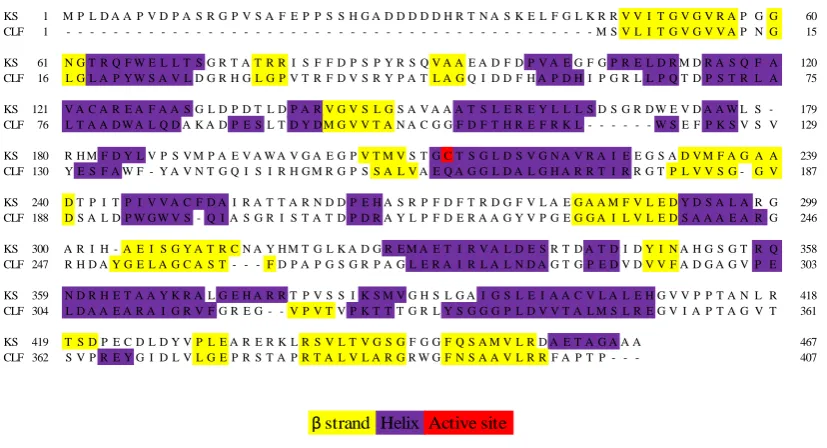
Figure 6 Protein sequences of ketosynthase (KS) and chain length factor (CLF),
from the actinorhodin minimal system. ... 21
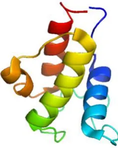
Figure 7 Solution NMR structure of the apo acyl carrier protein 44 from the
actinorhodin minimal system, clearly indicating the four helices. ... 24
Figure 8 Solution NMR structures of apo-ACP 44 (PDB: 2K0Y) (A and C) and
holo-ACP 43 (PDB: 2K0X) (B and D). ... 28
Figure 9 Solution NMR structures of malonyl-ACP 7 (PDB: 2KG8) and octanoyl
ACP 47 (PDB: 2KGC) showing the growing cavity between helices II and III with the size of the polyketide chain.94 ... 29
Figure 10 Structures of acetyl-ACP 40 and the N-acetylcysteamine (NAC) ester
derivative 53 showing the priming of a ketosynthase with the acetyl groups from both 40 and 53. ... 41
Figure 11 Malonyl-coenzyme A (CoA) 15 and nonhydrolysable malonyl-CoA
analogues 55-57 used for probing the biosynthesis of polyketides from type III PKSs. ... 44
Figure 12 Small molecule chain termination probes used for the probing of PKS
biosynthetic pathways, in particular in S. lasaliensis to generate a library of complex analogues.155, 156 ... 48
Figure 13 Common fragmentation pathways of proteins and peptides.179 ... 55
Figure 14 A comparison between the ‘natural’ 4’-phosphopantetheinyl arm and its
xv carrier protein. 66 is the ‘active’ malonyl carba(dethia) N-acetyl cysteamine probe.. ... 69
Figure 15 Multicyclic photocleavable protecting groups for carboxylic acids.. ... 73
Figure 16 Simple aromatic photolabile protecting groups for carboxylic acids ... 74
Figure 17 A selection of nitrobenzyl type photolabile groups to protect
carboxylates. ... 76
Figure 18 A comparison of the two probes synthesised in this work. ... 82
Figure 19 UV/Vis spectrum for the nonhydrolysable photolabile malonyl N-acetyl
cysteamine probe. ... 86
Figure 20 Irradiation of the nonhydrolysable photolabile NAC analogue 65 with
365 nm, 1000 W from 0 to 4 hours (A to E, hourly), showing the generation of decarboxylated product. ... 89
Figure 21 Irradiation of the nonhydrolysable photolabile NAC analogue 65 with
365 nm, 1000 W for 0 (A and C) and 1 (B and D) hours, at two different concentrations (0.1 mM (A and B) and 1 mM (C and D)), showing the increased photolysis yield of the decarboxylated product 129 at higher concentrations .. 90
Figure 22 SDS-PAGE results showing expression of the Pantothenate Kinase
(PanK or CoaA), the phosphopantetheine adenylyltransferase (PPAT or CoaD) and the dephosphocoenzyme A kinase (DPCK or CoaE) from E. coli. ... 103
Figure 23 SDS-PAGE analyses showing expression of the apo acyl carrier protein
44 (~11 kDa) from the actinorhodin minimal system, and the phosphopantetheinyl transferase, Sfp, (~28 kDa) from B. subtilis. ... 106
Figure 24 A: (Parent) mass spectrum of (hexahistidine-tagged) holo-acyl carrier
protein (ACP) 43 from the actinorhodin minimal system. B: Collisionally activated dissociation (CAD) of the 12+ parent ion of holo-ACP 43, generating a singly charged, characteristic, pantetheine ion 80. C: (Parent) mass spectrum of 4,5-dimethoxy-2-nitrobenzyl (DMNB) malonyl carba(dethia) ACP analogue. ACP is from the actinorhodin minimal system. D: CAD of the 12+ parent ion of DMNB malonyl carba(dethia) ACP analogue, generating a singly charged, characteristic, pantetheine ion 81. ... 108
Figure 25 LC-MS chromatogram showing the protonated and sodiated EIC for the
xvi
Figure 26 LC-MS chromatograms showing the photolysis of the DMNB NAC
analogue 65 to 129 in the absence (A) and in the presence of the ketosynthase and chain length factor from the actinorhodin minimal system (B) with 365 nm, 1000W for 4 hours.. ... 112
Figure 27 Irradiation of the nonhydrolysable photolabile NAC analogue 65 with
365 nm, 1000 W for 0 mins (A), 10 mins (B), 30 mins (C), 1 hour (D), 2 hours (E) and 5 hours (F), showing the release of the active probe 66 after just 10 mins, and its stability after 5 hours. ... 113
Figure 28 The effect of delayed addition of active probe 66 to the actinorhodin 2
minmal system on the production of SEK4 41 and SEK4b 42. ... 116
Figure 29 SEK4/4b 41/42 production in the actinorhodin minimal system
monitored by absorbance at 293 nm. ... 118
Figure 30 EIC from LC-MS analyses of the DMNB NAC analogue 65 hydrolysing
and subsequent decarboxylating to 129 after ethyl acetate extraction of the media in the presence (A to D) and in the absence (E) of S. lasaliensis ACP12, with a comparison of the different supplementation strategies. ... 120
Figure 31 EIC from LC-MS analyses of the extracts from feeding experiments with
the DMNB NAC analogue 65 to S. lasaliensis ACP12 mutant. Traces shown are the protonated and sodiated ions of compound 131, a ‘trapped’ intermediate
from lasalocid A 1 biosynthetic pathway, off-loaded from ACP10 and further reduced by KR11. ... 121
Figure 32 EIC from LC-MS analyses of the extracts from feeding experiments with
the DMNB NAC analogue 65 to S. lasaliensis ACP12 mutant. Trace shown is compound 132, a ‘trapped’ intermediate from lasalocid A 1 biosynthetic pathway, offloaded from ACP11. ... 122
Figure 33 EIC from LC-MS analyses of the extracts from feeding experiments with
the DMNB NAC analogue 65 to S. lasaliensis ACP12 mutant. Trace shown is the doubly charged ion of echinomycin 52, a natural product produced by a non-ribosomal peptide synthetase (NRPS) in S. lasaliensis. ... 123
Figure 34 EIC from LC-MS analyses of the DMNB NAC analogue 65 showing its
hydrolysis and subsequent decarboxylation to 129 after ethyl acetate extraction of liquid media in the presence of S. lasaliensis ACP12. ... 124
Figure 35 EIC from LC-MS analyses of the extracts from feeding experiments with
xvii Trace shown is compound 131, a ‘trapped’ intermediate from lasalocid A 1
biosynthetic pathway, off-loaded from ACP10 and further reduced by KR11. .125
Figure 36 EIC from LC-MS analyses of the extracts from feeding experiments with
the DMNB NAC analogue 65 to S. lasaliensis ACP12 mutant in liquid media. Trace shown is compound 132, a ‘trapped’ intermediate from lasalocid A 1
biosynthetic pathway, off-loaded from ACP11. . ... 128
Figure 37 EIC from LC-MS and MS/MS analyses of the extracts from feeding
experiments with the DMNB NAC analogue 65 to S. lasaliensis ACP12 mutant in liquid media. Trace shown and MS/MS is compound 135, a ‘trapped’
intermediate from lasalocid A 1 biosynthetic pathway, off-loaded from ACP8 with subsequent epoxidation and cyclisation. ... 130
Figure 38 EIC from LC-MS and MS/MS analyses of the extracts from feeding
experiments with the DMNB NAC analogue 65 to S. lasaliensis ACP12 mutant in liquid media. Trace shown and MS/MS is compound 136, a ‘trapped’
intermediate from lasalocid A 1 biosynthetic pathway, off-loaded from ACP8, with subsequent epoxidation, cyclisation and oxidation. ... 131
Figure 39 S. coelicolor M510 after 5 days growth supplemented with 1 mM, 5 mM
and 10 mM nonhydrolysable photolabile NAC probe 65 showing growth inhibition with increasing concentration. ... 133
Figure 40 EIC from LC-MS analyses of the DMNB NAC analogue 65 showing its
hydrolysis and subsequent decarboxylation to 129 after ethyl acetate extraction at the end of (A) day 2, (B) day 3, (C) day 4 and (D) day 5 of growth on solid media in the presence of S. coelicolor M510. ... 135
Figure 41 EIC from LC-MS analyses of shunt products of the actinorhodin minimal
system, SEK4/4b 41/42, (shown underneath) produced by S. coelicolor M510 in the presence of DMNB NAC analogue 65 extracted on (A) day 2, (B) day 3, (C) day 4, (D) day 5 and absence of DMNB NAC analogue 65 extracted on (E) day 2, (F) day 3, (G) day 4 and (H) day 5. ... 136
Figure 42 EIC from LC-MS analyses of the DMNB NAC analogue 65 showing its
xviii
Figure 43 EIC from LC-MS analyses of shunt products of the actinorhodin minimal
system, SEK4/4b 41/42, (shown underneath) produced by S. coelicolor M510 in the presence (A-G) and absence (H-N) of DMNB NAC analogue 65, extracted on day 5, following (A and H) no irradiation, (B and I) 1 hour, (C and J) 2 hours, (D and K) 3 hours of irradiation on day 3, and (E and L) 1 hour, (F and M) 2 hours, and (G and N) 3 hours of irradiation on day 4. ... 140
Figure 44 A sample of potential intermediates off-loaded from the actinorhodin 2
biosynthetic pathway by the DMNB NAC analogue 65. . ... 141
Figure 45 Irradiation of the photolabile acyl carrier protein (ACP) analogue 63 in
TrisCl (50 mM), KCl (20 mM), MgCl2 (10 mM) at pH 8, with 365 nm, 1000 W
for 0 (A), 2 (B), and 4 (C) hours, showing the synthesis of the decarboxylated product 144 by TOF-MS. ... 146
Figure 46 Comparison of the ratio of acetyl 40 to holo 43 (not
hexahistadine-tagged) acyl carrier protein (ACP) from the actinorhodin minimal system following 2 hours of incubation (A) with and (B) without the actinohorhodin ketosynthase-chain length factor (KS-CLF). ... 148
Figure 47 Comparison of the ratios of malonyl 7, acetyl 40 and holo43 acyl carrier
proteins (ACPs) from the actinorhodin minimal system following 2 hours of incubation (A) with and (B) without the actinorhodin 2 ketosynthase-chain length factor (KS-CLF). . ... 149
Figure 48 FTICR-MS analyses showing the irradiation of the nonhydrolysable
photolabile acyl carrier protein (ACP) analogue 63 with 365 nm, 1000 W, without the ketosynthase chain length factor (KS-CLF), analysed immediately after 0 (A), 2 (B), and 4 (C) hours, showing the synthesis of the deprotected malonyl product 64 and limited decarboxylated product 144. ... 151
Figure 49 FTICR-MS analyses showing the irradiation of the nonhydrolysable
photolabile acyl carrier protein (ACP) analogue 63 with 365 nm, 1000 W, in the presence of the ketosynthase chain length factor (KS-CLF), analysed immediately after 0 (A), 2 (B), and 4 (C) hours. ... 152
Figure 50 FTICR-MS time-dependant analysis showing a readily uncaged sample
xix
Figure 51 FTICR-MS analysis of active acyl carrier protein (ACP) probe 64
incubated in the actinorhodin minimal system (1:1 ratio, protected ACP probe
63 to malonyl-ACP 7) showing an off-loaded diketide 145. ... 159
Figure 52 FTICR-MS analysis of active acyl carrier protein (ACP) probe 64
incubated in the actinorhodin minimal system (1:10 ratio, protected ACP probe
63 to labelled malonyl-ACP 7b) showing an off-loaded labelled tetraketide 147.161
Figure 53 FTICR-MS analysis of ACP probe 64 incubated in the actinorhodin
minimal system (1:4 protected ACP probe 63 to labelled malonyl-ACP 7b), showing a captured labelled linear pentaketide 148. ... 164
Figure 54 FTICR-MS analysis of the ACP probe 64 incubated in the actinorhodin
minimal system (5:1 protected ACP probe 63 to malonyl-ACP 7) showing a captured putative cyclised pentaketide 154. ... 165
Figure 55 FTICR-MS analysis of the ACP probe 64 incubated in the actinorhodin
minimal system (5:1 protected ACP probe 63 to labelled malonyl-ACP 7b), showing a captured putative cyclised dehydrated pentaketide 155. ... 166
Figure 56 FTICR-MS analysis of ACP probe 64 incubated in the actinorhodin
minimal system (1:4 protected ACP probe 63 to malonyl-ACP 7), showing a captured linear hexaketide 149. ... 169
Figure 57 FTICR-MS analysis of ACP probe 64 incubated in the actinorhodin
minimal system (5:1 protected ACP probe 63 to malonyl-ACP 7), showing a captured cyclised hexaketide 156. ... 170
Figure 58 FTICR-MS analysis of ACP probe 64 incubated in the actinorhodin
minimal system (1:1 protected ACP probe 63 to malonyl-ACP 7), showing a captured cyclised dehydrated hexaketide 157. ... 171
Figure 59 Example of a region of a spectrum with a very intense peak (protected
ACP probe 63 at charge state 8+) with the inset showing the auto-correlation of the region indicated, confirming the 8+ charge state. ... 174
Figure 60 A blank region of a spectrum with the inset showing the auto-correlation
of the region indicated, confirming that no isotopic distributions were detected, and this region of the spectrum is, indeed, noise. ... 175
Figure 61 Spectrum of a detected cyclised hexaketide 156 of charge state 8+ with
xx
Figure 62 EIC of the peptide containing the active site serine generated by trypsin
digestion of the apo and holo acyl carrier proteins 44 and 45 from the actinorhodin minimal system. ... 177
Figure 63 LC-MS chromatogram showing the EIC for the isomers SEK4 41 and
SEK4b 42 produced by the actinorhodin minimal system. The (A and C)) unlabelled and (B and D) 13C labelled actinorhodin minimal system was activated by the addition of final enzyme, holo-ACP 43, then (A and B) 30 seconds and (C and D) 5 minutes later ‘active’ ACP probe 64, irradiated for 4 hours in the home built UVA light source, was added. ... 180
Figure 64 LC-MS chromatograms showing EIC of holo-ACP 43 (red trace) and
DMNB protected carba(dethia) malonyl-ACP analogue 63 (blue trace) expressed in E. coli BAP1 cells by supplementing with (A and C) 1 mM and (B and D) 10 mM DMNB protected carba(dethia) malonyl pantetheine analogue 61
(shown) at the same time as induction with Isopropyl β-D-1-thiogalactopyranoside (IPTG) at optical densities 600 nm of (A and B) 0.6 and (C and D) 0.3. ... 183
Figure 65 Structures of lasalocid A 1 and iso-lasalocid A. ... 191
Figure 66 Structure of the nonhydrolysable photolabile carba(dethia) pantetheine
61. ... 193
Figure 67 Example peak displaying the increased resolving power before (top) and
after (bottom) phasing. . ... 195
Figure 68 A) Mass spectrum of collisionally activated dissociation (CAD) of
[M+Na]+ parent ion 61. B) Mass spectrum of CAD of [M+Li]+ parent ion 61. Inset: assigned fragmentation diagram. ... 197
Figure 69 Mass spectra of: A) electron induced dissociation (EID) of [M+Na]+
parent ion 61; B) EID of [M+Li]+ parent ion 61; C) EID of [M+Cs]+ parent ion
61. ... 198
Figure 70 Fragmentation diagrams showing all the assigned fragments for 61. ... 203
Figure 71 Adduction of lithium to the nonhydrolysable photolabile malonyl
xxi
Figure 72 (A, D, G) Mass spectra showing caesium adduction to the
nonhydrolysable photolabile malonyl carba(dethia) acyl carrier protein (ACP)
63 by sample preparation with 1 mM Cs2CO3. ... 208
Figure 73 The effect of adduction of lithium to the nonhydrolysable photolabile
malonyl carba(dethia) acyl carrier protein (ACP) 63 on the yield of cleaved pantetheine analogue 81 (m/z 524.2239) and the phosphopantetheine analogue
169 (m/z 622.2008) via bond cleavage under collisionally activated dissociation (CAD) ... 210
Figure 74 Adduction of lithium to acetyl acyl carrier protein (ACP) 40 shows
improved yield of cleaved acetylated pantetheine 171 (m/z 303.1373) via phosphodiester bond cleavage under collisionally activated dissociation (CAD). ... 212
Figure 75 The effect of adduction of lithium to malonyl acyl carrier protein (ACP)
7 on the yield of cleaved malonyl pantetheine 173 (m/z 347.1271) via phosphodiester bond cleavage under collisionally activated dissociation (CAD). ... 213
Figure 76 The effect of adduction of lithium or caesium to acetoacetyl-ACP 167,
on the yield of cleaved acetoacetyl pantetheine 175 (m/z 345.1479) via phosphodiester bond cleavage under collisionally activated dissociation (CAD). ... 215
Figure 77 The effect of lithium adduction to myristoyl-ACP 168 on the yield of
cleaved myristoyl pantetheine 177 (m/z 471.32511) via phosphodiester bond cleavage under collisionally activated dissociation (CAD). ... 217
Figure 78 The effect of lithium adduction to myristoyl-ACP 168 on the yield of
cleaved myristoyl phosphopantetheine 178 (m/z 569.3020) via phosphodiester bond cleavage under collisionally activated dissociation (CAD). ... 218
Figure 79 The effect of caesium adduction to four different ACPs 63, 40, 167, and
7 on fragmentation pathways with collisionally activated dissociation (CAD). ... 219
Figure 80 Cleavage coverage maps showing b and y ions resulting from
xxii
Figure 81 Example spectrum showing b (top) and y (bottom) ions resulting from
collisionally activated dissociation (25V) of acetyl-ACP 40 with lithium adduction. ... 225
Figure 82 Cleavage coverage maps showing b and y ions resulting from
collisionally activated dissociation (35V) of malonyl-ACP 7 with lithium (A, red), with caesium (C, green) and without alkali (B, blue) adduction. ... 226
Figure 83 Cleavage coverage maps showing b and y ions resulting from
collisionally activated dissociation (30V) of acetoacetyl-ACP 167 with lithium (A, red), with caesium (C, green) and without alkali (B, blue) adduction. .... 227
Figure 84 Cleavage coverage maps showing b and y ions resulting from
collisionally activated dissociation (25V) of myristoyl-ACP 168 with (A, red) and without (B, blue) lithium adduction. ... 228
Figure 85 Cleavage coverage maps showing b and y ions resulting from
collisionally activated dissociation (28V and 18V) of nonhydrolysable photolabile malonyl carba(dethia) ACP analogue 63 with lithium (A, red), with caesium (C, green) and without alkali (B, blue) adduction. ... 229
Figure 86 Cleavage coverage maps only showing phosphorylated b and y ions
resulting from collisionally activated dissociation (25V) of acetyl-ACP 40 with (A, red) and without (B, blue) lithium adduction. ... 231
Figure 87 Cleavage coverage maps only showing phosphorylated b and y ions
resulting from collisionally activated dissociation (35V) of malonyl-ACP 7 with lithium (A, red), with caesium (C, green) (Note: none detected) and without alkali (B, blue) adduction. . ... 232
Figure 88 Cleavage coverage maps only showing phosphorylated b and y ions
resulting from collisionally activated dissociation (30V) of acetoacetyl-ACP
167 with lithium (A, red), with caesium (C, green) and without alkali (B, blue) adduction. . ... 233
Figure 89 Cleavage coverage maps only showing phosphorylated b and y ions
resulting from collisionally activated dissociation (25V) of myristoyl-ACP 168
with (A, red) and without (B, blue) lithium adduction. ... 234
Figure 90 Cleavage coverage maps only showing phosphorylated b and y ions
xxiii lithium (A, red), with caesium (C, green) (Note: none detected) and without alkali (B, blue) adduction. ... 235
Figure 91 Cleavage coverage maps only showing ‘b’ and ‘y’ ions with the
acetyl-phosphopantetheinyl arm bound, resulting from collisionally activated dissociation (25V) of acetyl-ACP 40 with (A, red) and without (B, blue) lithium adduction. ... 236
Figure 92 Cleavage coverage maps only showing ‘b’ and ‘y’ ions with the
malonyl-phosphopantetheinyl arm bound, resulting from collisionally activated dissociation (35V) of malonyl-ACP 7 with lithium (A, red), with caesium (C, green) (Note: none detected) and without alkali (B, blue) adduction. ... 237
Figure 93 Cleavage coverage maps only showing ‘b’ and ‘y’ ions with the
acetoacetyl-phosphopantetheinyl arm bound, resulting from collisionally activated dissociation (30V) of acetoacetyl-ACP 167 with lithium (A, red), with caesium (C, green) and without alkali (B, blue) adduction. . ... 238
Figure 94 Cleavage coverage maps only showing ‘b’ and ‘y’ ions with the
myristoyl-phosphopantetheinyl arm bound, resulting from collisionally activated dissociation (25V) of myristoyl-ACP 168 with (A, red) and without (B, blue) lithium adduction. . ... 239
Figure 95 Cleavage coverage maps only showing ‘b’ and ‘y’ ions with the
nonhydrolysable photolabile malonyl carba(dethia)-phosphopantetheinyl arm bound, resulting from collisionally activated dissociation (28V and 18V) of nonhydrolysable photolabile malonyl carba(dethia) ACP analogue 63 with lithium (A, red), with caesium (C, green) (Note: none detected) and without alkali (B, blue) adduction. ... 240
Figure 96 Potential next generation photolabile NAC analogues for off-loading of
xxiv
List of Schemes
Scheme 1 Illustration of different polyketide synthases (PKSs) ... 4
Scheme 2 Polyketide chain extension mechanism for the actinorhodin 2 type II
minimal system... 5
Scheme 3 Examples of common oxygenases found in type II polyketides synthases. 16
Scheme 4 Biosynthesis of actinorhodin 2 from acetyl 17 and malonyl-coenzyme A
15. . ... 19
Scheme 5 Biosynthesis of shunt products, SEK4/4b 41 and 42 and mutactin 45,
from the actinorhodin 2 biosynthetic pathway. ... 25
Scheme 6 Conversion of apo-ACP 44 to holo-ACP 43 with Coenzyme A 46 and a
phosphopantetheinyl transferase. ... 27
Scheme 7 Enzymatic preparation of Coenzyme A analogues via corresponding
pantetheine analogues with the aid of enzymes that biosynthesise Coenzyme A
46 in E. coli. ... 32
Scheme 8 Chemical degradation of 14C labelled 6-methylsalicylic acid (6-MSA) 50
demonstrating the incorporation of four acetates.4, 130 ... 34
Scheme 9 General synthesis of N-acetylcysteamine β-ketothioester (SNAC)
analogues via coupling to carbonyldiimidazole (CDI). ... 42
Scheme 10 In vivo off-loading of polyketide intermediates from the model system,
6-deoxyerythronolide B 51 synthase, with synthetic chain terminators. T ... 45
Scheme 11 Off-loading of intermediates from the lasalocid A 1 synthase, with
synthetic chain terminators from feeding experiments in S. lasaliensis.22, 155, 15647
Scheme 12 Generation of a pantetheine (Pant) ejection ion 58 or a phospho-Pant
ejection ion 59 from the corresponding ACPby CAD or IRMPD. ... 60
Scheme 13 Primary project aim ... 65
Scheme 14 Secondary project aim ... 67
Scheme 15 Mechanism of photolysis of the 4,5-dimethoxy-2-nitrobenzyl (DMNB)
group 107 protecting a carboxylic acid group. ... 79
Scheme 16 Synthesis of a nonhydrolysable photolabile malonyl N-acetyl
cysteamine analogue 65. ... 83
Scheme 17 ‘Protected’ 4,5-dimethoxy-2-nitrobenzyl malonyl carba(dethia) ACP
analogue 63 and its product after irradiation to the ‘active’ malonyl
xxv
Scheme 18 Products of the actinorhodin minimal system, SEK4 41 and SEK4b 42,
via the synthesis of an octaketide chain. ... 94
Scheme 19 Overview of the planned chemoenzymatic synthesis of a
nonhydrolysable photolabile malonyl acyl carrier protein 63 from pantothenic acid 60. ... 96
Scheme 20 Previous preparation of a nonhydrolysable malonyl carba(dethia)
coenzyme A analogue 71 by Tosin and co-workers. ... 97
Scheme 21 Synthesis of a nonhydrolysable 4,5-dimethoxy-2-nitrobenzyl malonyl
carba(dethia) pantetheine analogue with acetyl protection of the diol 73. ... 98
Scheme 22 Final synthesis of a nonhydrolysable 4,5-dimethoxy-2-nitrobenzyl
malonyl carba(dethia) pantetheine analogue 61. ... 99
Scheme 23 Enzymatic synthesis of a nonhydrolysable 4,5-dimethoxy-2-nitrobenzyl
malonyl carba(dethia) coenzyme A analogue 62 from a corresponding, chemically synthesised, pantetheine analogue 61. ... 104
Scheme 24 Enzymatic preparation of a nonhydrolysable
4.5-dimethoxy-2-nitrobenzyl malonyl carba(dethia) acyl carrier protein analogue 63 from the corresponding chemoenzymatically synthesised coenzyme A analogue 62.. .. 107
Scheme 25 A) In vivo generation of the active probe 66 from the methyl ester
protected probe 130.155 B) In vivo generation of the active probe 66, via endogenous esterases, or photolysis, from the 4,5-Dimethoxy-2-Nitrobenzyl (DMNB) protected probe 65. ... 127
Scheme 26 Lasalocid A 1 biosynthetic pathway from S. lasaliensis showing
previously ‘trapped’ intermediates generated by the incubation with NAC analogues. . ... 129
Scheme 27 Lasalocid A 1 biosynthetic pathway from S. lasaliensis showing newly
putative ‘trapped’ intermediates 135 and 136 generated by the incubation with DMNB NAC analogue 65 in an ACP12 deletion mutant following UV irradiation. ... 142
Scheme 28 Investigation of the actinorhodin minimal type II polyketide synthase by
means of a protein probe 63. ... 155
Scheme 29 Capture of putative pentaketide intermediates from the actinorhodin
minimal system... 162
Scheme 30 Capture of putative hexaketide intermediates from the actinorhodin
xxvi
Scheme 31 Capture of putative intermediates from the actinorhodin minimal system.
xxvii
List of Tables
Table 1 Overview of notable fragmentation techniques, the instruments they are
typically coupled with and their mode of action. ... 54
Table 2 Reaction conditions trialled for the synthesis of the nonhydrolysable
photolabile malonyl N-acetyl cysteamine analogue 65 from 128, shown. ... 84
Table 3 Conditions tested for the photolysis of a nonhydrolysable photolabile
malonyl N-acetyl cysteamine probe 65. ... 87
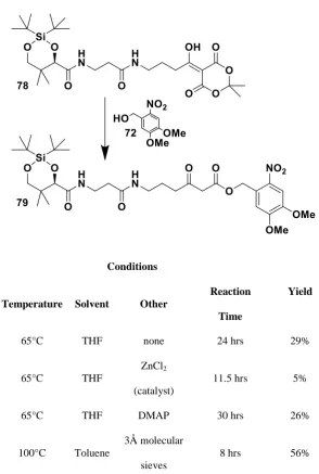
Table 4 Reaction conditions trialled for the synthesis of 79 from 78, shown. ... 100
Table 5 Reaction conditions trialled for the deprotection 79. ... 101
Table 6 Intermediates from the actinorhodin minimal system trapped by the
carba(dethia) malonyl-ACP probe 64 were detected by FTICR-MS direct injection. ... 157
Table 7 Comparison of lithiated and non-lithiated ACP samples... 244
Table 8 SDS-PAGE recipes ... 260
Table 9 LC-MS gradient for the analysis of the nonhydrolysable photolabile SNAC
analogue 65 photolysis experiments ... 268
Table 10 UPLC-MS gradient for the analysis of proteins ... 268
Table 11 UPLC-MS gradient for the analysis of acyl carrier proteins ... 269
Table 12 UPLC-MS gradient for the analysis of cell and enzyme reaction extracts ....
... 270
Table 13 UPLC-MS gradient for the analysis of trypsin digested acyl carrier
xxviii
Abbreviations and Definitions
Abbreviations Definitions
6Deb 6-deoxyerythronolide B 6-MSA 6-methylsalicylic acid
A(+) Monoisotopic mass and the following series for that ion Ac2O Acetic anhydride
ACP Acyl carrier protein
AcpH Acyl carrier protein hydrolase
act Actinorhodin
A-pHP 3-acetamide-para-hydroxyphenacyl Aqmoc Anthroquinon-2-ylmethoxycarbonyl Arg Arginine
AT Acyl transferase
ATP Adenosine triphosphate
Bhc 6-bromo-7-hydroxycoumarin-4-yl)methyl BLAST Basic Local Alignment Search Tool
CAD or CID Collisionally Activated/Induced Dissociation cAMP Cyclic adenosine monophosphate
CDA Calcium-dependent antibiotic
Chc (6-chloro-7-hydroxycoumarin-4-yl)methyl CLF Chain length factor
CM Coumarin-4-ylmethyl
CMCM (7-carboxymethoxycoumarin-4-yl)methyl CoA Coenzyme A
C-pHP 3-carboxyl-para-hydroxyphenacyl Cryo-EM Cryo-electron microscopy
D(NB) Di(nitrobenzyl)oxycarbonyl Da Daltons
DCM Dichloromethane
xxix
Abbreviations Definitions
DH Dehydratase DHK Dihydrokalafungin
DIPEA N,N-Diisopropylethylamine DMAP 4-Dimethylaminopyridine DMB 3,5-dimethoxybenzyl
DMDMB α,α-dimethyl-3,5-dimethoxybenzyl DMNB 4,5-dimethoxy-2-nitrobenzyl
DMNPE 4,5-dimethoxy 1-(2-nitrophenyl)ethyl DMNPP 2-(4,5-Dimethoxy-2-nitrophenyl)propyl DMO-pHP 3,5-dimethoxy-para-hydroxyphenacyl DNB 2,6-dinitrobenzyl
DNPT 2,4-dinitrophylthio
DPCK Dephosphocoenzyme A kinase ECD Electron capture dissociation
EDC 1-Ethyl-3-(3-dimethylaminopropyl)carbodiimide EIC Extracted ion chromatogram
EID Electron induced dissociation ER Enoyl reductase
ESI Electrospray ionisation ETD Electron transfer dissociation Ex. Extracted
FAD Flavin adenine dinucleoside FAS Fatty acid synthase
FID Free induction decay
FRET Fluorescence resonance energy transfer FT Fourier transform
FTICR Fourier transform ion cyclotron resonance FT-MS Fourier transform-mass spectrometry FWHM Full width at half maximum
xxx
Abbreviations Definitions
Glu Glutamine
GT Glycosyltransferase
HATU N,N,N′,N′-Tetramethyl-O-(7-azabenzotriazol-1-yl)uronium hexafluorophosphate
HCM (7-hydroxycoumarin-4-yl)methyl His Histidine
HNVDS ((2-hydroxy-3-naphthy)vinyl)-diisopropylsilyl HOBt Hydroxybenzotriazole
HPLC High performance liquid chromatography HR High resolution
HSDIS (hydroxystyryl)diisopropylsilyl ICR Ion cyclotron resonance
InsP3 1,4,5-triphosphate
IPTG Isopropyl β-D-1-thiogalactopyranoside IRMPD Infrared multiphoton dissociation KR Ketoreductase
KS Ketosynthase LB Lysogeny broth
LC Liquid chromatography LED Light emitting diode Leu Leucine
m/z Mass to charge ratio
MALDI Matrix assisted laser desorption ionisation
MCAT Malonyl-CoA: holo acyl carrier protein transacylase MCM (7-methoxycoumarin-4-yl)methyl
MDNB 4,5-methylenedioxy nitrobenzyl MeCN Acetonitrile
MeOH Methanol
xxxi
Abbreviations Definitions
MS/MS Tandem mass spectrometry MT Methyltransferase
MudPIT Multidimensional Protein Identification Technology MW Molecular weight
NAC N-acetylcysteamine
NADPH Nicotinamide adenine dinucleotide NB Nitro benzyl
Ni-NTA Nickel-nitrilotriacetic acid NMR Nuclear magnetic resonance NRPS Non-ribosomal peptide synthase NVOC 6-nitroveratroyloxycarbonyl
OASIS Orthogonal Active Site Identification System
OD600nm Optical Density at 600 nm
ORF Open reading frame PanK Pantothenate kinase Pant Pantetheine
pBB para-bromobenzyl PCP Peptidyl carrier protein PDB Protein database Phe Phenylalanine
Phmoc Phenanthren-9-ylmethoxycarbonyl
pHP para-hydroxyphenacyl PKS Polyketide synthase
pMOB para-methoxybenzyl
Pmoc Pyren-1-ylmethoxycarbonyl
pNB Para-nitrobenzyl PPant 4’-phosphopantetheine
PPAT Phosphopantetheine adenyltransferase ppm Parts per million
xxxii
Abbreviations Definitions
PTM Post translational modification QIT Quadrupole ion trap
Red Undecylprodigiosin RF Retention factor RP Reverse phase
SAM S-Adenosyl methionine
SDS-PAGE Sodium dodecyl sulphate polyacrylamide gel electrophoresis Ser Serine
SNAC N-acetylcysteamine β-ketothioester TBAF Tetra-n-butylammonium fluoride
tBhc 3,6,8-tribromo-7-hydroxycoumarin-4-ylmethyl TBS tert-butyldimethylsilyl
TCSSD Thiochrome S,S-dioxide TE Thioesterase
TFA Trifluoroacetic acid THF Tetrahydrofuran Thr Threonine
TLC Thin layer chromatography TOF Time of flight
UPLC Ultra pressure liquid chromatography UV Ultra violet
UV/Vis Ultra violet/Visible
xxxiii
Abstract
Polyketide natural products are a major source of pharmaceutical and agricultural compounds. Their biosynthesis is highly complex and the elucidation of intermediate steps is highly desirable to the scientific community for microorganism engineering purposes.
In this work, novel photoactivatable ‘chain termination’ probes were prepared as tools for the off-loading and capture of biosynthetic intermediates from polyketide synthases (PKSs); novel methods to analyse polyketide biosynthetic intermediates
via FTICR-MS were also investigated.
The chemical probes herein reported are nonhydrolysable analogues of ACP-bound malonate used in polyketide biosynthesis for carbon chain elongation. This research focused on the preparation of a chemoenzymatically modified acyl carrier protein that, upon activation via UV irradiation, should compete with discrete ‘natural’ acyl
carrier proteins to capture biosynthetic intermediates from challenging type II polyketide iterative assemblies. Promising preliminary results for the use of this tool
in vitro were obtained in the form of putative actinorhodin ACP-bound intermediates observed and characterised by FTICR-MS. Moreover, a 4,5-dimethoxy-2-nitrobenzyl group was also prepared and successfully employed for trapping biosynthetic intermediates from the in vivo assembly of the antibiotic lasalocid A.
2
1. Introduction
1.1. Polyketide natural products and their biosynthesis
Polyketides are a broad family of pharmaceutically and industrially important natural products.1-5 They are produced by bacteria, fungi, plants, and other organisms, and display a plethora of chemical structures and functions. Examples include lasalocid A 1, a coccidiostat biosynthesised from the soil bacterium Streptomyces lasaliensis6; actinorhodin 2, a complex aromatic antibiotic from Streptomyces. coelicolor7; phloroglucinol 3, a small aromatic metabolite from Pseudomonas fluorescens that displays a range of activities including antibacterial action8; resveratrol 4, an antioxidant found in grapes commonly reported as being responsible for the so-called ‘French paradox’9
; and lovastatin 5, a cholesterol lowering compound, and precursor to the drug simvastatin, biosynthesised by the fungus Aspergillus terreus10
(Figure 1). Polyketides also include many anticancer and antitumour agents,
insecticides, antifungals and antiviral compounds.
3
1.1.1. Fundamentals of polyketide biosynthesis
Polyketides are biosynthesised by polyketide synthase (PKS) enzymes. PKSs are roughly categorised into three types per the organisation of their domains and their catalytic activities (Scheme 1). While type I and type III PKSs are single proteins comprising multiple domains/catalytic activities, type II PKSs consist of discrete enzymes or domains that act iteratively to produce complex aromatic compounds.
11-13
Type I PKSs can be either ‘modular’ or iterative. Modular PKSs biosynthesise polyketides in a step-wise manner, utilising ‘modules’: these are groups of enzymes or domains which catalyse loading, elongation or termination of the polyketide chain where each domain is used only once. Conversely, iterative type PKSs use each of their domains multiple times. Type III PKSs are always iterative, but differ from type II in that coenzyme A (CoA) is the acyl carrier, whereas an acyl carrier protein (ACP) is used in all other types of PKS.
4
Scheme 1 Illustration of different polyketide synthases (PKSs): From top to bottom: Type I modular PKSs use modules for single rounds of chain extension and modification, as exemplified by the first two modules of the lasalocid A PKS which are each responsible for
one round of polyketide chain extension. Type I iterative PKSs use their domains iteratively, as shown, for the biosynthesis of Terreic acid 614 via the precursor 6-methylsalicylic acid (6-MSA) 50, showing the multiple usage of each domain. Type II PKS such as that responsible for the biosynthesis of actinorhodin 2. Type III PKSs such as that responsible for the biosynthesis of resveratrol 4, use coenzyme A (CoA) as the acyl carrier. KS = ketosynthase, AT = acyl transferase, ACP = acyl carrier protein, DH = dehydratase, KR = ketoreductase,
5
Scheme 2 Polyketide chain extension mechanism for the actinorhodin 2 type II minimal system. ACP = acyl carrier protein, KS = ketosynthase, CLF = chain length factor.
Acyl transferase (AT) domains are present in type I PKSs; they are responsible for selectively loading ACPs with malonyl starter and extender units provided by CoAs. ATs can either be cis- or trans-. The former are paired with a specific ACP, whereas the latter can transfer extender units to one or more ACPs.15 The 6-deoxyerthyronolide B synthase (DEBS) contains a cis-AT, and examples that harbour trans-ATs include PKSs that biosynthesise leinamycin16, bryostatin17 and disorazole15, 18. However, this domain is not necessary in all PKSs as some ACPs can self-malonate, such as in the biosynthesis of actinorhodin 2.19
Further product complexity is achieved by the action of reductive enzymes throughout chain extension. Ketoreductase (KR) domains stereoselectively reduce a β-keto group to a hydroxyl group, using nicotinamide adenine dinucleotide
6 The acyl carrier protein (ACP) plays a key role in shuttling the growing polyketide chains to different domains, and provides the starter unit to the ketosynthase (KS) for chain extension to begin (See section 1.2.1 for further detail).
Thioesterase (TE) domains catalyse the hydrolysis or cyclisation of advanced thioester intermediates, releasing the fully biosynthesised polyketide from the PKS. However, TEs are not always present in PKSs as spontaneous hydrolysis or cyclisation can occur to release the compound from the PKS.
‘Tailoring’ enzymes perform further polyketide structural modifications; they are often post-PKS enzymes, and include methyltransferases, cyclases, aromatases and glycosyltransferases, amongst many others. Product tailoring modifications are often crucial to their bioactivity. For example, the glycosylation of erythromycin is essential for its antibiotic action.21 The tailoring can also take place on the enzyme bound intermediates such as the epoxidation of the late stage intermediates leading to the polyether rings in lasalocid A 1.22
7 Generating analogues of natural products requires a similar long chemical synthetic process: if it were possible to produce these analogues by bioengineering organisms to introduce the favourable modifications this would cut the time and cost of the process, as well as make them environmentally more acceptable by avoiding the use of industrial chemicals. Currently, bioengineering of PKSs is in its early stages and understanding the fundamentals of how these enzymes function is crucial towards achieving this goal.
1.2. Type II polyketides and their biosynthesis
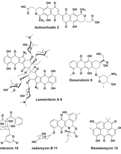
At this point, gram-positive actinomycetes are the only known organisms that biosynthesise polyketides by type II polyketide synthases.12 As outlined in section 1.1.1, type II PKSs differ from other PKSs in that the enzymes involved are discrete, and are always iterative enzymes. The products of type II PKSs tend to be highly aromatic due to the facility of the polyketide chain to undergo cyclisation and aromatisation prior to tailoring steps. Actinorhodin 2 (the polyketide of main interest in this thesis), lomaiviticin 8, doxorubicin 9, enterocin 10, jadomycin B 11, and resistomycin 12 are all examples of polyketides synthesised by type II PKSs (Figure 2).
8
Figure 2 Examples of polyketides biosynthesised by type II iterative polyketide synthases.
1.2.1. Biosynthesis of type II polyketides
Polyketide chain initiation and elongation in type II PKSs is carried out by the ‘minimal system’. This consists of a ketosynthase (KS) and a chain length factor (CLF), also called KSα and KSβ, and an acyl carrier protein (ACP) domain. 4, 24
9 and CLF co-express as a heterodimer complex. The crystal structure of the KS-CLF from the actinorhodin 2 PKS has been solved, showing the growing polyketide chain located at the heterodimer interface, in an amphipathic tunnel.25 The KS contains an active site cysteine, and mass spectrometry has shown that intermediates are bound to this cysteine, and not elsewhere in the KS-CLF complex.25
The ACP provides the KS with the starter and extender units for polyketide chain formation, and also transports the growing polyketide chain to the KS-CLF, linked via a 4’-phosphopantetheinyl (PPant) arm bound to an active site serine.26 The PPant arm binds all these different substrates as a thioester by employing its terminal thiol moiety (Scheme 2).
It is believed that the CLF is crucial for the initial decarboxylation of malonyl-ACP
7 to load an acetyl group onto the KS, and also to initiate the decarboxylative Claisen condensation that begins chain elongation.27 Subsequently, malonyl-ACP 7 is iteratively used for polyketide chain extension as previously described (Scheme 2).
The most common starter unit for type II PKSs are acetate units, as those used for example in the biosynthesis of actinorhodin 228. However, some PKSs have been found that use other starter units, such as propionate (doxorubicin 929 and lomaiviticin 830), and benzoate (enterocin 1031). The alternative starter units are synthesised by fatty acid synthases (FASs) or by type I PKSs, as in frenolicin 1332
and hedamycin 1433 biosynthesis, respectively. A third loading mechanism uses an AT domain to directly load starter units from CoA, as has been established for tetracycline biosynthesis.12, 34
10 the ACP with a malonyl group. The ACP can either self-malonylate19 or malonyl-CoA: holo acyl carrier protein transacylases (MCATs) can transfer the malonyl group from malonyl-CoA 15 to the ACP. It has been found that MCATs are not essential for polyketide synthesis in vitro,19 however, their importance in vivo is unknown. In vivo gene knock-outs of MCATs are not possible due to their crucial function in primary metabolism (fatty acid biosynthesis) in the cell.35
1.2.1.1. Ketosynthase-chain length factor – structure-function relationship
The minimal system, alone, is partially able to control the polyketide chain cyclisation by growing the chain within the KS-CLF complex and ‘folding it’ in a specific manner, however, additional cyclisations can occur.36, 37 Shunt products are, thus far, the only clues to the mechanisms by which the polyketide chains are synthesised in type II PKSs.
An X-ray crystal structure with the acetate starter unit bound to the KS-CLF of the actinorhodin minimal system has been published.25 They show a 17Ǻ long channel where the 4’-phosphopantetheinyl arm of the ACP would reside once in contact with
the KS-CLF, and saw an acetyl group bound to the active site cysteine. The crystal structure of the KS from the R1128 16 biosynthetic pathway has also been solved with acetyl-CoA 17, showing a 20Ǻ long channel that places the acetyl group of CoA against the key residues, asparagine, histidine and the active site cysteine that form the catalytic triad (Figure 3).38
11 minimal system, that the length of the polyketide chain is the controlling factor, and not the number of condensations.25
Figure 3 Exemplary type II ketosynthase crystal structures. Left: The ketosynthase (KS) dimeric structure from the R1128 16 biosynthetic pathway, shows the 20Ǻ tunnel where acetyl-Coenzyme A 17 is bound. (PDB: 1MZJ)39 Right: The ketosynthase chain length factor (KS-CLF) from the actinorhodin 2 biosynthetic pathway, with acetyl bound to the active site cysteine (Cys169), indicated by an arrow. (PDB: 1TQY)25 Inset: magnification of the
KS-CLF active site cysteine showing the bound acetyl group and 17Ǻ long channel where the 4’-phosphopantetheinyl cofactor is thought to reside.
12 achieved both in vivo and in vitro, showing promise for further engineering of type II PKSs.40 However, the KS-CLF does not appear to be the sole determinant of chain length, on two occasions the presence of the cyclase or aromatase enzymes has been demonstrated to influence the chain length.41, 42 This would seem to indicate that a larger PKS complex is formed from the discrete enzymes of type II PKSs, and that the nature of the product is influenced by the interactions taking place amongst the different protein components.
1.2.1.2. Acyl carrier proteins: structure-function relationship
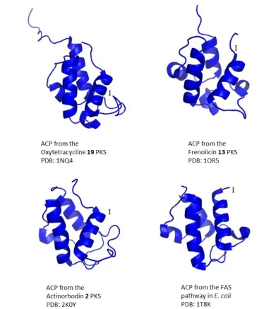
13
Figure 4 Exemplary structures of acyl carrier proteins (ACPs) from type II polyketide synthases (PKSs). Top left: NMR structure of the ACP from the oxytetracycline 19 PKS. (PDB: 1NQ4)45 Top right: NMR structure of the ACP from the Frenolicin 13 PKS. (PDB: 1OR5)44 Bottom left: NMR structure of the ACP from the actinorhodin 2 PKS. (PDB: 2K0Y)46 Bottom right: Crystal structure of ACP from the fatty acid pathway (FAS) in E. coli, containing Zn2+ ions and imidazole ions deriving from the crystallisation conditions.
[image:47.595.113.505.91.529.2]