warwick.ac.uk/lib-publications
Original citation:
Lv, Hua-nan, Rebrov, Evgeny V., Gao, Peng-zhao, Ma, Rui-xue, Lu, Zhou-li and Xu, Jia. (2016) Controllable synthesis of one-dimensional isolated Ni 0.5 Zn 0.5 Fe 2 O 4 microtubes for application as catalyst support in RF heated reactors. Ceramics International, 42 (6). pp. 7793-7802.
Permanent WRAP URL:
http://wrap.warwick.ac.uk/94090
Copyright and reuse:
The Warwick Research Archive Portal (WRAP) makes this work by researchers of the University of Warwick available open access under the following conditions. Copyright © and all moral rights to the version of the paper presented here belong to the individual author(s) and/or other copyright owners. To the extent reasonable and practicable the material made available in WRAP has been checked for eligibility before being made available.
Copies of full items can be used for personal research or study, educational, or not-for-profit purposes without prior permission or charge. Provided that the authors, title and full bibliographic details are credited, a hyperlink and/or URL is given for the original metadata page and the content is not changed in any way.
Publisher’s statement:
© 2016, Elsevier. Licensed under the Creative Commons Attribution-NonCommercial-NoDerivatives 4.0 International http://creativecommons.org/licenses/by-nc-nd/4.0/
A note on versions:
The version presented here may differ from the published version or, version of record, if you wish to cite this item you are advised to consult the publisher’s version. Please see the ‘permanent WRAP url’ above for details on accessing the published version and note that access may require a subscription.
Controllable synthesis of one-dimensional isolated Ni
0.5Zn
0.5Fe
2O
4microtubes for application as catalyst support in RF heated reactors
Hua-nan Lv1, Evgeny V. Rebrov2,3, Peng-zhao Gao1*, Rui-xue Ma1, Zhou-li Lu1, Jia Xu1
1 College of Materials Science and Engineering, Hunan University, Changsha, 410082, China 2 School of Engineering, University of Warwick, Coventry, CV4 7AL, UK
3 Department of Biotechnology and Chemistry, Tver State Technical University, 170026, Nab. A.Nikitina 22, Russia
Corresponding author: Peng-zhao Gao
Email: [email protected] Tel: +86 731 88822269; Fax: +86 731 88823554. (Peng-zhao Gao)
Abstract:
One-dimensional isolated Ni0.5Zn0.5Fe2O4 microtubes have been prepared via a template assisted sol-gel
method. Temperature dependence of the structural and magnetic properties was studied via XRD, N2
adsorption, SEM, TEM, and VSM. An increase in calcination temperature from 873 to 1273 K caused a
decrease in the specific surface area from 80.7 to 17.0 m2/g due to an increase of the grain size from 25.3
to 112 nm. All samples demonstrated anomalous coercivity behavior due to mechanical stresses acting
on their domain walls. The porous microtubes calcined at 1073 K have a mean external diameter of 3.7
m with a length-to-diameter ratio exceeding 12. The microtubes calcined at 973 K have the highest
coercivity of 88.1 Oe and demonstrated the largest specific heating rate of 4.36 W/g in a radiofrequency
field at 295 kHz.
Keywords: Ni-Zn ferrite; microtubes; magnetic properties; anomalous coercivity behavior; radio
2
1 Introduction
Recently, many researchers have studied the unusual morphologies of metal oxide, such as ordered
porous particles, rods, fibers, and hollow structures as the performances of them depend on their chemical
composition and surface properties as well as on their textural properties including morphology, surface
area, pore volume, and pore dimensions [1-4]. In particular, hollow metal oxide with tubular structures
have been extensively investigated because they offer advantages over other shapes, including a high
surface area, narrow pore size distribution, light weight, and facilitates mass diffusion [5]. Specially,
micro-tubes have important significance in the miniaturization of components and devices because of
their small diameter and high slenderness ratio [6,7], and it’s more suitable than nanotubes to load guest
species such as biomolecule, catalyzer, nanoparticles for its larger microns in diameter, so it has unique
applications in fields of drug delivery, catalysts loading and batteries and so on [8]. The currently used
techniques for preparing hollow metal oxide structures include template approach, dry or wet spinning,
electrospinning, and centrifugal spinning [8]. The template approach is very simple and cost-effective
compared to the other methods [9]. Inspired by nature, hollow metal and metal oxide structures have
already been produced employing butterfly wings [10], cotton fibers [11], cellulose acetate fibers [12]
and paper [13] as templates.
In recent years, low-cost Ni–Zn ferrites were applied in high-frequency device applications due to
their high values of chemical stability, magnetization, Curie temperature, permeability, and low power
losses at high frequencies [14, 15]. They have an inverse spinel structure formed by a nearly close packed
the formula (ZnxFe1-x)[Ni1-xFe1+x]O4, in which Zn2+ ions locate in A interstitial (tetrahedral) sites, Ni2+
ions in B (octahedral) sites [16], and Fe3+ ions in the spinel lattice involve both tetrahedral A and
octahedral B sites. The Ni–Zn ferrites have been prepared by co-precipitation, ball milling, spray
pyrolysis, combustion synthesis, hydrothermal synthesis, and sol–gel methods [17-22]. As the
microstructure and magnetic properties of ferrite materials are rather sensitive to the preparation method,
some of those methods are not commercially viable due to disadvantages such as complexity, long
synthesis time, or impurity penetration. Sol–gel is a flexible technique to adjust the properties of material
by optimizing synthesis parameters such as hydrolysis time, temperature, precursor concentration and
pH of the medium. The advantages of the sol–gel process include high purity of the resulting materials,
their chemical homogeneity, and high degree of control over particle and grain size. Furthermore, the
method can be easily scaled up to a large production scale which is crucial for the development of
chemical processes involving radio frequency (RF) induced heating [15].
Combination of spinel ferrites with catalyst nanoparticles allows a novel process intensified platform
for reactor system integration, in these composites, ferrites work as the susceptors of induction heating
to provide efficient RF heating due to the adjustable Curie temperatures and moderate magnetic losses
in the kHz range [23, 24], and catalyst loaded on the ferrites can work on some fine chemicals synthesis
[16, 25]. RF heating provides efficient, fast and uniform heat transfer into catalytic sites and flowing
fluid [26-29]. Base on the excellent textural properties, one-dimensional isolated spinel ferrites
micro-tubes may be a good candidate loading the relative catalyst for the reactor system integration, while
4
The goal of this study was to develop a method to prepare one dimensional isolated nickel zinc ferrite
microtubes(NZF microtubes) by a template assisted sol-gel synthesis using natural cotton fibers as
template. The influence of calcined temperature on the structural, magnetic and specific heating
properties in a radiofrequency field of 295 kHz was investigated, Also the tested temperature dependence
of magnetic and specific heating properties of the obtained NZF microtubes were studied.
2. Experimental process
2.1 Preparation of Ni0.5Zn0.5Fe2O4microtubes
Ni-Zn ferrite microtubes with a nominal composition of Ni0.5Zn0.5Fe2O4 were prepared by the template
assisted sol-gel synthesis [15, 30]. Solution A was prepared by dissolution of the corresponding metal
nitrates in ethanol (all from Aldrich Co., ACS grade). Citric acid was dissolved in ethanol in a separate
vessel to produce solution B which was added into solution A and the resulting mixture was stirred for 4
h. Then an ammonia solution was added dropwise till a pH of 2.3-2.5, the mixture was stirred for 24 h
and then it was absorbed by cotton fibers. These impregnated cotton fibers were dried in an oven at 353
K to get a Ni-Zn ferrite dry gel loaded by cotton fibers. The dry gel was calcined at 873, 973, 1073, 1173
or 1273 K for 1 h to produce the corresponding ferrite microtubes. The heating rate during calcination
was 5 K·min-1 from room temperature to the desired temperatures followed by a dwelling interval of 1 h
at that temperature [15]. The microtubes are labeled as NZF-T hereafter, where index T represents the
calcination temperature in K.
2.2 Characterization of Ni0.5Zn0.5Fe2O4microtubes
(X’Pert PRO) with nickel filtered Cu K radiation produced at 40 kV and 27.5 mA, at a scanning rate of
5o min-1 and a step of 0.02o. The Scherrer equation was used to calculate the crystal size (D, nm) [30].
The d(311) interplanar spacing was determined from the position of the (311) peak using the Bragg
equation. The lattice constant (a) was obtained using Bragg’s diffraction equation for a cubic lattice given
by Eq. 1 [31]:
𝑎 =
√ℎ2sin𝜃2+𝑘2+𝑙2ℎ𝑘l (1)
The morphology of the as-prepared samples was characterized by scanning electron microscopy (SEM,
JSM-6700F, Jeol, Oxford) equipped with an energy dispersive spectrometer (EDS).
The specific surface area of the as-prepared samples was determined by nitrogen adsorption at 77 K
on a Micromeritics NOVA 1000E nitrogen adsorption apparatus.
The magnetization curves of the as-prepared samples were measured by a vibrating sample
magnetometer (Princeton Measurements Corporation MicroMag 3900 VSM) equipped with a 2 Tesla
electromagnet at several temperatures in the range from 298 to 833 K. The saturation magnetization (Ms),
remnant magnetization (Mr), coercivity (Hc) and hysteretic losses were evaluated from the magnetization
curves.
Curie temperature (Tc) of the as-prepared samples was determined from the temperature dependence
of magnetic moment measured with an applied magnetic field of 1.5 T in the 373-1273K range, the
6
The RF induced heating properties of the as-prepared samples was measured at a external magnetic
field with a frequency of 295 kHz and intensity of 500Oe, and the results was given as the specific heating
rate(SHR) to a mixture of water and aimed samples. In these experiments, a slurry of ferrite sample (10
mg) in deionized water (80 mg) was placed in a quartz tube inserted along the center axis in a 50 mm RF
coil connected to an EasyHeat RF system (Ambrell) operated at a current of 200 A. The slurry
temperature was measured with a fiber optic sensor (FISO). The specific heating rate (SHR) was
calculated from the initial (linear) part of temperature vs time curves taken into account the specific heat
capacities of the ferrite and water in the slurry and their weight fractions (Eq. 2) [33].
𝑆𝐻𝑅 =𝑚1𝐶𝑝1+𝑚2𝐶𝑝2
𝑚1+𝑚2
𝑑𝑇
𝑑𝑡 (2)
where, m1 and m2 stand for the weight of water and nickel ferrite, respectively. 𝐶𝑝1 and 𝐶𝑝2 are the
specific heat capacity of water (4180 Jkg-1·K-1) and nickel ferrite (483 Jkg-1·K-1) [34], respectively.
𝑑𝑇
𝑑𝑡is the heating rate obtained from experiments.
3. Results and discussion
3.1 Phase indication of Ni0.5Zn0.5Fe2O4 microtubes
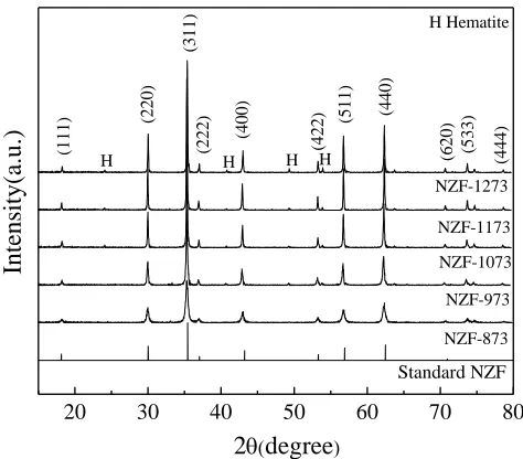
Figure 1 shows XRD spectra of Ni0.5Zn0.5Fe2O4 microtubes calcined at different temperatures. It can
be seen clearly that the spinel phase is formed in the entire temperature range studied (873-1273 K). The
samples calcined below 1173 K show a sharp diffraction peak at 35.54o 2 theta, which is ascribed to the
(311) plane. However, other peaks are rather wide which indicates that the obtained material has low
degree of crystallinity [15]. Also, a minor amount of impurity (hematite, JCPDF #87-1164, labeled as H
in Figure 1) is detected due to small discrepancies in molar ratios of metal nitrates or the segregation of
metal oxides during the drying step.
Please insert Figure 1 here
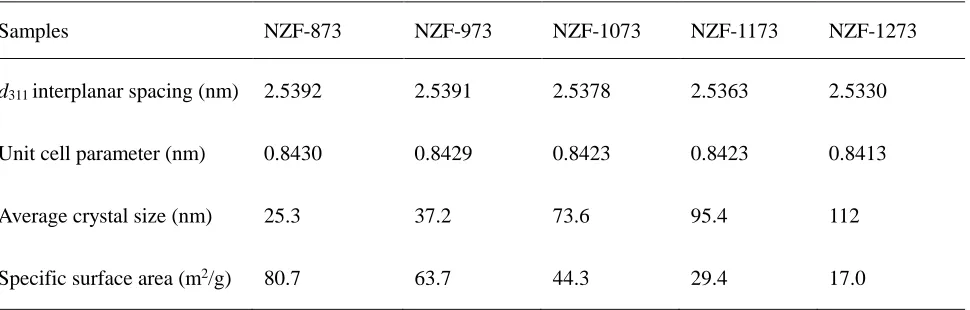
The unit cell parameter, interplanar spacing (d311), average crystal size and specific surface area of the
microtubes calcined at different temperatures are listed in Table 1. As the calcination temperature
increases from 873 to 1273 K, the d311 spacing and unit cell parameter approach the corresponding values
20 30 40 50 60 70 80
H (44
4 ) (5 3 3 ) (6 2 0 ) H H H Hematite NZF-1273 NZF-1173 NZF-1073 NZF-973 NZF-873 Standard NZF (4 4 0 ) (5 1 1 ) (4 2 2 ) (4 0 0 ) (2 2 2 ) (2 2 0 ) (1 1 1 ) (3 1 1 ) Intens ity( a.u. )
2degree
[image:8.612.187.424.314.522.2]H
8
for a bulk Ni0.5Zn0.5Fe2O4 material of 2.5330 and 0.8383 nm, respectively. The specific surface area
decreases by a factor of nearly five due to progressive aggregation of small crystallites into larger
particles. The average crystal size and specific surface area of the sample calcined at 1073 K are 73.6 nm
and 44.3 m2/g, respectively. The latter value is substantially higher than that previously reported for
nickel zinc ferrites [15]. Namely, a highly porous structure is obtained in the template-assisted sol-gel
method which greatly improves the specific surface area even it increases the average particle size at the
same time.
[image:9.612.68.553.380.535.2]Please insert Table 1 here
Table 1. Physical properties of Ni0.5Zn0.5Fe2O4 microtubes calcined at different temperatures.
Samples NZF-873 NZF-973 NZF-1073 NZF-1173 NZF-1273
d311 interplanar spacing (nm) 2.5392 2.5391 2.5378 2.5363 2.5330
Unit cell parameter (nm) 0.8430 0.8429 0.8423 0.8423 0.8413
Average crystal size (nm) 25.3 37.2 73.6 95.4 112
Specific surface area (m2/g) 80.7 63.7 44.3 29.4 17.0
3.2 Microstructure characterization and formation mechanism of Ni0.5Zn0.5Fe2O4 microtubes
Figure 2 shows characteristic SEM images of the template and Ni0.5Zn0.5Fe2O4 microtubes calcined at
different temperatures. It can be seen that one-dimensional isolated ferrite microtubes are obtained. Their
morphology and diameter as well as the ratio of length to diameter can be changed by increasing the
a vein-liked texture (Figure 2 (a, b)). The length of ferrite microtubes exceeds 40 m (Figure 2(c)). The
microtubes with a low degree of crystallinity are obtained after calcination at 873 K which agrees with
the XRD data. Their mean diameter is 6 m (Figure 2(d)). The microtubes with a mean diameter of 3.7
0.2 m and with a higher degree of crystallinity are obtained after calcination at 1073 K (Figure 2(e)).
They are formed by rather uniform nanoparticles with a size of 80 nm and they have a higher
length-to-diameter ratio of 12. After calcination at 1273 K, the microtubes with a mean length-to-diameter of 2.5 0.2 m
are obtained. These highly crystalline samples are formed by much larger particles with a mean size of
120 nm (Figure 2(f)).
Please insert Figure 2 here
10
Figure 2. SEM images of (a, b) cotton fiber template soaked with the nickel ferrite sol and then dried to form a gel,
(c, d) NZF-873; (e) NZF-1073; (f) NZF-1273 microtubes.
A characteristic feature of the fibers is the presence of small channels along the radial direction [35,
36]. Figure 3 (a) shows a characteristic SEM image of the template soaked with the nickel ferrite sol and
then dried to form a gel. The elemental analysis data are listed in Table 2. The elemental composition of
the template sample soaked with the nickel ferrite gel was determined at two different positions: near the
center (point A, Figure 3a) and near the surface (point B, Figure 3a) of a cotton fiber. From these data, it
can be concluded that the precursor sol penetrated about a few micron into the cotton fibers by capillary
action.
(c) (d)
(f) (e)
6.0 m
3.5 m
4.0 m
2.4 m
In order to study the structure transition during calcination, characteristic SEM images of the
microtubes calcined at different temperatures were taken (Figure 3(b-d)). As the temperature increases,
the template and dry gel absorbed in the surface layer are decomposedcausing an interfacial solid-state
reaction and diffusion which yields interconnected ferrite particles. The tube diameter decreases after
calcination at higher temperatures due to increased size of individual ferrite nanoparticles. One important
issue for the formation of perfect microtubes is that an initial continuous layer of dry gel is mandatory.
[image:12.612.89.526.336.681.2]Please insert Figure 3 here
Figure 3. SEM images of (a) cotton fiber template soaked with the nickel ferrite sol and then dried to form a gel, (b)
(c) (d)
(a) (b)
A
B
C
D
12
NZF-873; (c) NZF-1073; (d) NZF-1273 microtubes.
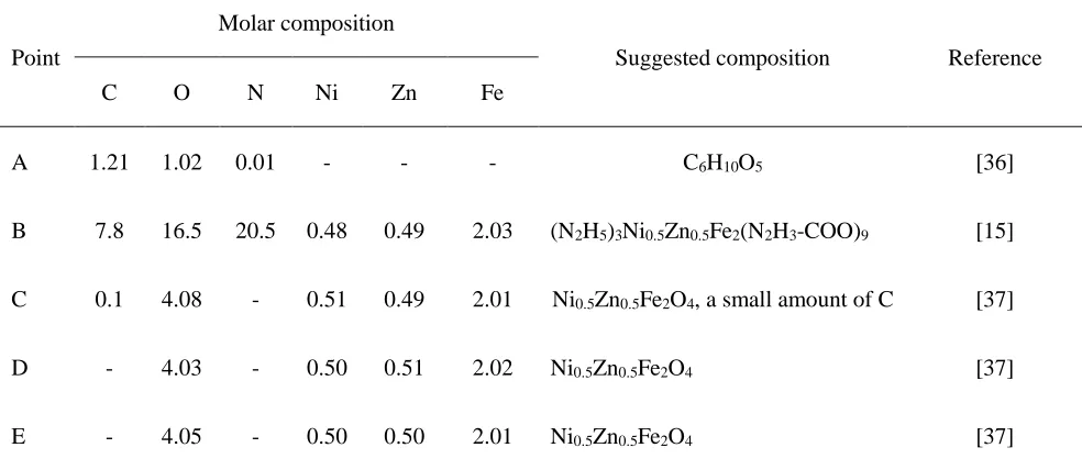
[image:13.612.60.552.195.409.2]Please insert Table 2 here
Table 2 Elemental analysis data for different parts of the samples shown in Figure 3.
Point
Molar composition
Suggested composition Reference
C O N Ni Zn Fe
A 1.21 1.02 0.01 - - - C6H10O5 [36]
B 7.8 16.5 20.5 0.48 0.49 2.03 (N2H5)3Ni0.5Zn0.5Fe2(N2H3-COO)9 [15]
C 0.1 4.08 - 0.51 0.49 2.01 Ni0.5Zn0.5Fe2O4, a small amount of C [37]
D - 4.03 - 0.50 0.51 2.02 Ni0.5Zn0.5Fe2O4 [37]
E - 4.05 - 0.50 0.50 2.01 Ni0.5Zn0.5Fe2O4 [37]
Based on the experiment in which the template soaked with the nickel ferrite gel was converted into
polycrystalline microtubes, it is possible to conclude that the reaction was surface mediated by template.
This was related to a controlled preferential adsorption of the precursors in a thin outer layer of the cotton
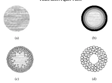
fibers and its very slow diffusion further inside the fibers during calcination. Figure 4 schematically
shows the main steps in the formation of Ni0.5Zn0.5Fe2O4 microtubes. In the first step, the sol penetrated
about a few microns into the cotton fibers by capillary action (Figure 4(a)). A higher amount of sol was
accumulated near the outer surface of the fibers (Figure 4 (b)). As a result, an uneven distribution of gel
particles along the radial direction of the fibers is obtained in the subsequent drying step (Figure 4 (c)).
more particles exist near the surface of the templates fibers, the interconnection and growth of individual
particles tends to form a relative dense structure, which transforms into tubular walls (Figure 4(d)). At
the same time, there exist little particles in the center of the fiber. They are weakly connected connected
to the walls and therefore they are removed when the template is removed during calcination.
Please insert Figure 4 here
(a) (b)
[image:14.612.117.478.246.515.2](c) (d)
Figure 4. Schematic diagram of formation of Ni0.5Zn0.5Fe2O4 microtubes (cross section view).
(a) cotton fiber template, (b) cotton fiber template soaked with the nickel ferrite sol, (c) cotton fiber template with
dry gel, (d) microtubes formed after calcination.
The individual nanoparticles can be seen in a TEM image (Figure 5). They have the shape of irregular
polyhedrons with a mean size between 20 and 120 nm. It should be mentioned that the observed particle
14
connecting necks between the two neighboring particles during calcination. No large aggregated
nanoparticles are observed confirming much higher degree of dispersion as compared with a
non-templated synthesis method [37]. Due to this fact, the specific area of the Ni0.5Zn0.5Fe2O4 microtubes was
considerably enhanced in this study.
[image:15.612.99.519.242.601.2]Please insert Figure 5 here
Figure 5. TEM images of Ni0.5Zn0.5Fe2O4 microtubes calcined at different temperatures
(a) NZF-873; (b) NZF-1073; (c) NZF-1273.
(a) (b)
3.3 Room temperature magnetic properties of Ni0.5Zn0.5Fe2O4 microtubes
A study of the magnetic properties was performed in order to determine if the samples possess specific
magnetic properties and to look for possible effects of the structural transition during calcination on the
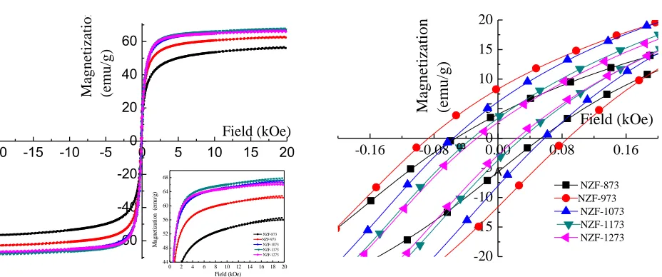
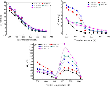
thermal variation of saturation magnetization and coercivity. Room temperature hysteresis loops of the
Ni0.5Zn0.5Fe2O4 microtubes calcined at different temperatures are shown in Figure 6 and their magnetic
parameters are listed in Table 3.
Please insert Figure 6 here
As the calcination temperature increases from 873 to 1273 K, the Curie temperature increases,
however the saturation magnetization first increases and then remains virtually the same in the samples
calcined above 1073 K. The remnant magnetization and coercivity monotonously decrease in the
973--20 -15 -10 -5 0 5 10 15 20
-60 -40 -20 0 20 40 60
0 2 4 6 8 10 12 14 16 18 20 44 48 52 56 60 64 68 NZF-873 NZF-973 NZF-1073 NZF-1173 NZF-1273 M ag n et iz at io n (e m u /g ) Field (kOe) Field (kOe) Ma gne ti za ti on (em u/g)
-0.16 -0.08 0.00 0.08 0.16
[image:16.612.77.541.333.527.2]-20 -15 -10 -5 0 5 10 15 20 NZF-873 NZF-973 NZF-1073 NZF-1173 NZF-1273 Field (kOe) Ma gne ti z a ti on (em u/g) B A
Figure 6. (a) Magnetization curves of Ni0.5Zn0.5Fe2O4 microtubes calcined at different temperatures, (b) hysteresis
16
1273 K range. The maximum value of saturation magnetization exceeds that of the bulk Ni-Zn ferrite
(56 emu/g) [38]. The saturation magnetization in nanoparticles is influenced by both the intrinsic
(composition, preferential site occupancy of the cations, exchange effect) and extrinsic factors
(microstructure and grain size) [39-41]. A relatively low value of saturation magnetization inNZF-873
can be explained by the existence of noncollinear spins at the surface. The exchange interaction between
the Ni-Zn ferrite and the hematite impurity decreases the saturation magnetization in samples calcined at
higher temperatures [40, 42]. At the same time, the increased mean crystal size increases saturation
magnetization. The combined effect of exchange interaction and crystal size levels out therefore the
saturation magnetization remains rather constant in the samples calcined above 1073 K.
[image:17.612.62.554.441.709.2]Please insert Table 3 here
Table 3. Magnetic properties of Ni0.5Zn0.5Fe2O4 microtubes calcined at different temperatures.
Sample NZF-873 NZF-973 NZF-1073 NZF-1173 NZF-1273
Saturation magnetization (emu/g) 56.6 62.8 67.8 67.1 66.6
Remnant magnetization (emu/g) 4.8 9.6 7.0 3.6 2.8
Coercivity (Oe) 56.9 88.1 56.6 31.4 26.1
Curie temperature (K) 532 534 535 537 549
p (erg/cm2) a 0.174 0.174 0.175 0.175 0.177
dcr (nm) b 27.3 22.2 19.1 19.5 20.1
MSO (emu/g) c 73.2 86.6 95.9 92.7 94.0
B (K-1.5) c 5.0510-5 5.6110-5 5.7410-5 5.9010-5 5.5910-5
P (W/g) d 2.54 4.35 3.02 1.66 1.37
a calculated by Eq. 4,
b calculated by Eq. 3,
c calculated by Eq. 6,
d calculated by Eq. 7
Additional information regarding magnetic structure of the samples can be obtained by comparing the
mean crystal size obtained by XRD with the critical magnetic domain size (dcr) which is related to the
transition from single to multidomain region [43]. The latter can be estimated by Eq. 3.
units) CGS (in 2 9 2 πM ε d s p
cr (3)
where Ms is the saturation magnetization (unit in G) and εp is the surface energy of the domain wall
calculated by Eq. 4 [15].
5 0 1
2 B C .
p a K T k ε
(4)
where kB is the Boltzmann constant (1.3810-16 erg·K-1), Tc the Curie temperature, a is the crystalline
lattice constant, and K1is the absolute value of magnetocrystalline anisotropy constant. The anisotropy
constant only slightly changes with composition at room temperature [15, 43], therefore the value
reported by Srinivas et al.(K1 = 1.74104 erg/cm3 [44]) was used in this study.
18
NZF-873 has a single domain structure, i.e. composed of a single magnetic domain, while all other
samples possess multidomain structure. The highest coercivity of 88.1 Oe is observed inNZF-973 as its
grain size (37.2 nm) is only slightly above the critical size (22.2 nm). In the multidomain region, the
coercivity decreases as the grain size increases from 37.2 to 120 nm (Table 1). The coercivity of
crystallites with a size larger than the critical size of single domain (dcr) is inversely proportional to the
grain size (D) [45] (Eq. 5).
𝐻𝐶3 = 𝜌
𝑐√𝐴𝐾𝑀 1
𝑠𝐷 (5)
where pc is a dimensionless factor, A is the exchange constant.
Larger grain tends to contain a greater number of domain walls. The magnetization or demagnetization
caused by domain wall movement requires less energy than that required by domain rotation. On contrast,
with the contribution to magnetization or demagnetization due to domain rotation, the wall movement
increases as the number of walls increases with increasing grain sizes [46]. Therefore, samples having
larger grains are expected to have lower coercivity, and vice versa (Eq. 6). This seems to be the main
reason for the changes in coercivity of the Ni–Zn ferrite microtubes.
As the calcination temperature increases, the Curie temperature of the microtube increases from 534
to 549 K (Table 3). A similar trend was observed by Sepelak et al. for nanostructured Mg ferrites [47].
These authors observed that Néel temperature (TN) increases from 632 to 648 K with increasing
TN with calcination temperature has also been observed in nanostructured NiFe2O4 [15].
3.4 Temperature dependence of magnetic properties of Ni0.5Zn0.5Fe2O4 microtubes
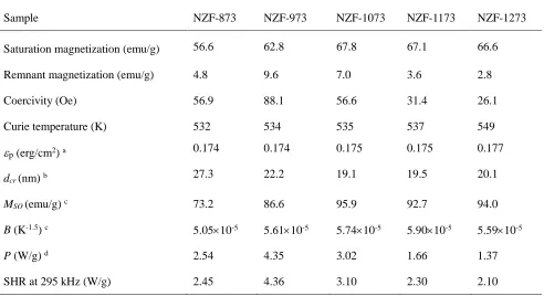
Figure 7 shows the temperature dependence of the saturation magnetization, remnant magnetization
and coercivity of the Ni0.5Zn0.5Fe2O4 microtubes. As the temperature increases, the saturation and
remnant magnetization monotonously decrease (Figure 7a and b). No remnant magnetization is observed
above the Curie temperature (Tc) in samples calcined at 873 and 973 K. This indicates that the samples
are superparamagnetic with a blocking temperature (TB) less than or equal to the TC. The samples calcined
above 1073 K demonstrated a non-zero remnant magnetization above their Curie temperature which is
due to their paramagnetic properties.
The tested temperature dependence of saturation magnetization is described by the Bloch law (Eq.6)
[48]:
Ms = MS0 (1-BT1.5) (6)
where MS0 is the saturation magnetization at T = 0 K, B is the Bloch constant [49]. The MS0 magnetization
increases with calcination temperature till 1073 K and then remains relatively constant (Table 3). For the
present microtubes, the range of prefactor B values of (5.1-5.9)×10-5 K-1.5 obtained from the fitting
procedure is close to the value of 7.5×10⁻⁵ K-1.5 obtained for isolated nanoparticles [15]. The similarity
20
common crystal structure, their close lattice parameters (0.841-0.843 nm in this study vs 0.840 nm in Ref
[15]) and their similar Curie temperatures.
The coercivity of all samples expresses a non-monotonous temperature dependence with a local
maximum in the range between 573 and 823 K, which is slightly above their Curie temperature (Figure
7c). The absolute values of coercivity of the NZF-1073 of 40 and 35 Oe measured at 373 and 423 K are
close to those of Ni-Zn ferrite nanoparticles calcined at 1073 K (42 and 39 Oe, respectively [15]). Below
the Curie temperature, the coercivity decreases with an increase in temperature in accordance with the
ferromagnetism theory as the degree of atomic thermal vibration increases [50]. Above the Curie
temperature, an internal induced magnetic field is formed via a preferred orientation of the magnetic
moments in the microtubes in the direction opposite to that of the applied magnetic field. The magnitude
of this effect is proportional to the magnetic susceptibility, which decreases with temperature resulting
in a decrease of coercivity [51].
However such coercivity behavior cannot be accounted for by exclusively considering the temperature
dependence. The characteristic internal stress patterns in the nearly free standing microtubes may also
result in the anomalous coercivity behavior since the internal stress induces anisotropy in the microtubes.
The presence of temperature-dependent mechanical stresses acting on the domain walls must be
considered as a cause of the observed behavior.
From the viewpoint of application of the obtained materials in catalytic reactors under radiofrequency
heating, it is important that they possess high specific surface area and demonstrate high specific heating
rate which allows to maintain desired temperature inside the reactor. As it was mentioned above, the
materials obtained at 873 and 973 K demonstrated the highest specific surface area. Their specific heating
300 400 500 600 700 800 900
-10 0 10 20 30 40 50 60 70 80
NZF-873 NZF-973
NZF-1073 NZF-1173
NZF-1273 Tested temperature(K) Ms (e mu/ g ) (a)
300 400 500 600 700 800
0 1 2 3 4 5 6 7 8 9
NZF-873 NZF-973
NZF-1073 NZF-1173
NZF-1273 (b) M r (e mu/ g )
Tested temperature (K)
300 400 500 600 700 800 0 20 40 60 80 100 120 140 160 180 200 220
240 NZF-873 NZF-973
NZF-1073 NZF-1173
NZF-1273
H c
(Oe
)
Tested temperature (K)
[image:22.612.79.533.71.430.2](c)
Figure 7. (a) Saturation magnetization, (b) remnant magnetization, (c) coercivity of Ni0.5Zn0.5Fe2O4
microtubes as a function of temperature. Symbols represent experimental data. Solid lines in (a) represent
22
rate is another important factor in evaluating their potential for catalytic applications as susceptors of RF
field [33, 51]. Hysteresis loss is the main mechanism of heating of Ni0.5Zn0.5Fe2O4 microtubes. The heat
released is proportional to the area between the two magnetization curves. Figure 8 shows hysteresis loss
of the microtubes as a function of tested temperature. Due to a non-zero saturation magnetization and a
moderate coercivity, the microtubes could be heated by RF heating in the temperature range above their
Curie temperature. This is a unique feature of these materials which was not observed in the nanoparticles
of the same phase composition.
The hysteresis loss can be approximated as a function of coercivity and saturation magnetization
following the approach developed in our previous study [15]:
P =C0∙Hc ∙Ms (7)
where C0 is a constant.
300 400 500 600 700 800 900 -1
0 1 2 3 4 5 6 7 8
H
yst
ere
si
s l
oss
(kJ
m
-3 )
Tested temperature(K)
[image:24.612.175.436.75.270.2]NZF-873 NZF-973 NZF-1073 NZF-1173 NZF-1273
Figure 8. Hysteresis loss of the ferrite microtubes as a function of tested temperature.
Symbols represent experimental data. Solid lines are the guide for the eye.
The P values and the corresponding specific heating rates are listed in Table 3. It can be seen that the
heating rate of three samples: NZF-873, NZF-973 and NZF-1073 can be predicted with a high degree of
accuracy from their coercivity and saturation magnetization. However, the coercivity drops by a factor
of 3 in NZF-1173 and NZF-1273 as compared to its maximum value observed in NZF-973, while their
saturation magnetization remains rather constant as it was discussed above. The heating rate decreases
only by a factor of 2. In other words, the heating rate determined from hysteresis loops becomes smaller
than that measured directly by calorimetry which proved that it was Néel relaxation that contributed to
the heating effect. The average crystal size inNZF-1173 and NZF-1273 (95.4 and 112 nm, respectively)
is much bigger than the critical domain size (20 nm) so these two samples have a multidomain structure
24
4. Conclusions
One dimensional isolated Ni0.5Zn0.5Fe2O4 microtubes with the length exceeding 40 m and with high
specific area and magnetic saturation have been prepared by a template-assisted sol-gel method. The
possible formation mechanism of Ni0.5Zn0.5Fe2O4 microtubes was proposed. The formation of microtubes
was attributed to the inhomogeneous infiltration of the precursor sol to a cotton fiber template. An
increase in calcination temperature caused the decrease of diameter and specific surface area of
microtubes while the average crystal size and the Curie temperature increased. The coercivity expressed
a non-monotonous temperature dependence caused by mechanical stresses acting on the magnetic
domain walls. While hysteresis loss was the main heating mechanism in all samples, Néel relaxation
contributed to the heating effect in the samples with the mean particle size above 95 nm. The sample
with a mean particle size of 37 nm demonstrated the best combination of specific surface area and the
largest specific heating rate in RF field of 295 kHz.
5. Acknowledgements
The financial support from the Science and Technology Planning Project of Hunan Province, China
(2012WK3023), the Royal Academy of Engineering for research exchanges with China and India–Major
Award (2011-2012), the European Research Council (project 279867, “RF-enhanced microprocessing
for fine chemicals synthesis using catalysts supported on magnetic nanoparticles, RFMiFiCS”), and the
References
[1] B. Boury, R.G. Nair, S.K. Samdarshi, T. Makiabadia, P.H. Mutina, Non-hydrolytic synthesis of hierarchical TiO2
nanostructures using natural cellulose materials as both oxygen donors and template, New J. Chem. 36 (2012)
2196-200.
[2] V.F. Solovyov, L.-J. Wu, M.W. Rupich, S. Sathyamurthy, X. Li, Q. Li, Two-stage epitaxial growth of
vertically-aligned SnO2 nano-rods on (001) ceria, J. Cryst. Growth 408 (2014) 107-111.
[3] A.K. Gain, L. Zhang, W. Liu, Microstructure and material properties of porous hydroxyapatite-zirconia
nanocomposites using polymethyl methacrylate powders, Mater. Design 67 (2015) 136-144.
[4] K.-J. Hwang, C.-H. Hwang, I.-H. Lee, T. Kim, S. Jin, J.-Y. Park, Synthesis and characterization of hollow metal
oxide micro-tubes using a biomaterial template, Biomass Bioenergy 68 (2014) 62-66.
[5] N. Bao, Z. Wei, Z. Ma. Si-doped mesoporous TiO2 continuous fibers: preparation by centrifugal spinning and
photocatalytic properties, J. Hazard. Mater. 174 (2010) 129-136.
[6] H. Wang, S. Zhang, T. Liu, Preparation and application of micro-tubes, Chem. World (China) 12 (2005) 52-56.
[7] Y. Ma, C. Xiong, W. Huang, J. Zhao, X. Li, Q. Fan, W. Huang, Preparation of carbon microtubes by
carbonizing
the fluff of chinar Tree and their application as supercapacitor electrodes, Chin. J. Inorg. Chem. 3 (2012)
546-550.
[8] Xia Yu, Preparation and properties of fluorescent microtubes based on polyoxometalates, Northeast Normal
University(China), Doctor degree thesis(in Chinese), 2013.11.
[9] P. Song, Q. Wang, Z. Zhang, Z. X. Yang, Synthesis and gas sensing properties of biomorphic LaFeO3 hollow fibers
templated from cotton, Sens. Actuators. B 147 (2010) 248-254.
[10] W. Peng, X. Hun, Di Zhang, Bioinspired fabrication of magneto-optic hierarchical architecture by Hydrothermal
process from butterfly wing. J. Magnet. Magnet. Mater. 304 (2006) 197–202.
[11] P. Song, Qi Wang, Z. Yang, Biomorphic synthesis and gas response of In2O3 microtubules using cotton fibers as
templates, Sens. Actuators. B, 168 (2012) 421-428.
[12] C. Zeng, P. Li, L. Zhang, Preparation of magnetic nickel hollow fibers with a trilobe structure using cellulose
26
[13] J. Li, F.-L. Kwong, H. L. N. Dickon, Synthesis of a biomorphic molybdenum trioxide templated from paper, J. Am.
Ceram. Soc. 91 (2008) 1350-1353.
[14] Z. Peng, X. Fu, H. Ge, Z. Fu, C. Wang, L. Qi, H. Miao, Effect of Pr3+ doping on magnetic and dielectric properties
of Ni–Zn ferrites by one-step synthesis, J. Magnet.Magnet. Mater. 323 (2011) 2513–2518.
[15] P. Gao, X. Hua, V. Degirmenci, D. Rooney, M. Khraisheh, R. Pollard, R.M. Bowman, E. V. Rebrov., Structural and
magnetic properties of Ni1−xZnxFe2O4 (x=0, 0.5 and 1) nanopowders prepared by sol–gel method, J. Magnet.
Magnet. Mater. 348 (2013) 44-50.
[16] P. Gao, E.V. Rebrov, T.M.W.G.M. Verhoeven, J.C. Schouten, R. Kleismit, G. Kozlowski, J. Cetnar, Z. Turgut, G.
Subramanyam, Structural investigations and magnetic properties of sol–gel Ni0.5Zn0.5Fe2O4 thin films for
microwave heating, J. Appl. Phys.107 (2010)044317:1-7.
[17] S.J. Azhagushanmugam, N. Suriyanarayanan, R. Jayaprakash, Synthesis and characterization of nanocrystalline
Ni(0.6) Zn(0.4) Fe2O4 spinel ferrite magnetic material, Phys. Procedia 49 (2013) 44-48.
[18] Kh. Gheisari, Sh. Shahriari, S. Javadpour. Structure and magnetic properties of ball-mill prepared nanocrystalline
Ni–Zn ferrite powders at elevated temperatures, J. Alloys Compd. 552 (2013)146-151.
[19] A. Sutka, G. Strikis, G. Mezinskis, A. Lusis, J. Zavickis, J. Kleperis, D. Jakovlevs, Properties of Ni–Zn ferrite thin
films deposited using spray pyrolysis, Thin Solid Films 526 (2012) 65-69.
[20] V.V. Awati, S.M. Rathod, Sagar E. Shirsath, Maheshkumar L. Mane, Fabrication of Cu2+ substituted nanocrystalline
Ni–Zn ferrite by solution combustion route: Investigations on structure, cation occupancy and magnetic behavior,
J. Alloys Compd. 553 (2013) 157-162.
[21] U. Wongpratat, S. Meansiri, E. Swatsitang, Local structure and magnetic property of Ni1−xZnxFe2O4 (x = 0, 0.25,
0.50, 0.75, 1.00) nanoparticles prepared by hydrothermal method, Microelectronic Eng. 126 (2014) 19-26.
[22] Y. Liu, J.-J. Li, F.-F. Min, J.-Bo Zhu, M.-X. Zhang, Microwave-assisted synthesis and magnetic properties of
Ni1−xZnxFe2O4 ferrite powder, J Magnet Magnet Mater 354 (2014) 295-298.
[23] R. Benraba, H. Boukhlouf, A. Löfberg, A. Rubbens, R.-N. Vannier, E. BordesRichard, A. Barama, Nickel ferrite
spinel as catalyst precursor in the dry reforming of methane: synthesis, characterization and catalytic properties, J.
Nat. Gas. Chem. 21 (2012) 595–604.
Synth. 1 (2012) 19–32.
[25] E.V. Rebrov, P. Gao, T.M.W.G.M. Verhoeven, J.C. Schouten, R. Kleismit, Z. Turgut, G. Kozlowski, Structural and
magnetic properties of sol–gel Co2xNi0.5-xZn0.5-xFe2O4 thin films, J Magnet. Magnet. Mater. 323 (2011) 723–729.
[26] A. Ovenston, J.R. Walls, Generation of heat in a single catalyst pellet placed in an electromagnetic field for
endothermic reforming of hydrocarbons, J Chem. Soc. Faraday Transactions 1: Phys. Chem. in Condens. Phases
79 (1983) 1073–1084.
[27] S.I. Al-Mayman, S.M. Al-Zahrani, Catalytic cracking of gas oils in electromagnetic fields: reactor design and
performance, Fuel. Proces. Technol. 80 (2003) 169–182.
[28] P. Duquenne, A. Deltour, G. Lacoste, Application of inductive heating to granular media: temperature distribution
in a granular bed, Int. J. Heat and Mass Trans. 36 (1993) 2473–2477.
[29] T.K. Houlding, K. Tchabanenko, M.T. Rahman, E.V. Rebrov, Direct amide formation using radiofrequency heating,
Org. Biomol. Chem. 11 (2013) 4171–4177.
[30] P. Gao, B. Yan, D. Li, X. Gan, P. Li. W. Liu, Preparation and characterization of wire-liked NiFe2O4 nanoparticles
with high specific area and excellent magnetic properties via a template-assembled sol-gel method, Key Eng. Mater.
633 (2015) 26-30.
[31] A.V. Raut, D.V. Kurmude, D.R. Shengule, K.M. Jadhav, Effect of gamma irradiation on the structural and magnetic
properties of Co–Zn spinel ferrite nanoparticles, Mater. Res. Bull. 63 (2015) 123–128.
[32] Zhen-Fa Zi, Qiang-Chun Liu, Jian-ming Dai, Yan-kun Fu, Anomalous behavior of magnetic properties in CoFe2O4
ferrite nanoparticles (in Chinese). Sci Sin-Phys Mech Astron, 2012, 42: 242-248.
[33] T.K. Houlding, P. Gao, V. Degirmenci, K. Tchabanenko, E.V. Rebrov, Mechanochemical synthesis of
TiO2/NiFe2O4 magnetic catalysts for operation under RF field, Mater. Sci. Eng. B. 193 (2015) 175-180.
[34] K.B. Modi, S.J. Shah, N.B. Pujara, T.K. Pathak, N.H. Vasoya, I.G. Jhala, Infrared spectral evolution, elastic, optical
and thermodynamic properties study on mechanically milled Ni0.5Zn0.5Fe2O4 spinel ferrite, J. Mol. Struct. 1049
(2013) 250-262.
[35] Tayebeh Fattahi Meyabadi, Fatemeh Dadashian, Gity Mir Mohamad Sadeghi, Hamid Ebrahimi Zanjani Asl,
Spherical cellulose nanoparticles preparation from waste cotton using a green method, Powder Technol., 261 (2014)
28
[36]Y. Xiaojiao. Study on the microstructure and fracture mechanism of kapok fiber[D]. Donghua University(China),
Master degree thesis(in Chinese), 2015, 01
[37] B. Yan, P. Gao, Z. Lu, R. Ma, E.V. Rebrov, H. Zheng, Y. Gao. Effect of Pr3+ substitution on the microstructure,
specific surface area, magnetic properties and specific heating rate of Ni0.5Zn0.5PrxFe2-xO4 nanoparticles
synthesized via sol–gel method, J. Alloys Compd., 639(2015) 626-634.
[38] Q. Ma. Study on the preparation and magnetic properties of Ni1-xZnxFe2O4 nanoparticles and composite with Co3O4.
PhD thesis. Anhui University (China) (2013) 23-25.
[39] A.H Lu, E.L. Salabas, F. Schüth. Magnetic nanoparticles: Synthesis, protection, functionalization, and application,
Angew. Chem. Int. Ed. 46 (2007) 1222-1244.
[40] A.L. Xia, C.H. Zuo, L.J. Zhang, C.X. Cao, Y. Deng, W. Xu, M.F. Xie, S.L. Ran, C.G. Jin, X.G. Liu, Magnetic
properties, exchange coupling and novel stripe domains in bulk SrFe12O19/(Ni, Zn)Fe2O4 composites, J. Phys., D.
47 (2014) 415004.
[41] M.A. Gabal, Effect of Mg substitution on the magnetic properties of NiCuZn ferrite nanoparticles prepared through
a novel method using egg white, J. Magnet. Magnet. Mater. 321 (2009) 3144-3148.
[42] M. Younas, M. Atif, M. Nadeem, M. Siddique, M. Idrees, R. Grossinger, Colossal resistivity with diminished
tangent loss in Zn–Ni ferrite nanoparticles, J. Phys. D. 44 (2011) 345402
[43] I.Z. Rahman, T.T. Ahmed, A study on Cu substituted chemically processed Ni–Zn–Cu ferrites, J. Magnet. Magnet.
Mater. 290–291 (2005) 1576-1579.
[44] Ch. Srinivas, B.V.Tirupanyam, A.Satish, V.Seshubai, D.L.Sastry, O.F.Caltun, Effect of Ni2+ substitution on
structural and magnetic properties of Ni–Zn ferrite nanoparticles, J. Magnet. Magnet. Mater. 382(2015)15–19.
[45] D.S. Xue, G.Z. Chai, X.L. Li, X.L. Fan, Effects of grain size distribution on coercivity and permeability of
ferromagnets, J. Magnet. Magnet. Mater. 320 (2008) 1541–43.
[46] A.C.F.M. Costa, E. Tortella, M.R. Morelli, R.H.G.A. Kiminami, Synthesis, microstructure and magnetic properties
of Ni–Zn ferrites, J. Magnet. Magnet. Mater. 256 (2003) 174–182.
[47] V. Š epelák , D. Schultze , F. Krumeich , U. Steinike , K.D. Becker, Mechanically induced cation redistribution in
magnesium ferrite and its thermal stability, Solid State Ionics 141-142 (2001) 677–682.
on nano-cobalt ferrites, J. Alloys Compd. 653 (2015) 513-522.
[49] K. Maaz, A.Mumtaz, S.K.Hasanain, M.F.Bertino, Temperature dependent coercivity and magnetization of
nickelferrite nanoparticles, J. Magnet. Magnet. Mater. 322 (2010) 2199–2202
[50] E.H. Frei, S. Shtrikman, D. Treves. Critical size and nucleation field of ideal ferromagnetic particles. Phys Rev 106
(1957) 446–455.
[51] S. Chatterjee, V. Degirmenci, F. Aiouache, E.V. Rebrov, Design of a radio frequency heated isothermal