
ISSN Online: 2325-7083 ISSN Print: 2325-7075
DOI: 10.4236/crcm.2018.73022 Mar. 21, 2018 241 Case Reports in Clinical Medicine
Primary Vulvar Ewing Sarcoma in a
30-Year-Old Woman: A Case Report
Fatoumata Matokoma Sidibe
1, Hajar Ouahbi
1, Rajae Kanab
1, Layla Tahiri
2,
Fatim Zhara El Mrabet
1, Samia Arifi
1, Hind El Fatemi
2, Nawfel Mellas
11Medical Oncology Department, HASSAN II University Hospital, Fez, Morocco 2Pathology Department, HASSAN II University Hospital, Fez, Morocco
Abstract
Primary Ewing sarcoma (ES) and primitive neuroectodermal tumor (PNET) are considered as Ewing sarcoma family of tumors (ESFT), characterized by chromosomal translocation t(11; 22) (q24; q12) leading to a chimeric tran-script EWS-FLI1 in 85% of cases. It typically involves the soft tissues of the chest wall, pelvis, paravertebral region, abdominal wall, retroperitoneal region and extremities in children, adolescents and young adults. It rarely occurs in the female genital tract. We report an extremely rare case of advanced vulvar Ewing sarcoma/PNET of the vulva confirmed by Fluorescence In Situ Hy-bridization (FISH) in a 30-year-old woman. The patient was treated by 6 cy-cles of chemotherapy followed by radiotherapy with favourable outcome.
Keywords
Ewing Sarcoma/Primitive Neuroectodermal Tumor, Vulva, Advanced Disease, Multimodal Treatment
1. Introduction
Primary Ewing sarcoma (ES) and primitive neuroectodermal tumor (PNET) are considered Ewing sarcoma family of tumors (ESFT), characterized by chromo-somal translocation t(11; 22) (q24; q12) leading to a chimeric transcript EWS- FLI1 (a member of ETS gene) in 85% of cases [1]. In 5% - 10% of cases EWS gene is fused with other members of Erythroblast Transformation-Specific (ETS) gene (ERG, ETV 1, ETV 4 and FEV). ES/PNET is an aggressive malignant round cell tumor yet chemosensitive [2]. They commonly affect bones and especially diaphysis of longs bones. Extraskeletal ES/PNET can arise anywhere in the body, including soft tissue, skin and visceral organs [3]. However, they have been How to cite this paper: Sidibe, F.M.,
Ou-ahbi, H., Kanab, R., Tahiri, L., El Mrabet, F.Z., Arifi, S., El Fatemi, H. and Mellas, N. (2018) Primary Vulvar Ewing Sarcoma in a 30-Year-Old Woman: A Case Report. Case Reports in Clinical Medicine, 7, 241-248. https://doi.org/10.4236/crcm.2018.73022
Received: December 17, 2017 Accepted: March 18, 2018 Published: March 21, 2018
Copyright © 2018 by authors and Scientific Research Publishing Inc. This work is licensed under the Creative Commons Attribution International License (CC BY 4.0).
http://creativecommons.org/licenses/by/4.0/
DOI: 10.4236/crcm.2018.73022 242 Case Reports in Clinical Medicine rarely found in the female genital tract, with occasional reports of tumors arising in the vulva, vagina, cervix, uterine corpus, broad ligament, and ovary [4].
Here, we present a case of 30-year-old woman with a confirmed ES arising in the vulva and we report a systematic review of the published literature regarding primary vulvar ES.
2. Case Report
A 30-year-old woman, with a history of two spontaneous vaginal deliveries at term, presented with progressive left vulvar swelling since 10 months.
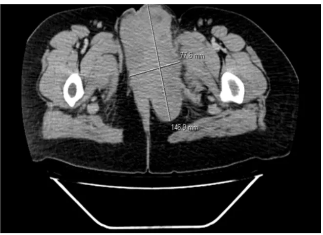
Physical examination showed a “non-cystic”, painless, mobile lesion, 15 × 12 cm in size. She had no further symptoms or lesions. Abdominal and pelvic computed tomography (CT) showed a mixed mass in the left vulva, 146 mm × 78 mm in size, which invades by contiguity the vagina, anal margin, low rectum, obturator muscle, ischion and pubis (Figure 1).
Blood test and chest CT were normal and bone scan showed ischion and pubis fixation. The extensive surgical resection of the lesion was not recommended due to patient’s young age, histological type, size and extension of the vulvar tumor. She underwent core biopsy.
Microscopic sections with hematoxylin and eosin staining (HES) displayed a malignant round cell tumor with monomorphic cells arranged in a diffuse man-ner, containing fine chromatin, scanty indistinct cytoplasm, and inconspicuous nucleoli (Figure 2).
[image:2.595.209.540.443.684.2]Immunohistochemestry showed diffuse cytoplasmic membranous staining with CD99, suggesting ESFT diagnosis (Figure 3). In parallel, muscle markers
DOI: 10.4236/crcm.2018.73022 243 Case Reports in Clinical Medicine Figure 2. Microscopically (Hematoxylin and eosin staining ×20) showing monomorphic small round cells arranged in a diffuse manner, containing fine chromatin, scanty indis-tinct cytoplasm, and inconspicuous nucleoli.
Figure 3. Immunohistochemical study usinganti-CD99 monocal antibody (EPR3097Y) and showing cytoplasmic membranous diffuse staining with CD99
(desmin, myogenin), CD45, the neuroendocrine markers (Synaptophysine and-chromogranin A), epithelial markers (EMA, CKAE1/AE3), and Bcl2 were nega-tive.
Finally, in molecular cytogenetic analyse, fluorescent in-situ hybridization (FISH) showed 100% rearrangement of EWSR1 on chromosome 21 of 100 ana-lyzed nuclei, confirming the diagnosis of primary vulvar ES/PNET.
Patient received combination chemotherapy with Doxorubicine 20 mg/m2/
Day (D), D1-3, Vincristine 2 mg D1, ifosfamide 2000 mg/m2/D, D1-D3 and
uromitexan every three weeks. After six cycles, CT scan evaluation showed a 46% reduction of vulvar mass according to RECIST (Response Evaluation Crite-ria In Solid Tumor) 1.1 criteCrite-ria [5].
[image:3.595.208.542.298.464.2]DOI: 10.4236/crcm.2018.73022 244 Case Reports in Clinical Medicine receives monthly biphosphonate for the local bone extension. We regularly fol-lowed her with quarterly physical examination, blood test and chest, abdominal and pelvic CT scan. Since then, she had a stable disease with persistent mass of 41 × 30 mm in size after 22 months of follow up.
3. Discussion
Ewing sarcoma (ES) and primitive neuroectodermal tumor (PNET) family were regarded as distinct in the past. Recently, studies have shown that the small round-cell tumor seen in both tumor types share common phenotypic and mo-lecular features, supporting the concept of a single tumor category [6]. ES/PNET are characterized by the fusion of the ESW gene on chromosome 22q12 with various members of the Erythroblast Transformation-Specific (ETS) gene family (FLI1, ERG, ETV1, ETV4 and FEV). The fusion with ESW and FLI1 on chro-mosome 11 and corresponding chromosomal translocation t(11; 22) (q24; q12) is present in 85% of patients with ES/PNET [7].
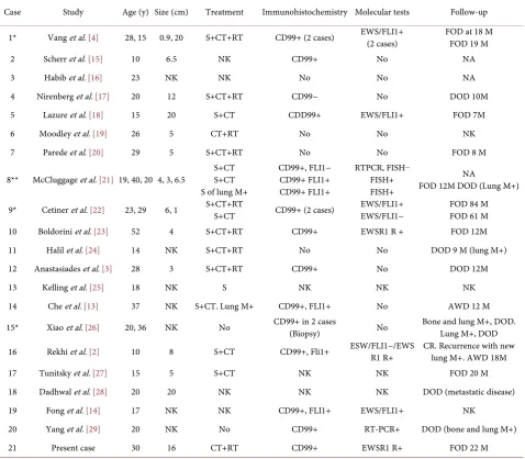
ES/PNET are an uncommon high-grade malignant neoplasm that may affect both skeletal and extraskeletal sites. Extraskeletal Ewing sarcoma (EES) is an unusual aggressive tumor with poor prognosis. It involves soft tissues of the chest wall, extremities, paravertebral and retroperitoneal regions, pelvis and ab-domen, skin, visceral organs, head and neck [8] [9] [10] [11] but rarely occurs in the female genital tract. In this last location, EES has extremely low incidence in the vulva with nearly 25 such documented cases in the literature (Table 1). Here, we report the twenty sixth case of primary vulvar EES in a 30-year-old woman who was treated with multiagent chemotherapy and definitive radiation. We also summarized the clinicopathologic details and management of previously de-scribed cases of vulvar EES together with our case.
Vulvar EES occurs in young women and women of reproductive age (as the case of our patient), median age is 20 years and mean age is 24 years ranging from 10 - 52 years (Table 1). In general, patients present with painless vulvar swelling.
DOI: 10.4236/crcm.2018.73022 245 Case Reports in Clinical Medicine Table 1. Clinicopathologic features of reported cases of vulvar ES/PNET.
Case Study Age (y) Size (cm) Treatment Immunohistochemistry Molecular tests Follow-up
1* Vang et al. [4] 28, 15 0.9, 20 S+CT+RT CD99+ (2 cases) EWS/FLI1+ (2 cases) FOD at 18 M FOD 19 M
2 Scherr et al. [15] 10 6.5 NK CD99+ No NA
3 Habib et al. [16] 23 NK NK No No NA
4 Nirenberg et al. [17] 20 12 S+CT+RT CD99− No DOD 10M
5 Lazure et al. [18] 15 20 S+CT CDD99+ EWS/FLI1+ FOD 7M
6 Moodley et al. [19] 26 5 CT+RT No No NK
7 Parede et al. [20] 29 5 S+CT+RT No No FOD 8 M
8** McCluggage et al. [21] 19, 40, 20 4, 3, 6.5 S+CT S+CT S of lung M+
CD99+, FLI1− CD99+ FLI1+ CD99+ FLI1+
RTPCR, FISH−
FISH+ FISH+
NA
FOD 12M DOD (Lung M+)
9* Cetiner et al. [22] 23, 29 6, 1 S+CT+RT S+CT CD99+ (2 cases) EWS/FLI1+ EWS/FLI1− FOD 84 M FOD 61 M
10 Boldorini et al. [23] 52 4 S+CT+RT CD99+ EWSR1 R + FOD 12M
11 Halil et al. [24] 14 NK S+CT+RT No No DOD 9 M (lung M+)
12 Anastasiades et al. [3] 28 3 S+CT+RT CD99+ No DOD 12M
13 Kelling et al. [25] 18 NK S NK NK NK
14 Che et al. [13] 37 NK S+CT. Lung M+ CD99+, FLI1+ No AWD 12 M
15* Xiao et al. [26] 20, 36 NK No CD99+ in 2 cases (Biopsy) No Bone and lung M+, DOD. Lung M+, DOD
16 Rekhi et al. [2] 10 8 S+CT CD99+, Fli1+ ESW/FLI1−/EWSR1 R+ CR. Recurrence with new lung M+. AWD 18M
17 Tunitsky et al. [27] 15 5 S+CT NK NK FOD 20 M
18 Dadhwal et al. [28] 20 20 NK NK NK DOD (metastatic disease)
19 Fong et al. [14] 17 NK NK CD99+, FLI1+ EWS/FLI1+ NK
20 Yang et al. [29] 20 NK No CD99+ RT-PCR+ DOD (bone and lung M+)
21 Present case 30 16 CT+RT CD99+ EWSR1 R+ FOD 22 M
CT: chemotherapy, RT: Radiotherapy, S: surgery, NK: not known, NA: not available, FOD: Free of disease, DOD: died of disease, AWD: alive of disease, RT-PCR: Reverse transcriptase polymerase chain reaction, FISH: Fluorescence in situ hybridization, EWSR1 R+: EWSR1 rearrangement positive, M: month, Y: Years, M+: metastases, *: 2 cases, **: 3 cases.
These main morphological, histological, immunohistochemical and cytoge-netic features were all found in our reported case. In parallel, we excluded the differential diagnosis in immunohistochemistry staining as lymphoblastic lym-phoma, small cell neuroendocrine carcinoma, rhabdomyosarcoma, carcinosar-coma and epithelial carcinoma.
Treatment of vulvar EES includes complete surgical excision of the lesion, chemotherapy, and, occasionally, radiation therapy (Table 1).
Only one patient in the reported case (case 6) received chemotherapy follow by radiation therapy without surgery, as in our case. She developed chest metas-tases within a short period of time and subsequently died.
DOI: 10.4236/crcm.2018.73022 246 Case Reports in Clinical Medicine 1.1 [5], surgery was not attempted due to initial very advanced disease and the high risk of R1 resection (microscopic margin invasion).
Except in two cases of Cetiner and al, which had long follow-up (more than 4 and 7 years), others reported cases have been limited (ranging 7 - 20 months in 12 cases or died at short time in 4) or no long term follow up data (six cases). Thus, the best way of treating vulvar EES is difficult to predict but it appears clear that multimodal treatment is better.
Our patient received chemotherapy, and then radiotherapy. About 22 months after diagnosis, she is alive and no evidence of progressive disease.
4. Conclusions
Primary vulvar ES/PNET is a rare malignancy that requires early diagnosis and treatment. Multimodal treatment including surgery, chemotherapy, and/or ra-diotherapy is required.
Because of the rare reported cases in literature, it is difficult to draw any con-clusion about tumor behaviour, epidemiology or standard management. Studies of more cases of primary vulvar ES/PNET with longer follow-up periods are needed to clarify its clinicopathologic features and its treatment.
Disclosure
No author has any potential conflict of interest.
References
[1] Aurias, A., Rimbaut, C., Buffe, D., Dubousset, J. and Mazabraud, A. (1983) Transloca-tion of Chromosome 22 in Ewing’s Sarcoma. Comptes Rendus des Seances de l'Academie des Sciences—Serie III, 296, 1105-1107.
[2] Rekhi, B., Chinnaswamy, G., Vora, T., Shah, S. and Rangarajan, V. (2015) Primary Ewing Sarcoma of Vulva, Confirmed with Molecular Cytogenetic Analysis: A Rare Case Report with Diagnostic and Treatment Implications. Indian Journal of Pa-thology & Microbiology, 58, 341-344. https://doi.org/10.4103/0377-4929.162869 [3] Anastasiades, E.N., El Abiad, S.A. and Chouairy, J.C. (2012) Ewing Sarcoma/Primitive
Neuroectodermal Tumor (PNET) of the Vulva. Case Report and Review of the Li-terature.Le Journal Medical Libanais, 60, 113-116.
[4] Vang, R., Taubenberger, J.K., Mannion, C.M., Bijwaard, K., Malpica, A., Ordonez, N.G., Tavassoli, F.A. and Silver, S.A. (2000) Primary Vulvar and Vaginal Extraos-seous Ewing’s Sarcoma/Peripheral Neuroectodermal Tumor: Diagnostic Confirma-tion with CD99 Immunostaining and Reverse Transcriptase-Polymerase Chain Re-action. International Journal of Gynecological Pathology, 19, 103-109.
https://doi.org/10.1097/00004347-200004000-00002
[5] Eisenhauer, E.A., Therasse, P., Bogaerts, J., Schwartz, L.H., Sargent, D., Ford, R., Dancey, J., Arbuck, S., Gwyther, S., Mooney, M., Rubinstein, L., Shankar, L., Dodd, L., Kaplan, R., Lacombe, D. and Verweij, J. (2009) New Response Evaluation criTe-ria in Solid Tumours: Revised RECIST Guideline (Version 1.1). European Journal of Cancer, 45, 228-247. https://doi.org/10.1016/j.ejca.2008.10.026
Neuroec-DOI: 10.4236/crcm.2018.73022 247 Case Reports in Clinical Medicine todermal Tumor with Cranial Metastasis. Journal of the Chinese Medical Associa-tion, 72, 332-335. https://doi.org/10.1016/S1726-4901(09)70381-8
[7] Folpe, A.L., Hill, C.E., Parham, D.M., O’Shea, P.A. and Weiss, S.W. (2000) Immu-nohistochemical Detection of FLI-1 Protein Expression: A Study of 132 Round Cell Tumors with Emphasis on CD99-Positive Mimics of Ewing’s Sarcoma/Primitive Neuroectodermal Tumor. The American Journal of Surgical Pathology, 24, 1657-1662. https://doi.org/10.1097/00000478-200012000-00010
[8] Dagher, R., Pham, T.A., Sorbara, L., Kumar, S., Long, L., Bernstein, D., Mackall, C., Raffeld, M., Tsokos, M. and Helman, L. (2001) Molecular Confirmation of Ewing Sarcoma. Journal of Pediatric Hematology/Oncology, 23, 221-224.
https://doi.org/10.1097/00043426-200105000-00009
[9] Carvajal, R. and Meyers, P. (2005) Ewing’s Sarcoma and Primitive Neuroectodermal Family of Tumors. Hematology/Oncology Clinics of North America, 19, 501-525. https://doi.org/10.1016/j.hoc.2005.03.004
[10] Gaona-Luviano, P., Unda-Franco, E., González-Jara, L., Romero, P. and Medina- Franco, H. (2003) Primitive Neuroectodermal Tumor of the Vagina. Gynecologic Oncology, 91, 456-458. https://doi.org/10.1016/S0090-8258(03)00500-6
[11] Khoury, J.D. (2005) Ewing Sarcoma Family of Tumors. Advances in Anatomic Pa-thology, 12, 212-220. https://doi.org/10.1097/01.pap.0000175114.55541.52
[12] Chokoeva, A.A., Tchernev, G., Cardoso, J.C., Patterson, J.W., Dechev, I., Valkanov, S., Zanardelli, M., Lotti, T. and Wollina, U. (2015) Vulvar Sarcomas: Short Guide-line for Histopathological Recognition and Clinical Management. Part 2. International Journal of Immunopathology and Pharmacology, 28, 178-186. https://doi.org/10.1177/0394632015575977
[13] Che, S.M., Cao, P.L., Chen, H.W., Liu, Z. and Meng, D. (2013) Primary Ewing’s Sarcoma of Vulva: A Case Report and a Review of the Literature. Journal of Obstet-rics and Gynaecology Research, 39, 746-749.
https://doi.org/10.1111/j.1447-0756.2012.02019.x
[14] Fong, Y.E., López-Terrada, D. and Zhai, Q.J. (2008) Primary Ewing Sarcoma/ Peri-pheral Primitive Neuroectodermal Tumor of the Vulva. Human Pathology, 39, 1535-1539. https://doi.org/10.1016/j.humpath.2008.01.013
[15] Scherr, G.R., d’Ablaing 3rd, G. and Ouzounian, J.G. (1994) Peripheral Primitive Neuroectodermal Tumor of the Vulva. Gynecol Oncol, 54, 254-258.
https://doi.org/10.1006/gyno.1994.1204
[16] Habib, K., Finet, J.F., Plantier, F., et al. (1992)A Rare Lesion of the Vulva. Archives d'Anatomie et de Cytologie Pathologiques, 40, 158-159.
[17] Nirenberg, A., Ostör, A.G., Slavin, J., et al. (1995) Primary Vulvar Sarcomas. Inter-national Journal of Gynecological Pathology, 14, 55-62.
https://doi.org/10.1097/00004347-199501000-00010
[18] Lazure, T., Alsamad, I.A., Meuric, S., et al. (2001) Primary Uterine and Vulvar Ew-ing’s Sarcoma/Peripheral Neuroectodermal Tumors in Children: Two Unusual Lo-cations. Annales de Pathologie, 21, 263-266.
[19] Moodley, M. and Jordaan, A. (2005) Ewing’s Sarcoma of the Vulva. A Case Report. International Journal of Gynecological Cancer, 15, 117-118.
https://doi.org/10.1111/j.1525-1438.2005.00330.x
[20] Paredes, E., Duarte, A., Couceiro, A., et al. (1995) A Peripheral Neuroectodermal Tumor of the Vulva. Acta Medica Portuguesa, 8, 161-163.
DOI: 10.4236/crcm.2018.73022 248 Case Reports in Clinical Medicine Flanagan, A.M. and Fisher, C. (2007) Ewing Family of Tumours Involving the Vul-va and Vagina: Report of a Series of Four Cases. Journal of Clinical Pathology, 60, 674-680. https://doi.org/10.1136/jcp.2006.040931
[22] Cetiner, H., Kir, G., Gelmann, E.P., et al. (2009) Primary Vulvar Ewing Sarco-ma/Primitive Neuroectodermal Tumor. A Report of 2 Cases and Review of the Li-terature. International Journal of Gynecological Cancer, 19, 1131-1136.
https://doi.org/10.1111/IGC.0b013e3181acae33
[23] Boldorini, R., Riboni, F., Cristina, S., Allegrini, S., Valentini, S., Muscara, M. and Ruspa, G. (2010) Primary Vulvar Ewing’s Sarcoma/Primitive Neuroectodermal Tumor in a Post-Menopausal Woman: A Case Report. Pathology, Research and Practice, 206, 476-479.
[24] Halil, S., Kucuk, M., Arvas, M., Aydin, O. and Calay, Z.Z. (2011) Peripheral Primi-tive Neuroectodermal Tumor (PNET) of the Vulva: A Case Report. European Jour-nal of Gynaecological Oncology, 32, 117-118.
[25] Kelling, K., Noack, F., Altgassen, C., Kujath, P., Bohlmann, M.K. and Hoellen, F. (2012) Primary Metastasized Extraskeletal Ewing Sarcoma of the Vulva: Report of a Case and Review of the Literature. Archives of Gynecology and Obstetrics, 285, 785-789. https://doi.org/10.1007/s00404-011-2011-x
[26] Xiao, C., Zhao, J., Guo, P., Wang, D., Zhao, D., Ren, T., et al. (2014) Clinical Analy-sis of Primary Primitive Neuroectodermal Tumors in the Female Genital Tract. International Journal of Gynecological Cancer, 24, 404-409.
https://doi.org/10.1097/IGC.0000000000000082
[27] Tunitsky-Bitton, E., Uy-Khroh, M.J., Michener, C. and Tarr, M.E. (2015) Primary Ewing Sarcomapresenting a Vulvar Mass in an Adolescent: Case Report and Review of Literature. Journal of Pediatric and Adolescent Gynecology, 28, e179-e183. https://doi.org/10.1016/j.jpag.2015.04.004
[28] Dadhwal, V., Bahadur, A., Gupta, R., Bansal, S. and Mittal, S. (2010) Peripheral Neuroectodermal Tumor of the Vulva: A Case Report. Journal of Lower Genital Tract Disease, 14, 59-62. https://doi.org/10.1097/LGT.0b013e3181b0f8f3

![(E) 2 [2 (2 Thienyl)vinyl] 1H 1,3 benzimidazole](data:image/gif;base64,R0lGODlhAQABAIAAAP///wAAACH5BAEAAAAALAAAAAABAAEAAAICRAEAOw==)