ORIGINAL RESEARCH
Gender Differences in Language and
Motor-Related Fibers in a Population of Healthy Preterm
Neonates at Term-Equivalent Age: A Diffusion
Tensor and Probabilistic Tractography Study
Y. Liu T. Metens J. Absil V. De Maertelaer D. Bale´riaux P. David V. Denolin B. Van Overmeire F. Avni P. Van Bogaert A. Aeby
BACKGROUND AND PURPOSE: Sex differences in white matter structure are controversial. In this MR imaging study, we aimed to investigate possible sex differences in language and motor-related tracts in healthy preterm neonates by using DTI and probabilistic tractography.
MATERIALS AND METHODS: Thirty-eight preterm neonates (19 boys and 19 girls, age-matched), healthy at term-equivalent age and at 12 months were included. TBV was measured individually. Probabilistic tractography provided tract volumes, relative tract volumes (volume normalized to TBV), FA, MD, and⬜in the SLF, in the TRs, and in the CSTs. Data were compared by using independent ttests, and Bonferroni corrections were performed to adjust for multiple comparisons.
RESULTS: We showed that healthy preterm boys had larger TBV than girls. However, girls had statistically significantly larger relative tract volumes than boys bilaterally in the parieto-temporal SLF, and in the left CST. Moreover, in the left parieto-temporal SLF, a trend toward lower MD and⬜was observed in females.
CONCLUSIONS:Structural sex differences were found in preterm neonates at term-equivalent age in both sides of the parieto-temporal SLF and in the left CST. Further studies are necessary to investigate whether these structural differences are related to later sex differences in language skills and handedness or to the effect of prematurity.
ABBREVIATIONS:ATR⫽anterior thalamic radiation; CST⫽corticospinal tract; DTT ⫽diffusion tensor tractography; FA⫽fractional anisotropy; GA⫽gestational age; MD⫽mean diffusivity; PTR⫽posterior thalamic radiation; SENSE⫽sensitivity encoding; SLF ⫽superior longitudinal fasciculus; STR⫽superior thalamic radiation; TBV⫽total brain volume; TR⫽thalamic radiation; //⫽longitudinal diffusivity;⬜⫽transverse diffusivity
S
ubstantial interest in sex differences in neural structures has been generated in recent years by observations of sex differences in cognitive functions.1-3 A male advantage forspatial abilities has been widely observed in humans and other animals,4whereas a female advantage has been seen for verbal
abilities such as verbal fluency and verbal memory in adult life.5-7 This difference also has been found in children,
with girls having better language development at an early age7-10 and boys experiencing more frequent language
impairments.11,12
Therefore, postmortem pathologic and in vivo quantitative brain imaging studies have been looking for differences
be-tween males and females. In adults, several studies have shown that men have larger (by⬃10%) brains than women.13-15
In-terestingly, these differences are already present in chil-dren16-18and neonates.19In adults, the regional volumetric
gray matter distribution patterns tend to show an enlargement in females when adjusting for brain size.14,20-24In children, findings of sex differences in relative gray matter volume have shown enlargement in females, most prominently in the tem-poral and parietal cortices.25,26
Studies on sex effects on global and regional WM are con-troversial; both significant22,27,28and nonsignificant interac-tions29,30have been reported. It is possible that the measured
WM volumes, as determined from conventional MR imaging, reflect changes in macrostructure only, and may not be sensi-tive to WM microstructure.31,32Such microstructural changes
are within the reach of DTI, an MR imaging technique that allows studying the in vivo microstructure and the volume of the major WM tracts. DTI assesses and quantifies water diffu-sion at a microstructural level, given that water diffuses more easily in the direction of the fibers than orthogonally.33-35
Dif-fusion indices, such as FA, MD, and//and⬜, allow us to
indirectly quantify brain microstructure.36,37Results for sex differences in diffusion indices in adults, either global or re-gional, have been inconsistent. One study showed no sex dif-ference,38whereas others showed significant sex differences,
but only when focusing on predefined brain regions, such as the frontal lobe or the corpus callosum.39-42Nevertheless, it Received February 9, 2011; accepted after revision March 28.
From the Departments of Radiology (Y.L., T.M., J.A., D.B., P.D., F.A.), Neonatology (B.V.O.), and Pediatric Neurology (P.V.B., A.A.) and Laboratoire de Cartographie Fonctionnelle du Cerveau (P.V.B., A.A.), ULB-Hoˆpital Erasme, Brussels, Belgium; Imaging Diagnosis Center (Y.L.), Shanghai Children’s Medical Center, Shanghai, China; Department of Biostatistics and Medical Computer Science (V.De M.), Faculty of Medicine, ULB, Brussels, Belgium; and Philips Healthcare Benelux (V.D.), Brussels, Belgium.
This work was supported by grants from the Fonds Xe´nophilia (ULB) and the Fond de la Recherche Scientifique of Belgium (grant 1.5.149.10).
Please address correspondence to Yan Liu, Department of Radiology, ULB-Hoˆpital Erasme, 808 Lennik St, 1070 Brussels, Belgium; e-mail: [email protected]
Indicates open access to non-subscribers at www.ajnr.org
Indicates article with supplemental on-line table. http://dx.doi.org/10.3174/ajnr.A2690
PEDIATRICS
ORIGINAL
should be noted that all these studies by using either ROI anal-ysis or voxel-based morphometric techniques have an error related to anatomic ambiguity in the ROI definition, WM seg-mentation, and other postprocessing steps such as spatial nor-malization and smoothing.22,43These methods focus on
pre-defined brain regions but not on specific WM tracts. DTT provides a 3D reconstruction of specific WM tracts and is able to overcome these confounding effects. Moreover, to our knowledge, no diffusion imaging studies have yet investigated whether sex differences are present in neonates.
In this study, we investigated, by using DTI and DTT, whether sex-related differences were present in the language and motor related fibers in healthy preterm neonates at term-equivalent time.
Materials and Methods
Subjects
Among preterm neonates born between June 2005 and June 2009 who underwent brain MR imaging to detect lesions related to premature
birth,4478 preterm neonates with acceptable (see below) DTI were
studied. The inclusion criteria for normality were as follows: 1):
nor-mal head circumference at birth (⬎5th and⬍95th percentiles), 2)
5-minute Apgar score⬎6, 3) lack of evidence for congenital infection
or multiple congenital anomaly syndrome, 4) normal structural brain MR imaging as assessed by 2 board-certified neuroradiologists (D.B., P.D.), and 5) normal physical and neurologic examination at term-equivalent age and at 12 months corrected for GA as assessed by a board-certificated neuropediatrician (A.A.). On the basis of these cri-teria, 28 neonates were excluded. Furthermore, 12 normal neonates were excluded to obtain sex groups of equal sample size, that were matched for GA at birth and corrected GA at the time of MR imaging. Thirty-eight healthy preterm neonates (19 boys and 19 girls) were finally included in this study (Table). The study was approved by the ethics committee of our institution (reference P2004/207 and P2009/ 234), and informed written parental consent was obtained for each participant.
MR Imaging Data Acquisition
MR imaging data were acquired by using a 1.5T magnet (Achieva; Philips, Best, the Netherlands) equipped with an 8-channel SENSE head coil. The following sequences were acquired for all subjects: 1) sagittal 3D T1-weighted gradient-echo images, 2) coronal T2-weighted turbo-spin-echo images, 3) spin-echo echo-planar images
(DTI): TR/TE⫽5888/92 ms, FOV⫽220⫻220 mm2, 32
noncol-linear diffusion-sensitizing gradient directions with diffusion
sensi-tivity ofb⫽600 s/mm2and a 2⫻2 mm2in-plane resolution,
accel-eration factor (SENSE) of 2.2, section thickness⫽2.3 mm, and the
scanning time for DTI acquisition of 3 minutes 40 seconds. No sedation was used, and the neonates were spontaneously asleep, positioned in a vacuum immobilization pillow to minimize body and head movements. Ear-muffs were placed to minimize noise exposure. Oxygen saturation and electrocardiography were moni-tored throughout the acquisition.
Data Postprocessing
Data analysis was performed by using FSL software.45
Image Preparation
Image artifacts due to eddy current distortions and head movements were minimized by registering the DTI from 32 directions to the B0
images.46DTI images corresponding to directions with motion
arti-facts was excluded from further data processing. DTI was considered
as acceptable when⬍5 directions had to be excluded. Extraction of
the brain parenchyma from scalp and skull was performed with the FSL Brain Extraction Tool; any small errors identified in the masks
were manually corrected.14Maps of the diffusion indices were
ob-tained by using FSL Diffusion Toolbox.47
Probabilistic Tractography
The bundles were reconstructed in each subject by a single
investiga-tor (Y.L.) by using multitensor probabilistic tractography.31Seed
masks and waypoint masks were generated on color-coded FA maps, placed carefully by one radiologist (Y.L.) and checked by a second
radiologist (D.B.).48The SLF was separately tracked into 2 parts48,49:
the frontoparietal SLF and the parieto-temporal SLF. For the fronto-parietal SLF, a seed mask covered the frontal WM and the waypoint mask covered the frontoparietal WM; for the parieto-temporal SLF, the seed mask was the same as the waypoint mask of the frontoparietal
SLF, and the waypoint mask covered the temporal lobe.48The TRs
were studied separately in 4 subradiations48: ATR, the motor and
sensory STR, and the PTR. A seed mask was positioned in the bottom of the thalamus and a waypoint mask was positioned in the anterior limb of the internal capsule for the ATR; in the precentral gyrus for the motor STR; in the postcentral gyrus for the sensory STR, and in the occipital lobe for the PTRs. The CST was isolated as a whole, by using a seed mask positioned in the cerebral peduncle and a waypoint mask
in the precentral gyrus.50
The original tracts were normalized by the total number of
sam-ples going from the seed mask to the target mask.51Finally, the
ob-tained connectivity distributions were thresholded with a probability
of 2%.48,52,53
We calculated the TBV by measuring the volume of the voxels
located in the brain mask.14,54To assess tract macrostructure, tract
volumes and relative tract volumes (defined as the ratio between in-dividual tract volume and TBV) were computed. The microstructure
of the tracts was evaluated with diffusion indices (FA, MD,//, and
⬜) by using FSL maths.48
Statistical Analyses
All variables were analyzed with the SPSS software (SPSS, Chicago, Illinois). A 1-sample Kolmogorov-Smirnov test was performed to de-tect a possible departure from normality of our variables. Sex-related differences in the TBV, the volumes, the relative volumes, and the
diffusion indices (FA, MD,//, and⬜) of each tract were analyzed by
using attest for independent samples. Adjustment for multiple
com-parisons was performed by using the Bonferroni correction,55
statis-Gestational age at birth and at the time of MRI scan
Gestational Age (wk) Independentt
Test,P
Mean⫾SD Range Median At birth
F 30.1⫾2.5 26.0–34.4 30.7 .992
M 30.1⫾2.0 26.4–33.7 30.6
At the time of MRI
F 37.6⫾1.3 35.4–40.0 37.4 .413
M 38.1⫾2.2 36.0–42.0 37.1
tical significance was reached whenP⬍.004. A trend toward
signifi-cance was reported whenP⬍.05.
Results
Brain Volume
The TBV of the 38 participants ranged from 367 to 614 cm3 (mean⫾SD, 438⫾52 cm3). TBV values in males (461⫾59
cm3) were 10.7% larger than those in females (414⫾30 cm3; P⫽.004).
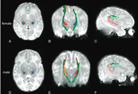
Sex Differences in Principal WM Tracts
WM tracts related to sensorimotor and language functions are shown in Fig 1.
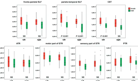
Relative tract volumes were statistically significantly larger in females than in males (Fig 2) bilaterally in the parieto-tem-poral SLF (left,P⬍.001; right,P⬍.001) and in the left CST (P⬍.001). Moreover, trend toward larger tract volumes (On-line Table 1) was found bilaterally in the parieto-temporal SLF (left,P⫽.034; right,P⫽.011).
A trend toward lower MD (P⫽.041) and⬜(P⫽.033) in females was observed in the left parieto-temporal SLF (On-line Table).
Discussion
In this in vivo brain MR imaging study, we investigated sex differences in the TBV and WM tracts with DTI probabilistic
tractography in the language and motor networks in a popu-lation of healthy preterm neonates scanned at term-equivalent age. We found, like other studies in neonates19 and in
adults,14,23that at term-equivalent age healthy preterm male
neonates had larger TBV than females. The original findings of our study were that female neonates had larger relative tract volumes bilaterally in the parieto-temporal SLF and in the left CST, with a trend toward lower MD and⬜in the left parieto-temporal SLF after Bonferroni correction.
Previous studies have shown that the temporal cortex is larger in females than in males. This has been demonstrated in children by using structural imaging,25,26and also in adults
through pathologic studies showing larger planum tempo-rale56and Heschl gyrus,57and a greater attenuation of neu-rons58in females. Given that the parieto-temporal SLF is
sup-posed to transmit auditory information from the superior temporal gyrus to the inferior parietal lobe, we suggest that our results may reflect an early established difference in favor of female neonates in the number or size of axons in these lan-guage-related regions.
The SLF is one of the slowest maturing WM tracts, being not yet myelinated at birth.59,60Lower MD and⬜are proba-bly caused by a decrease in brain water content and an increase in membrane attenuation, and they suggest an advanced pre-myelination stage characterized by proliferation and matura-tion of oligodendrocytes.61,62Therefore, we propose that this
[image:3.594.52.536.45.375.2]microstructural sex difference might be caused by an ad-vanced maturation in the left parieto-temporal SLF in female neonates.
The finding of different tract relative volumes with no sig-nificant difference in diffusion indices is a feature with no straightforward interpretation. In the right parieto-temporal SLF, a larger relative tract volume associated with a trend to-ward larger tract volume in females was found in the absence of difference in diffusion indices: this might possibly reflect macrostructural changes (more axons at the same myelination stage). In the CST, a larger relative tract volume in females was observed together with no significant difference in either tract volume or diffusion indices, suggesting a similar maturation and number of axons in a smaller female brain. In other pub-lished series, differences in tract volumes were not always as-sociated with differences in diffusion indices.33,48,63,64
More-over, we used probabilistic tractography, which does not directly rely on diffusion index values, but on the uncertainty orientation of the distribution function, enabling it to prog-ress across regions with principal direction uncertainty and through regions with crossing fibers. Therefore, in probabilis-tic tractography, volume measurement is not directly linked to diffusion indices.51
Language acquisition and processing have shown sex-re-lated differences in infants as young as 2 years old.8,10,65
Mac-coby and Jacklin66reported that girls outperformed boys
dur-ing preschool and early years in articulation, length of sentences, verbal fluency, grammar, and spelling. In giving the California Verbal Learning Test to children between 5 and 16 years old, girls were found to use more semantic clustering, to recall and recognize more items, and to relate words together
more as a recall aid than did boys.67In addition, language
impairments have been found to occur more frequently in boys than in girls.11,12Because of its implication in language function, we suggest that the sex effect on parieto-temporal SLF relative tract volume and microstructure might explain the more rapid development of language skills hitherto re-ported in females.
Although we could not evidence a significant sex effect on the volume of the CST, we showed that the relative volume of the left CST is larger in females. Interestingly, studies in adults have already shown, after adjusting for the TBV, an increased volume21and gray matter concentration14in the precentral
gyri in females. A relatively larger left CST in females might explain why meta-analyses showed more right-handed fe-males than fe-males in the general population.68However, the relationship between handedness and asymmetry in the adult brain is not established, because some studies have found such relationship69,70but others have not.71
Sex differences in the volume and microstructure of certain WM tracts, as observed in this study, might result from genetic factors as well as from effects of sex steroids on brain develop-ment, both factors being known to affect regional tissue com-position.72-74
Another hypothesis is that our results may have been influ-enced by the effect of prematurity. Indeed, even if the normal-ity of our preterm population was based on robust structural and clinical criteria, as in previous studies,48,52we cannot ex-clude the possibility that the sex related differences observed in the language and motor networks may have been caused by subtle cerebral lesions, because certain studies seem to suggest that preterm males may be more sensitive to brain injuries
[image:4.594.58.531.44.322.2]than females.75,76Therefore, it would be of interest to
investi-gate whether these sex differences are also present in healthy term neonates.
Because the first years of life are perhaps the most dynamic phase of postnatal brain development, with rapid development of a wide range of cognitive and motor functions,77the link between
structural sex differences at term-equivalent age with later func-tional differences should be interpreted with great caution. Lon-gitudinal studies combining cognitive evaluation with structural and functional imaging may provide insights into the structure-function relationship in sex differences.
Another limitation of our study is that the reproducibility of mask placement was not assessed. Nevertheless, mask place-ments were checked by 2 radiologists and in probabilistic trac-tography, by using the approach of normalization, the size of seed and target masks can be ignored.51
Conclusions
In this DTI and probabilistic tractography study on healthy pre-term neonates, we demonstrated that sex differences are present in language and motor-related tracts at term-equivalent age. Fur-ther studies are needed to investigate wheFur-ther these structural differences are related to later sex differences in language skills and handedness or to the effect of prematurity.
Acknowledgments
We are grateful to Doni Tamblyn for assistance in language editing.
Disclosures: Viviane De Maertelaer.Research Support (including provision of equipment or materials):University of Brussels; Vincent Denolin.Consultant:Advice on sequence pa-rameters and data analysis.
References
1. Weissa EM, Kemmlera G, Deisenhammerb EA, et al.Sex differences in cogni-tive functions.Pers Individ Dif2003;35:863–75
2. Kimura D.Sex and Cognition.Cambridge, Massachusetts: MIT Press; 1999 3. Kimura D, Harshman RA.Sex differences in brain organization for verbal and
non-verbal functions.Prog Brain Res1984;61:423– 41
4. Jones CM, Braithwaite V. A., Healy SD.The evolution of sex differences in spatial ability.Behav Neurosci2003;117:403–11
5. Caplan PJ, Crawford M, Hyde JS, et al.Gender Differences in Human Cognition.
New York: Oxford University Press; 1997
6. Halpern DF.Sex Differences in Cognitive Abilities.Hillsdale, New Jersey: L. Erl-baum Associates; 1992
7. Hyde JS, Linn MC.Gender differences in verbal ability: a meta analysis. Psy-chol Bull1988;104:53– 69
8. Bornstein MH, Haynes OM.Vocabulary competence in early childhood: mea-surement, latent construct, and predictive validity.Child Dev1998;69:654 –71 9. Lung FW, Shu BC, Chiang TL, et al.Predictive validity of Bayley scale in lan-guage development of children at 6 –36 months.Pediatr Int2009;51:666 – 69 10. Reilly S, Wake M, Bavin EL, et al.Predicting language at 2 years of age: a
prospective community study.Pediatrics2007;120:1441– 49
11. Law J, Boyle J, Harris F, et al.Prevalence and natural history of primary speech and language delay: findings from a systematic review of the literature.Int J Lang Communi Disord2000;35:165– 88
12. Robinson RJ.Causes and associations of severe and persistent specific speech and language disorders in children.Dev Med Child Neurol1991;33:943– 62 13. Leonard C, Towler S, Welcome S, et al.Size matters: cerebral volume
influ-ences sex differinflu-ences in neuroanatomy.Cereb Cortex2008;18:2920 –31 14. Luders E, Narr KL, Thompson PM, et al.Mapping cortical gray matter in the
young adult brain: effects of gender.Neuroimage2005;26:493–501 15. Peters M.Sex differences in human brain size and the general meaning of
differences in brain size.Can J Psychol1991;45:507–22
16. Giedd JN, Castellanos FX, Rajapakse JC, et al.Sexual dimorphism of the devel-oping human brain. Prog Neuropsychopharmacol Biol Psychiatry 1997; 21:1185–201
17. Reiss AL, Abrams MT, Singer HS, et al.Brain development, gender and IQ in children: a volumetric imaging study.Brain1996;119:1763–74
18. Wilke M, Krageloh-Mann I, Holland SK.Global and local development of gray and white matter volume in normal children and adolescents.Exp Brain Res
2007;178:296 –307
19. Gilmore JH, Lin W, Prastawa MW, et al.Regional gray matter growth, sexual dimorphism, and cerebral asymmetry in the neonatal brain.J Neurosci
2007;27:1255– 60
20. Allen JS, Damasio H, Grabowski TJ, et al.Sexual dimorphism and asymmetries in the gray-white composition of the human cerebrum. Neuroimage
2003;18:880 –94
21. Goldstein JM, Seidman LJ, Horton NJ, et al.Normal sexual dimorphism of the adult human brain assessed by in vivo magnetic resonance imaging.Cereb Cortex2001;11:490 –97
22. Good CD, Johnsrude I, Ashburner J, et al.Cerebral asymmetry and the effects of sex and handedness on brain structure: a voxel-based morphometric anal-ysis of 465 normal adult human brains.Neuroimage2001;14:685–700 23. Gur RC, Turetsky BI, Matsui M, et al.Sex differences in brain gray and white
matter in healthy young adults: correlations with cognitive performance. J Neurosci1999;19:4065–72
24. Nopoulos P, Flaum M, O’Leary D, et al.Sexual dimorphism in the human brain: evaluation of tissue volume, tissue composition and surface anatomy using magnetic resonance imaging.Psychiatry Res2000;98:1–13
25. Sowell ER, Peterson B, Kan E, et al.Sex differences in cortical thickness mapped in 176 healthy individuals between 7 and 87 years of age.Cereb Cortex
2007;17:1550 – 60
26. Sowell ER, Trauner DA, Gamst A, et al.Development of cortical and subcorti-cal brain structures in childhood and adolescence: a structural MRI study.Dev Med Child Neurol2002;44:4 –16
27. Blanton RE, Levitt JG, Peterson JR, et al.Gender differences in the left inferior frontal gyrus in normal children.Neuroimage2004;22:626 –36
28. Hsu JL, Leemans A, Bai CH, et al.Gender differences and age-related white matter changes of the human brain: a diffusion tensor imaging study. Neuro-image2008;39:566 –77
29. De Bellis MD, Keshavan MS, Beers SR, et al.Sex differences in brain maturation during childhood and adolescence.Cereb Cortex2001;11:552–57
30. Smith CD, Chebrolu H, Wekstein D, et al.Age and gender effects on human brain anatomy: a voxel-based morphometric study in healthy elderly. Neuro-biol Aging2007;28:1075– 87
31. Behrens TE, Johansen-Berg H, Woolrich MW, et al.Non-invasive mapping of connections between human thalamus and cortex using diffusion imaging. Nat Neurosci2003;6:750 –57
32. Dubois J, Hertz-Pannier L, Dehaene-Lambertz G, et al.Assessment of the early organization and maturation of infants’ cerebral white matter fiber bundles: a feasibility study using quantitative diffusion tensor imaging and tractogra-phy.Neuroimage2006;30:1121–32
33. Hu¨ppi PS, Dubois J.Diffusion tensor imaging of brain development.Semin Fetal Neonatal Med2006;11:489 –97
34. Hu¨ppi PS, Maier SE, Peled S, et al.Microstructural development of human newborn cerebral white matter assessed in vivo by diffusion tensor magnetic resonance imaging.Pediatr Res1998;44:584 –90
35. Jellison BJ, Field AS, Medow J, et al.Diffusion tensor imaging of cerebral white matter: a pictorial review of physics, fiber tract anatomy, and tumor imaging patterns.AJNR Am J Neuroradiol2004;25:356 – 69
36. Berman JI, Mukherjee P, Partridge S, et al.Quantitative diffusion tensor MRI fiber tractography of sensorimotor white matter development in premature infants.Neuroimage2005;27:862–71
37. Ulug˘ AM, van Zijl PC.Orientation-independent diffusion imaging without tensor diagonalization: anisotropy definitions based on physical attributes of the diffusion ellipsoid.J Magn Reson Imaging1999;9:804 –13
38. Sullivan EV, Adalsteinsson E, Hedehus M, et al.Equivalent disruption of re-gional white matter microstructure in ageing healthy men and women. Neu-roreport2001;12:99 –104
39. Schmithorst VJ, Holland SK, Dardzinski BJ.Developmental differences in white matter architecture between boys and girls. Hum Brain Mapp
2008;29:696 –710
40. Silveri MM, Rohan ML, Pimentel PJ, et al.Sex differences in the relationship between white matter microstructure and impulsivity in adolescents.Magn Reson Imaging2006;24:833– 41
41. Szeszko PR, Vogel J, Ashtari M, et al.Sex differences in frontal lobe white matter microstructure: a DTI study.Neuroreport2003;14:2469 –73 42. Westerhausen R, Kreuder F, Dos Santos Sequeira S, et al.Effects of handedness
and gender on macro- and microstructure of the corpus callosum and its subregions: a combined high-resolution and diffusion tensor MRI study. Cogn Brain Res2004;21:418 –26
43. Deichmann R, Good CD, Josephs O, et al.Optimization of 3-D MP-RAGE sequences for structural brain imaging.Neuroimage2000;12:112–27 44. Woodward LJ, Anderson PJ, Austin NC, et al.Neonatal MRI to predict
neuro-developmental outcomes in preterm infants. N Engl J Med 2006; 17:727–29
45. Smith SM, Jenkinson M, Woolrich MW, et al.Advances in functional and structural MR image analysis and implementation as FSL.Neuroimage
46. Counsell SJ, Dyet LE, Larkman DJ, et al.Thalamo-cortical connectivity in chil-dren born preterm mapped using probabilistic magnetic resonance tractog-raphy.Neuroimage2007;34:896 –904
47. Bassi L, Ricci D, Volzone A, et al.Probabilistic diffusion tractography of the optic radiations and visual function in preterm infants at term-equivalent age.Brain2008;131:573– 82
48. Liu Y, Bale´riaux D, Kavec M, et al.Structural asymmetries in motor and lan-guage networks in a population of healthy preterm neonates at term-equiva-lent age: a diffusion tensor imaging and probabilistic tractography study. Neuroimage2010;51:783– 88
49. Makris N, Kennedy DN, McInerney S, et al.Segmentation of subcomponents within the superior longitudinal fascicle in humans: a quantitative, in vivo, DT-MRI study.Cereb Cortex2005;15:854 – 69
50. Wakana S, Caprihan A, Panzenboeck MM, et al.Reproducibility of quantita-tive tractography methods applied to cerebral white matter.Neuroimage
2007;36:630 – 44
51. Johansen-Berg H, Behrens T. Diffusion MRI. London, United Kingdom: Elsevier; 2009:333–52, 434 –36
52. Aeby A, Liu Y, De Tie`ge X, et al.Maturation of thalamic radiations between 34 and 41 weeks gestation: a combined voxel-based study and probabilistic trac-tography using diffusion tensor imaging. AJNR Am J Neuroradiol
2009;30:1780 – 86
53. Powell HW, Parker GJ, Alexander DC, et al.Hemispheric asymmetries in lan-guage-related pathways: a combined functional MRI and tractography study. Neuroimage2006;32:388 –99
54. Choi CH, Lee JM, Koo BB, et al.Sex differences in the temporal lobe white matter and the corpus callosum: a diffusion tensor tractography study. Neu-roreport2010;21:73–77
55. Campbell MJ, Machin D.Medical Statistics.London, United Kingdom: John Wiley & Sons; 1999:148
56. Harasty J, Double KL, Halliday GM, et al.Language-associated cortical regions are proportionally larger in the female brain.Arch Neurol1997;54:171–76 57. Rademacher J, Morosan P, Schleicher A, et al.Human primary auditory cortex
in women and men.Neuroreport2001;12(8):1561– 65
58. Witelson SF, Glezer II, Kigar DL.Women have greater density of neurons in posterior temporal cortex.J Neurosci1995;15:3418 –28
59. Kinney HC, Brody BA, Kloman AS, et al.Sequence of central nervous system myelination in human infancy: II. Patterns of myelination in autopsied in-fants.J Neuropathol Exp Neurol1988;47:217–34
60. Thompson PM, Giedd JN, Woods RP, et al.Growth patterns in the developing brain detected by using continuum mechanical tensor maps. Nature
2000;404:190 –93
61. Dubois J, Dehaene-Lambertz G, Perrin M, et al.Asynchrony of the early
mat-uration of white matter bundles in healthy infants: quantitative landmarks revealed non-invasively by diffusion tensor imaging.Hum Brain Mapp2008; 29:14 –27
62. Neil JJ, Miller J, Mukherjee P, et al.Diffusion tensor imaging of normal and injured developing human brain: a technical review. NMR Biomed
2002;15:543–52
63. Dubois J, Hertz-Pannier L, Cachia A, et al.Structural asymmetries in the infant language and sensori-motor networks.Cereb Cortex2009;19:414 –23 64. Thompson DK, Inder TE, Faggian N, et al.Characterization of the corpus
callosum in very preterm and full-term infants utilizing MRI.Neuroimage
2011;15:479 –90
65. Hindmarsh GJ, O’Callaghan MJ, Mohay HA, et al.Gender differences in cog-nitive abilities at 2 years in ELBW infants.Early Hum Dev2000;60:115–22 66. Maccoby EE, Jacklin CN.The Psychology of Sex Differences.Stanford, California:
Stanford University Press; 1974
67. Kramer JH, Kaplan E, Delis DC, et al.Developmental sex differences in verbal learning.Neuropsychology1997;11:577– 84
68. Sommer IE, Aleman A, Somers M, et al.Sex differences in handedness, asym-metry of the planum temporale and functional language lateralization.Brain Res2008;1206:76 – 88
69. Herve´ PY, Leonard G, Perron M, et al.Handedness, motor skills and matura-tion of the corticospinal tract in the adolescent brain.Hum Brain Mapp
2009;30:3151– 62
70. Rademacher J, Bu¨rgel U, Geyer S, et al.Variability and asymmetry in the hu-man precentral motor system. A cytoarchitectonic and myeloarchitectonic brain mapping study.Brain2001;124:2232–58
71. Westerhausen R, Huster RJ, Kreuder F, et al.Corticospinal tract asymmetries at the level of the internal capsule: is there an association with handedness? Neu-roimage2007;37:379 – 86
72. Geschwind N, Galaburda AM.Cerebral lateralization. Biological mechanisms, associations, and pathology: III. A hypothesis and a program for research. Arch Neurol1985;42:634 –54
73. Thompson M, Cannon TD, Narr KL, et al.Genetic influences on brain struc-ture.Nat Neurosci2001;4:1253–58
74. Toga AW, Thompson PM.Genetics of brain structure and intelligence.Annu Rev Neurosci2005;28:1–23
75. Lauterbach MD, Raz S, Sander CJ.Neonatal hypoxic risk in preterm birth infants: the influence of sex and severity of respiratory distress on cognitive recovery.Neuropsychology2001;15:411–20
76. Nunez JN, McCarthy MM.Sex differences and hormonal effects in a model of preterm infant brain injury.Ann NY Acad Sci2003;1008:281– 84