ORIGINAL RESEARCH
PATIENT SAFETY
Temporal Bone CT: Improved Image Quality and Potential for
Decreased Radiation Dose Using an Ultra-High-Resolution Scan
Mode with an Iterative Reconstruction Algorithm
S. Leng, F.E. Diehn, J.I. Lane, K.K. Koeller, R.J. Witte, R.E. Carter, and C.H. McCollough
ABSTRACT
BACKGROUND AND PURPOSE: Radiation dose in temporal bone CT imaging can be high due to the requirement of high spatial resolution. In this study, we assessed whether CT imaging of the temporal bone by using an ultra-high-resolution scan mode combined with iterative reconstruction provides higher spatial resolution and lower image noise than a z-axis ultra-high-resolution mode.
MATERIALS AND METHODS: Patients with baseline temporal bone CT scans acquired by using a z-axis ultra-high-resolution protocol and a follow-up scan by using the ultra-high-resolution–iterative reconstruction technique were identified. Images of left and right temporal bones were reconstructed in the axial, coronal, and Poschl planes. Three neuroradiologists assessed the spatial resolution of the following structures: round and oval windows, incudomallear and incudostapedial joints, basal turn spiral lamina, and scutum. The paired z-axis ultra-high-resolution and ultra-high-resolution–iterative reconstruction images were displayed side by side in random order, with readers blinded to the imaging protocol. Image noise was compared in ROIs over the posterior fossa.
RESULTS:We identified 8 patients, yielding 16 sets of temporal bone images (left and right). Three sets were excluded because the patient underwent surgery between the 2 examinations. Spatial resolution was comparable (Poschl) or slightly better (axial and coronal planes) with ultra-high-resolution–iterative reconstruction than with z-axis ultra-high-resolution. A pairedttest indicated that noise was signifi-cantly lower with ultra-high-resolution–iterative reconstruction than with z-axis ultra-high-resolution (P⬍.001), with a mean noise reduction of 37% (range, 18%– 49%).
CONCLUSIONS: The ultra-high-resolution–iterative reconstruction scan mode has similar or slightly better resolution relative to the z-axis ultra-high-resolution mode for CT of the temporal bone but significantly (P⬍.01) lower image noise, which may enable the dose to be reduced by approximately 50%.
ABBREVIATIONS:IR⫽iterative reconstruction; UHR⫽ultra-high-resolution; zUHR⫽z-axis ultra-high-resolution
S
ince the introduction of multidetector techniques, CT has be-come a major diagnostic technique for temporal bone imag-ing because its high spatial resolution is well-suited to the task of visualizing the fine anatomic structures of the middle and inner ear.1-5To improve spatial resolution, different approaches havebeen introduced. One of these is the use of an attenuating comb filter to reduce the detector aperture in both fan and cone angle directions, which is referred to as the z-axis ultra-high-resolution
(zUHR) technique.6This technique, in combination with a flying
focal spot technique, provides nominal image thickness thinner than the detector cell size at the isocenter.6,7
Due to the requirement for high spatial resolution, the ra-diation dose in temporal CT can be high, especially with the zUHR technique because its dose efficiency is reduced as pho-tons passing through the patient are blocked from the detector by the comb filters in both fan and cone angle directions.8A
recent focus of CT imaging has been to reduce patient exposure to ionizing radiation, following the as low as reasonably achievable principle.9-13However, the consequent reduction
in photons can adversely affect image quality and present a great challenge when imaging small, anatomically complex structures embedded in attenuating bone, such as those of the middle and inner ear. Iterative reconstruction (IR) is a prom-ising reconstruction technique that is superior to standard fil-tered back-projection reconstructions and theoretically can be
Received November 3, 2014; accepted after revision February 16, 2015. From the Departments of Radiology (S.L., F.E.D., J.I.L, K.K.K., R.J.W., C.H.M.) and Health Sciences Research, Division of Biomedical Statistics and Informatics (R.E.C.), Mayo Clinic, Rochester, Minnesota.
Paper previously presented at: Annual Meeting of the Radiological Society of North American, December 1– 6, 2013; Chicago, Illinois.
used to improve resolution at standard radiation doses or to maintain current resolution by using a reduced radiation dose.14-17
Recently, a new technique combining a deconvolution tech-nique and an IR algorithm, referred to as ultra-high-resolution (UHR)-IR, has been introduced to improve dose efficiency of the zUHR mode. Phantom studies demonstrated that this technique improved dose efficiency by removing the comb filter along the cone (z) direction.8In this study, we retrospectively reviewed
temporal bone CT examinations in patients who had baseline studies by using the standard zUHR technique and follow-up ex-aminations by using UHR-IR to determine whether UHR-IR pro-vided improved resolution and lower noise than zUHR in the clinical setting, which could enable reductions in dose.
MATERIALS AND METHODS
Patient Enrollment and CT Scans
This retrospective study was approved by our institutional review board and was Health Insurance Portability and Accountability Act– compliant. Patients with temporal bone CT scans acquired by using a zUHR protocol who underwent a follow-up scan by using the UHR-IR technique were identified by searching the electronic medical records. Patients who had not provided autho-rization for research were excluded from this study. Temporal bones in which inner ear surgery was performed between the 2 examinations were also excluded.
Baseline scans were acquired on a 64-section CT scanner (Sen-sation 64; Siemens, Forchheim, Germany) by using the zUHR mode (12⫻0.3 mm collimation), with a tube potential of 120 kV, 400 effective mAs, 1-second rotation time, and 0.8 helical pitch. The automatic exposure control was off, and the volume CT dose index was 88 mGy. Images were reconstructed by using a standard filtered back-projection algorithm with a special kernel designed for the UHR mode (U70). Images were reconstructed with 0.4-mm section thickness at 0.3-mm increments. Both the z-axis and in-plane flying focal spot were used for data acquisition.6,7
The follow-up UHR-IR scans were conducted on a 128-sec-tion CT scanner (Somatom Defini128-sec-tion Flash; Siemens) by using the UHR scan mode (16⫻0.6 mm collimation), with a tube potential of 120 kV, 375 effective mAs, 1-second rotation time, and 0.8 helical pitch. The automatic exposure control was off, and the volume CT dose index was 82 mGy. Images were recon-structed by using an IR algorithm (sinogram-affirmed iterative reconstruction, SAFIRE; Siemens) with a special kernel designed for the UHR mode (V80). The strength of the IR algorithm was set at 3 on a scale of 1 (least noise reduction) to 5 (most noise reduc-tion). The thinnest available image section thickness (0.5 mm) was used, with an increment of 0.3 mm.
For both original and follow-up examinations, images of the left and right temporal bones were reconstructed in the axial, coronal, and Poschl planes, as per our routine clinical protocol.
Assessment of Spatial Resolution
Image quality was independently assessed by 3 fellowship-trained neuroradiologists experienced in temporal bone image interpre-tation, with a focus on differences in image sharpness (spatial resolution) between the 2 techniques. All images were reviewed
on a calibrated monitor used for clinical diagnosis located inside a darkened room, with ambient light⬍10 lux. Baseline and fol-low-up images of the same patient and same side of the head in the 3 planes were displayed side by side in a randomized order, with the readers blinded to reconstruction parameters. The 3 neurora-diologist reviewers assessed the spatial resolution on each of the axial, coronal, and Poschl planes, focusing on the following struc-tures: round window, incudomallear joint, and basal turn spiral lamina (axial plane); oval window and scutum (coronal plane); and the basal turn spiral lamina and incudostapedial joint (Poschl plane). Readers compared the spatial resolution of the displayed image on the left with that displayed on the right, and another investigator (not a reader) determined post hoc which image was zUHR and which was UHR-IR to apply the following grading scale to the UHR-IR images (relative to the zUHR images) for each structure: 1⫽inferior resolution with degraded visualiza-tion, 2⫽slightly inferior resolution without affecting visualiza-tion, 3⫽equivalent, 4⫽ slightly superior resolution without affecting visualization, 5⫽ superior resolution with improved visualization.
Image Noise Measurement
Image noise was measured as the SD of CT numbers inside a circular ROI placed on the axial images. The ROI size was approx-imately 0.4 cm2and was placed over the posterior fossa area, with
locations matched as closely as possible between those of the zUHR and UHR-IR scans.
Statistical Analysis
The Wilcoxon signed rank test was performed to compare the scores of image resolution between UHR-IR and zUHR for the aforementioned individual structures (round window, incu-domallear joint, oval window, incudostapedial joint, spiral lamina in the basal turn, and scutum) and reconstruction planes (axial, coronal, and Poschl). For the comparison of spatial resolution in reconstruction planes, the averaged score of structures in the plane was used. A 2-tailed pairedttest was used to compare image
[image:2.594.302.533.49.189.2]noise between the 2 techniques. With both tests,P⬍.01 was considered a sta-tistically significant difference. The dif-ference in image noise between the 2 techniques was calculated, and dose re-duction was estimated on the basis of the relationship between image noise and radiation dose (ie, radiation dose is in-versely proportional to the square of im-age noise in CT).
RESULTS
Study Sample
We identified 8 patients (2 male and 6 female; age ranges, 16 –75 years of age) who had initial examinations with the zUHR technique and underwent a fol-low-up scan by using the new UHR-IR technique. Images from the left and right side of each patient provided 16 sets of temporal bone CT images. Three of these datasets were excluded because the patient had inner ear surgery be-tween the 2 examinations; this change left 13 sets of images included in the final data analysis. The median time interval between examinations was 12 months (range, 1–34 months). For the initial ex-aminations, the principal indications were the following: hearing loss (n⫽4; 2 conductive and 2 sensorineural), in-flammatory disease (n ⫽ 2), trauma (n⫽1), and skeletal dysplasia (n⫽1). For the follow-up examinations, the in-dications were the following: postopera-tive evaluation (n⫽3; 1 internal audi-tory canal decompression, 1 cochlear implant, and 1 ossicular reconstruc-tion), follow-up or ruling out inflamma-tory disease (n⫽3), further evaluation of bilateral fractures (n⫽1), and fol-low-up of a presumed mastoid heman-gioma (n⫽1).
Spatial Resolution
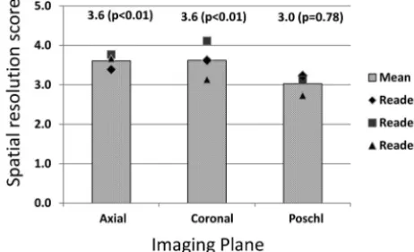
Spatial resolution scores showed that the 3 readers found the UHR-IR images to be of significantly higher quality (P⬍.01, Wil-coxon signed rank test) than the zUHR images in the axial and coronal planes but not in the Poschl plane (Fig 1). Spa-tial resolution scores of individual struc-tures showed that readers found the UHR-IR images to be of significantly higher quality (P ⬍ .01, Wilcoxon signed rank test) for the round window, incudomallear joint, oval window and scutum (Table). UHR images had
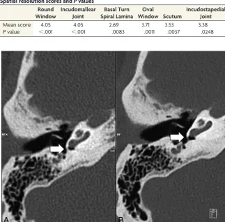
FIG 2.Comparison of spatial resolution of the round window. Representative axial CT images of the round window of the same patient scanned with the zUHR technique (A) and UHR-IR technique (B). The UHR-IR technique produced superior spatial resolution and lower image noise.
FIG 3. Comparison of the spatial resolution of the incudomallear joint. Representative axial images of the incudomallear joint of the same patient scanned with the zUHR technique (A) and UHR-IR technique (B). The UHR-IR technique produced superior spatial resolution and lower image noise.
Spatial resolution scores andPvalues Round
Window
Incudomallear Joint
Basal Turn Spiral Lamina
Oval
Window Scutum
Incudostapedial Joint
Mean score 4.05 4.05 2.69 3.71 3.53 3.38
[image:3.594.54.378.50.371.2] [image:3.594.55.376.432.686.2]higher quality than zUHR images for the incudostapedial joint, but the difference was not statistically significant (Table). For the basal turn spiral lamina, UHR images had a lower quality than zUHR images (Table). The superior sharpness produced by the UHR-IR technique can be seen in representative images at the round window and incudomallear joint (Figs 2 and 3).
Noise and Potential Dose Reduction
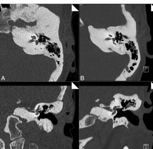
In all cases, images obtained with UHR-IR had lower noise than those obtained with zUHR (all,P⬍.01; pairedttest; Fig 4). In representative images (Fig 5), the UHR-IR and zUHR techniques produced similar sharpness, but the UHR-IR images had much
lower image noise in both the axial and coronal planes. The mean noise reduction by using UHR-IR, relative to zUHR, was 37% (range, 18%– 49%). This translated to a dose reduction potential of 61% (range, 33%–74%).
DISCUSSION
Reduction in the ionizing radiation dose has been a major focus of technology development in diagnostic CT in recent years.9-11,14,18
Temporal bone CT requires a high radiation dose relative to other diagnostic CT examinations; for example, temporal bone CT by using the standard technique at our institution (zUHR) results in a volume CT dose index of 88 mGy, compared with 38 – 69 mGy for a routine head CT examination. Given that radiation reduc-tion can adversely affect image quality, techniques that allow dose reduction without sacrificing image resolution would be exceed-ingly useful in this area of CT imaging.
In this study, radiation dose reduction was achieved by using 2 techniques: 1) the deconvolution technique, and 2) iterative re-construction. The deconvolution technique allows removal of the comb filter along the z-direction compared with zUHR, which substantially improves the dose efficiency by increasing the num-ber of photons detected by the detector. Most important, the spa-tial resolution was preserved, which was substanspa-tially better than that in other scanners without the comb filter techniques.6
Itera-tive reconstruction has the potential to reduce image noise and radiation dose compared with standard filtered back-projection reconstruction algorithms in many studies,14-17including in the
temporal bone.13The amount of dose
reduction highly depends on the specific vendor, scanner platform and imaging task. Dose reduction in this study was a combination of these 2 techniques, similar to that demonstrated in the previous phantom studies by compar-ing filtered back-projection–zUHR, IR-zUHR, and IR-UHR.8
It is important to maintain the spa-tial resolution while reducing image noise and radiation dose, especially in temporal bone CT, in which high spatial resolution is critical. In this study, we found that the UHR-IR scan mode by using a z-deconvolution technique pro-duced resolutions similar to or slightly better than those produced by the zUHR mode, but with significantly (37%) lower image noise. This reduced image noise could potentially allow UHR-IR to be used to reduce the dose by⬎50% on the basis of the relationship between im-age noise and radiation dose in CT. This study was performed by using the IR-UHR technique on the second-genera-tion dual-source scanner (Flash) and compared to the same patients’ prior ex-aminations on older scanners. By com-paring scans from the same patients, we avoided the potential compounding
fac-FIG 4. Lower image noise in images acquired with UHR-IR. Image noise was measured at the posterior fossa in axial images from each of the 13 datasets by using the zUHR and UHR-IR techniques.
[image:4.594.53.287.202.331.2] [image:4.594.57.373.379.687.2]tor caused by patient-to-patient variation. The IR-UHR tech-nique has also been implemented on the third-generation dual-source scanner, and dose reduction was reported by comparing scans of different patients randomly assigned to the first-, sec-ond-, and third-generation dual-source scanners.19Given the
im-proved source and detector technologies, more dose reduction may be achievable by using the third-generation dual-source scanner.
There are several limitations to this study. The first is the small patient cohort, due to the limited number of patients scanned with both zUHR and UHR-IR. However, image noise was lower with UHR-IR for each individual case, and the pairedt test showed this difference to be statistically different. Another limi-tation is that the retrospective methodology did not permit stan-dardization of the time interval between examinations. The third limitation of this study is that the zUHR examinations were per-formed on a different scanner platform from that of the UHR-IR examinations. This was because prior examinations were only available on a different scanner platform and because the new scanner was only recently available. The amount of dose reduc-tion could be potentially less if the zUHR examinareduc-tions were per-formed on the same new scanner platform.
CONCLUSIONS
This study demonstrated that UHR-IR reduces image noise by ⬎30% while providing similar or better spatial resolution than the existing zUHR technique. This may enable a substantial re-duction in radiation dose without a corresponding loss of resolu-tion. This is a considerable achievement for temporal bone CT, which currently requires one of the highest doses in clinical CT imaging. These preliminary findings need to be corroborated with further studies performed with a reduced radiation dose.
ACKNOWLEDGMENTS
The authors thank Thomas Vrieze, Mike Bruesewitz, Sally Rein-hart, and Christine Tipka for helping collect patient data and Kris-tina Nunez and Naomi Ruff for their assistance in manuscript preparation.
Disclosures: Cynthia H. McCollough—UNRELATED:Grants/Grants Pending: Sie-mens,*Comments: Dr McCollough reports grants from Siemens, outside the sub-mitted work. *Money paid to the institution.
REFERENCES
1. Lane JI, Lindell EP, Witte RJ, et al.Middle and inner ear: improved depiction with multiplanar reconstruction of volumetric CT data.
Radiographics2006;26:115–24
2. Lane JI, Witte RJ.Temporal Bone: An Imaging Atlas.Berlin: Springer-Verlag; 2009
3. Purcell DD, Fischbein NJ, Patel A, et al.Two temporal bone com-puted tomography measurements increase recognition of malfor-mations and predict sensorineural hearing loss. Laryngoscope 2006;116:1439 – 46
4. Swartz JD, Loevner LA.Imaging of the Temporal Bone.New York: Thieme Medical; 2008
5. Noble JH, Dawant BM, Warren FM, et al.Automatic identification and 3D rendering of temporal bone anatomy. Otol Neurotol 2009;30:436 – 42
6. Flohr T, Stierstorfer K, Su¨ß C, et al.Novel ultrahigh resolution data acquisition and image reconstruction for multi-detector row CT.
Med Phys2007;34:1712
7. Flohr TG, Stierstorfer K, Ulzheimer S, et al.Image reconstruction and image quality evaluation for a 64-slice CT scanner with z-flying focal spot.Med Phys2005;32:2536 – 47
8. McCollough CH, Leng S, Sunnegardh J, et al.Spatial resolution im-provement and dose reduction potential for inner ear CT imaging using a z-axis deconvolution technique.Med Phys2013;40:061904 9. McCollough C, Chen G, Kalender WA, et al.Achieving routine
sub-mSv CT scanning: report from the summit on management of radi-ation dose in CT.Radiology2012;264:567– 80
10. McCollough CH, Primak AN, Braun N, et al.Strategies for reducing radiation dose in CT (PMC 2743386). Radiol Clin North Am 2009;47:27– 40
11. Yu L, Liu X, Leng S, et al.Radiation dose reduction in CT: techniques and future perspective.Imaging Med2009;1:65– 84
12. Nauer C, Rieke A, Zubler C, et al.Low-dose temporal bone CT in infants and young children: effective dose and image quality.AJNR Am J Neuroradiol2011;32:1375– 80
13. Niu Y, Mehta D, Zhang Z, et al.Radiation dose reduction in tempo-ral bone CT with iterative reconstruction technique.AJNR Am J Neuroradiol2012;33:1020 –26
14. Thibault JB, Sauer KD, Bouman CA, et al.A three-dimensional sta-tistical approach to improved image quality for multislice helical CT.Med Phys2007;34:4526 – 44
15. Winklehner A, Karlo C, Puippe G, et al.Raw data-based iterative reconstruction in body CTA: evaluation of radiation dose saving potential.Eur Radiol2011;21:2521–26
16. Singh S, Kalra MK, Hsieh J, et al.Abdominal CT: comparison of adaptive statistical iterative and filtered back projection recon-struction techniques.Radiology2010;257:373– 83
17. Silva AC, Lawder HJ, Hara A, et al.Innovations in CT dose reduction strategy: application of the adaptive statistical iterative reconstruc-tion algorithm.AJR Am J Roentgenol2010;194:191–99
18. National Council on Radiation Protection and Measurements.
Ionizing radiation exposure of the population of the United States. Bethesda: National Council on Radiation Protection and Measurements; 2009: Report 160