Original Article
Expression of ITGB1 predicts prognosis in colorectal
cancer: a large prospective study based on
tissue microarray
Qi-Zhi Liu1*, Xian-Hua Gao1*, Wen-Jun Chang2*, Hai-Feng Gong1, Chuan-Gang Fu1, Wei Zhang1, Guang-Wen
Cao2
1Department of Colorectal Surgery, Changhai Hospital, Second Military Medical University, Shanghai, China; 2
De-partment of Epidemiology, Second Military Medical University, Shanghai, China. *Equal contributors.
Received August 23, 2015; Accepted September 25, 2015; Epub October 1, 2015; Published October 15, 2015
Abstract: Background: ITGB1 is a heterodimeric cell-surface receptor involved in cell functions such as proliferation, migration, invasion and survival. The aim of this study was to assess ITGB1 expression in colorectal cancer and
cor-relate it with clinicopathological features, as well as to evaluate its potential prognostic significance. Materials and
methods: In this study, we examined the expression of ITGB1 using tissue microarrays containing analyzed speci-mens by immunohistochemistry. ITGB1 expression was further correlated with clinicopathological and prognostic
data. The prognostic significance was assessed using Kaplan-Meier survival estimates and log-rank tests. A multi -variate study with the Cox’s proportional hazard model was used to evaluate the prognostic aspects. Results: ITGB1
expression was present in 88.5% of the analyzed specimens. Significant differences in ITGB1 expression were found
between normal mucosa and carcinomas(P<0.001). High ITGB1 expression was associated with poor prognosis, and it independently correlated with shortened overall survival and disease-free survival in colorectal cancer pa-tients (P<0.001).More so,ITGB1 expression, bowel wall invasion, lymph node metastasis and distant metastasis were independent prognostic factors for overall survival.Additionally, significant differences in ITGB1 expression
were observed in adenomas and tumors from patients with familial adenomatous polyposis compared to normal colon mucosa (P<0.05) Conclusion: The results of this study indicate that ITGB1 overexpression in colorectal tumors is associated with poor prognosis, as well as aggressive clinicopathological features. Therefore, ITGB1 expression could be used as potential prognostic predictor in colorectal cancer patients.
Keywords: Colorectal cancer, prognosis, tissue microarray, ITGB1
Introduction
Colorectal cancer (CRC) is the third most com-mon malignant cancer worldwide [1]. Surgery is the primary method of treatment for CRC, but the high rate of recurrence and/or metastasis after surgery hinders a patient’s recovery, even with postoperative chemotherapy and/or radia-tion therapy [2, 3]. Currently, the gold standard for determining postoperative treatment and prognostication for CRC patients is clinicopath-ological TNM staging [4]. Nevertheless, the TNM stage offers little help in the treatment of an individual patient. In addition, it is not under-stood why only some patients respond to thera-py or have a good clinical outcome [5]. Therefore, understanding treatment failure and
proliferation, migration, invasion and survival [10-12]. ITGB1 functions as mediator of cell and extracellular matrix signaling in cell prolif-eration, apoptosis and survival [13]. It has been reported that high ITGB1 expression is associ-ated with poor prognosis of patients with inva-sive breast [14], lung [15] and pancreatic [16] cancer. However, its value as a prognostic marker, as well as its correlation with clinical significance, has been rarely studied in CRC patients.
Hence, we explored the expression of ITGB1, as well as examined its relationship with clinico-pathological features and prognosis, in CRC patients.
Material and methods
Patient’s selection
A total of 726 patients from Changhai Hospital (Second Military Medical University), including 56 normal patients without a tumor, 51 colon polyps patients, 582 CRC patients (stage I-IV), 16 familial adenomatous polyposis patients and 21 colorectal liver metastases patients, were recruited between 2001 and 2013 and included in this study. Patients were included/ excluded according to the following the criteria: (a) definitive pathological diagnosis of CRC or normal controls; (b) no anticancer treatment prior to surgery; (c) curative resection with the cut surface being free of cancer as confirmed by a pathologist; (d) availability of suitable par-affin-embedded tissues; and (e) complete clini -copathological and follow-up data. The study was approved by the medical ethical boards of the Changhai Hospital (Second Military Medical University) and patients’ informed consent was obtained. Clinicopathological characteristics, including sex, age, tumor stage, bowel wall inva-sion, lymph node metastasis, distant metasta-sis, tumor differentiation, survival, postopera-tive therapy, carcinoembryonic antigen (CEA) and carbohydrate antigen-199 (CA-199), were included in the records. Tumor stage was
deter-metastasis). Overall survival (OS) time was defined as the time from the date of surgery to the confirmed death date for dead patients or from the date of surgery to the date of the last follow-up for surviving patients [17].
Follow-up of patients after surgery
Selected patients were evaluated every 3 months during the first postoperative year, every 6 months during the following year, and afterwards once a year until September 30, 2013, which was the ending date of our study. Follow-up was completed by phone or mail. Dates of death/recurrence, cause of death and postoperative treatment were recorded simul-taneously. During the follow-up period all patients were monitored by CEA, CA-199, colo-noscopy, and chest X-ray for possible recur-rence. If recurrence was suspected, a comput-ed tomography scan (CT) of the abdomen or magnetic resonance imaging (MRI) or positron emission tomography (PET) was performed for further confirmation.
Implementation of tissue microarray and im-munohistochemistry
the arraying was complete, the tissue microar-ray (TMA) blocks were completely sectioned at a 4 μm thickness, yielding more than 80 slides from each block. Thus, five different tissue microarray blocks were constructed, with each block containing a total of 160 specimens. Finally, 800 samples were aligned in five differ -ent tissue microarray blocks. If pati-ents suf-fered from familial adenomatous polyposis (FAP) or presented with liver metastasis before surgery, three specimens (tumor, matching noncancerous mucosa and adenoma or liver metastatic tissue, respectively) were obtained from each patient.
Immunohistochemistry analysis was performed using a mouse anti-human ITGB1 (1:25 dilu-tion) monoclonal antibody (Abcam, ab3167, USA). Briefly, immunohistochemistry of tissue microarrays was carried out as follows: sec-tions were deparaffinized in xylene, rehydrated, and washed in phosphate buffered saline (PBS) for 10, 5 and 10 min, respectively. After appli-cation of endogenous peroxidase for 10 min and antigen retrieval at 98° for 25 min, slides were pre-incubated with blocking serum for 30 min, and then incubated with the ITGB1 mono-clonal antibody at 4° overnight. Subsequently, the sections were thoroughly rinsed with PBS, incubated with secondary antibodies, and treated with horseradish
peroxidase-conjugat-ed streptavidin. The immunohistochemical reaction was visualized with 3,3’-diaminobenzi-dine tetrahydrochloride and counterstained with hematoxylin.
Quantification of ITGB1 expression by immuno -histochemistry
The density of ITGB1-positive staining was eval-uated by two independent pathologists, with-out prior knowledge of the patient characteris-tics, using a Leica DMI3000 microscope (magnification of ×200). Positive ITGB1 staining in each photograph was evaluated in the cyto-sol as follows: the staining intensity was first scored (0 point, negative staining; 1 point, weak staining, light yellow; 2 points, moderate staining, yellowish brown; 3 points, strong staining, brown) and then the proportion of pos-itive cells was scored (0 point, 0-5% pospos-itive cells; 1 point, 5-25% positive cells; 2 points, 26-50% positive cells; 3 points, 51-75% posi-tive cells; 4 points, 76-100% posiposi-tive cells). The final score was obtained by multiplying the scores of staining intensity and percentage of positive cells for each specimen.
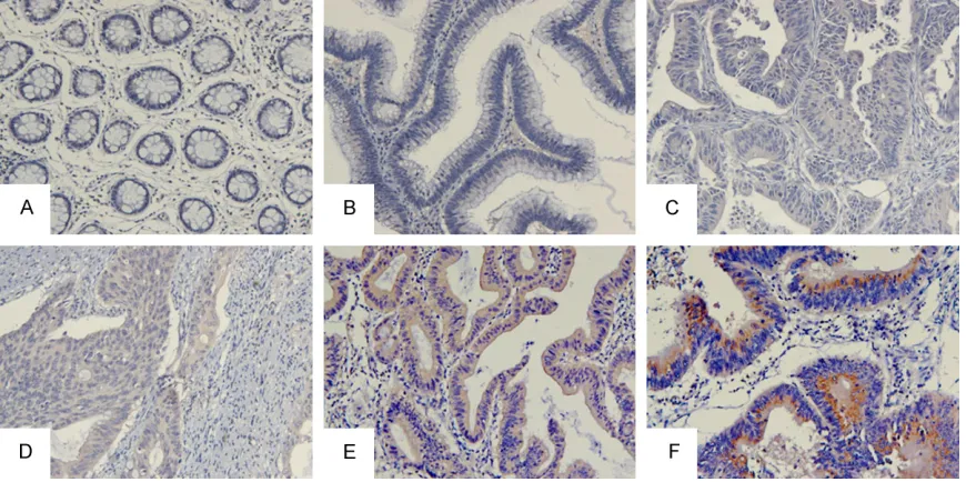
[image:3.612.90.525.72.289.2]For statistical purposes, specimens were divid-ed into four grades according to their overall scores: absent expression (-), 0 points; weak expression (+), a total of 1-4 points; moderate Figure 1. Representative cases of ITGB1 expression in normal mucosa, adenomas and different CRC tumor stages. Positive immunohistochemical staining for ITGB1 was present mainly in the cytoplasm of tumor cells. A. Absence of expression in normal mucosa scored as “-”; B. Absence of expression in adenoma scored as “-”; C. Low intensity
expression (++), 5-8 points; and strong expres-sion (+++), 9-12 points. All samples were ano-nymized and independently scored by two investigators. In case of disagreement, the slides were re-examined until the final consen -sus was reached.
Statistical analysis
The associations of ITGB1 expression with clini-copathological features were tested with the Kruskal-Wallis test. For the analysis of the train -ing set, the survival curves were estimated by the Kaplan-Meier method and compared by with the log-rank test. To determine the inde-pendence of our classifier to clinicopathologi -cal variables in predicting an individual’s risk of survival, we analyzed the validation set using univariate analysis followed by multivariate analysis in a Cox proportional-hazards model for prognostic predictors. All calculations were performed with SPSS statistical package ver-sion 17.0 (SPSS, Chicago, IL). P<0.05 (two-sid-ed) was considered statistically significant. Results
ITGB1 expression in mucosa and carcinoma
tissues of the patients at different stages ITGB1 expression was observed in 610 out of the 689 (88.5%) analyzed specimens including
The Kruskal-Wallis test was used to analyze the relationship between the ITGB1 expression in normal mucosa and adenomas and different tumor stages.Significant differences in ITGB1 expression were observed between carcino-mas of all stages compared to ITGB1 expres-sion in normal mucosa and adenomas (P<0.001). The ITGB1 expression was not sta-tistically significant between adenomas and stage I cancer patients, between stage II and II cancer patients, and between stage III and stage IV cancer patients (P>0.05) (Table 1).
ITGB1 expression in FAP patients
Among patients selected for this study, 16 FAP patients were included to examine the expres-sion of ITGB1 in their tumors, adenomas and matching non-cancerous mucosa. A significant difference in ITGB1 expression was found in tumors and adenomas of FAP patients com-pared to ITGB1 expression in matching normal mucosa (P<0.05), while no difference was found between ITGB1 expression in adenomas compared tumors in these patients (P>0.05) (Table 2; Figure 2).
ITGB1 expression in patients with liver metas -tasis
In addition, ITGB1 expression was examined in the normal mucosa and primary or metastatic Stage IV cancer 8 10 25 12 <0.001 <0.001 <0.001 0.002 0.474
aCompared with the “Normal mucosa” group; bCompared with the “Adenoma” group; cCompared with the “Stage I cancer”
[image:4.612.93.523.84.189.2]group; dCompared with the “Stage II cancer” group; eCompared with the “Stage III cancer” group.
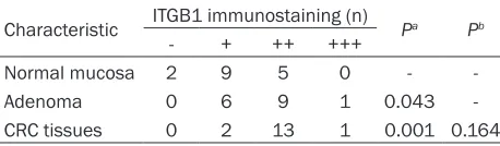
Table 2. Expression of ITGB1 in normal mucosa, ad-enomas and tumors of FAP patients
Characteristic ITGB1 immunostaining (n) Pa Pb
- + ++ +++
Normal mucosa 2 9 5 0 -
-Adenoma 0 6 9 1 0.043
-CRC tissues 0 2 13 1 0.001 0.164
aCompared with the “Normal mucosa” group; bCompared with the
“Adenoma” group.
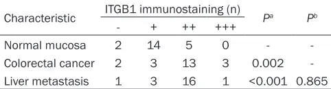
[image:4.612.91.320.261.328.2]tumors of 21 patients with liver metastasis before surgery. Significant differences in ITGB1 expression in colorectal cancer or metastatic liver tumors compared to the normal mucosa were observed (P<0.05), while there was no dif-ference in ITGB1 expression between colorec-tal and metastatic liver tumors (P>0.05) (Table 3; Figure 3).
ITGB1 expression and clinicopathological fea -tures of CRC patients
In order to evaluate the relationship between the ITGB1 expression and tumor biology, clini-copathologic features, including sex, age, bowel wall invasion, lymph node metastasis, distant metastasis, postoperative treatment, tumor differentiation, CEA and CA-199, and ITGB1 expression associations were investigated. Patients with high ITGB1 expression were more likely to exhibit aggressive clinicopathological features, such as lymph node metastasis, dis-tant metastasis, non-postoperative treatment and tumor differentiation (P<0.001, respective-ly). The details are shown in Table 4.
ITGB1 expression and overall and disease-free
survival of CRC patients
Kaplan-Meier analysis showed that patients with high ITGB1 expression (++ and +++) had a
included in the univariate analysis using the Cox proportional hazards model. All of these parameters were significant predictors for OS. To further explore their independent predictive effect for OS, a multivariate Cox proportional-hazards model was performed within the same parameters. It was found that bowel wall inva-sion [hazard ratio (HR), 2.012; 95% confidence interval (CI), 1.065-3.800; P=0.031], lymph node metastasis (HR, 1.929; CI, 1.468-2.534; P<0.001), distant metastasis (HR, 3.648; CI, 2.287-5.820; P<0.001) and ITGB1 expression (HR, 1.537; CI, 1.147-2.059; P=0.004) were independent prognostic factors for OS. However, CEA, CA-199 and postoperative treat-ment were not found to be independent prog-nostic indicators for OS (Table 5).
[image:5.612.88.523.71.179.2]From Table 5, we can find out that bowel wall invasion, lymph node metastasis, distant metastasis, ITGB1 expression, CEA and CA-199 are all risk factors for CRC patients. While, post-operative treatment (HR, 0.752; CI, 0.362-1.561; P=0.445) is a protective factor for CRC patients. Of all patients included, patients who expressed low ITGB1 expression and accepted postoperative treatment, including chemother-apy or/and radiotherchemother-apy showed better progno-sis than that of patients who expressed high expression and without postoperative treat-ment (P<0.001).
Figure 2. ITGB1 expression in normal mucosa, adenomas and tumor tissue from the same FAP patient. A. Normal
mucosa; B. Adenoma tissue; C. Colorectal cancer. Magnification, ×200.
Table 3. Expression of ITGB1 in normal mucosa and tumors of patients with colorectal liver metastasis Characteristic ITGB1 immunostaining (n) Pa Pb
- + ++ +++
Normal mucosa 2 14 5 0 -
-Colorectal cancer 2 3 13 3 0.002 -Liver metastasis 1 3 16 1 <0.001 0.865
aCompared with the “normal mucosa” group; bCompared with the
“colorectal cancer” group.
significantly lower DFS and OS compared to patients with low ITGB1 expression (+ and -) (Figure 4).
Univariate and multivariate analysis of predictive factors of OS in CRC patients
[image:5.612.90.331.256.321.2]Discussion
Integrins are heterodimeric cell-sur-face receptors consisting of α and β subunits, which integrate the extra-cellular matrix with the intraextra-cellular cytoskeleton to mediate cell adhe-sion, survival, differentiation and migration by a wide range of intracel-lular signaling pathways [13, 18, 19]. ITGB1 is the most important member of the integrin family because it facil-itates cell-cell and cell-extracellular matrix interactions to mediate the survival, differentiation, angiogene-sis, andinvasion of cancer cells [10, 20, 21].
ITGB1 also acts as a signal transduc-er in signaling pathways involved in the regulation of survival and prolif-eration through the PI3K/Akt and p130Cas/paxillin/JNK signaling path-ways [22, 23]. In addition, ITGB1 has been reported to mediate the resis-tance to chemotherapy and radiation by enhancing cell survival and inhibi-tion of apoptosis in several human cancers, and thus could be consid-ered as an important therapeutic tar-get for anti-cancer therapy [24-26]. Indeed, inhibition of ITGB1 has been shown to enhance radiotherapy effi -cacy and result in apoptosis in malig-nant breast cancer models [27, 28]. All of these findings suggest that ITGB1 may be of great clinical signifi -cance in -cancer patients.
At present, the potential effects of ITGB1 expression on clinical progno-Figure 3. ITGB1 expression in normal mucosa, metastatic liver tissue and CRC from the same patient with primary
[image:6.612.93.523.71.179.2]metastasis. A. Normal tissue; B. Metastatic liver tissue; C. Colorectal cancer. Magnification, ×200.
Table 4. Relationship between the ITGB1 immunostain-ing and clinicopathological characteristics of patients with colorectal cancer
Characteristic ITGB1 immunostaining (n) p
- + ++ +++
Sex (n) 0.479
Male 23 122 164 21
Female 23 96 114 19
Age (years) 0.348
<60 29 106 139 18
≥ 60 17 112 139 22
Bowel wall invasion 0.534
T1 1 3 5 0
T2 5 35 29 4
T3 38 180 241 35
T4 2 0 3 1
Lymph node metastasis <0.001
N0 29 154 100 21
N1 8 40 120 13
N2 9 24 58 6
Distant metastasis 0.019
M0 38 208 253 28
M1 8 10 25 12
Postoperative treatment <0.001
Yes 32 136 235 33
No 14 82 43 7
Tumor differentiation <0.001
Well 0 6 11 1
Moderate 24 192 246 37
Poor 9 13 15 2
Mucinous adenocarcinoma 13 7 6 0
Serum CEA 0.551
<5 ng/mL 31 135 168 25
≥5 ng/mL 15 83 110 15
Serum CA-199 0.307
<37 U/ml 41 182 229 32
[image:6.612.92.345.268.717.2]sis have been reported in breast cancer [14], ovarian cancer [29] and small-cell lung cancer [30, 31]. However, correlation of ITGB1 expres-sion and clinical prognosis in CRC patients on a large scale in China has been lacking. In the present study, we demonstrated that high ITGB1 expression was accompanied with aggressive clinicopathological features, includ-ing lymph node metastasis, liver metastasis and tumor differentiation, and advanced stag-es of CRC cancer (P<0.05). In addition, increased ITGB1 expression was closely associ-ated with decreased OS and DFS of CRC patients. More importantly, in our study, ITGB1 remained an independent factor associated with OS (HR, 1.537; CI, 1.147-2.059; P=0.004) after multivariate regression analysis. Therefore, based on these findings we can con
[image:7.612.97.518.72.252.2]-of refractory tumors and advanced metastatic disease [32]. More so, a recent study has shown that stimulation of the TLR4/MD2 com -plex by lipopolysaccharide activates PI3K/AKT signaling and promotes downstream ITGB1 function, thereby increasing the adhesiveness and metastatic capacity of CRC cells [33]. The results of our study indicated that ITGB1 expression could be used as a potential bio-marker to study the mechanism of tumor pro-gression, based on the differences in ITGB1 expression in normal mucosa compared to pri-mary liver metastasis tissue from the same patient. In addition, although ITGB1 is dispens-able for the initiation of ErbB2 tumor induction, it plays an important role in the metastatic phase of tumor progression [34]. Therefore, ITGB1 expression may be useful in the evalua-tion of the potential for tumor metastasis. Figure 4. Prognostic significance assessed using Kaplan-Meier survival estimates and log-rank tests stratified by ITGB1. High ITGB1 expression was associated with decreased OS and DFS. A. Kaplan-Meier survival curves showed a significantly decreased OS among patients with high ITGB1 expression (++ and +++) compared with patients with low expression (+ and -). P<0.001, log-rank test; B. Kaplan-Meier survival curves showed a significantly reduced DFS
among patients with high ITGB1 intensity scores compared with patients with low scores. P<0.001, log-rank test.
Table 5. Cox’s multivariate analysis for OS
Characteristic OS
HR 95% CI p
Bowel wall invasion (T1-T2 vs T3-T4) 2.012 1.065-3.800 0.031 Lymph node metastasis (No vs Yes) 1.929 1.468-2.534 <0.001
Distant metastasis (No vs Yes) 3.648 2.287-5.820 <0.001 ITGB1 (Low vs High) 1.537 1.147-2.059 0.004
CEA (<5 vs ≥5 ng/mL) 1.367 0.868-2.152 0.178
CA-199 (<37 vs ≥37 U/mL) 1.482 0.907-2.421 0.116 Postoperative treatment (No vs Treatment) 0.752 0.362-1.562 0.445 Abbreviation: OS: overall survival, HR: hazard ratio, CI: confidence interval.
clude that ITGB1 is an attrac-tive target for therapeutic strategies and a good predic-tor for clinical prognosis of CRC patients.
[image:7.612.94.377.348.476.2]which showed significant differences in ITGB1 expression in normal mucosa, adenomas and tumors of the same FAP patient, ITGB1 expres-sion could be a potential marker from clinical progression from adenoma through early stage carcinoma to advanced stage carcinoma for FAP patients. In addition ITGB1 expression could be considered as a therapeutic target for FAP treatment.
There were some limitations in our study. Although this study was initially based on a large number of samples, many of them were excluded due to lack of information regarding post-operation adjuvant therapy and/or clinico-pathological features.
In summary, using the tissue microarray meth-od, we demonstrated that ITGB1 expression could mediate cancer progression and distin-guish low- and high-risk patients after surgery in CRC. Nevertheless, the definite role of ITGB1, as well as its potential as a clinical marker of CRC, is still far from being unambiguously established. Thus, further studies are needed to fully understand its role in CRC development and progression.
Disclosure of conflict of interest
None.
Address correspondence to: Dr. Wei Zhang,
De-partment of Colorectal Surgery, Changhai Hospital,
Second Military Medical University, Shanghai
200433, People’s Republic of China. E-mail:
[email protected]; Dr. Guang-Wen Cao, Department of Epidemiology, Second Military Medical University, Shanghai 20043, People’s
Republic of China. E-mail: caoguangwen@yahoo. com
References
[1] Ferlay J, Shin HR, Bray F, Forman D, Mathers C and Parkin DM. Estimates of worldwide burden
of cancer in 2008. Globocan 2008. Int J Can-cer 2010; 127: 2893-917.
lon cancer in a national cohort study was
ad-versely affected by TNM stage, lymph node ra
-tio, gender, and old age. Int J Colorectal Dis
2011; 26: 1299-307.
[5] Nagtegaal ID, Gosens MJ, Marijnen CA, Rutten HJ, van de Velde CJ and van Krieken JH. Com -binations of tumor and treatment parameters are more discriminative for prognosis than the
present TNM system in rectal cancer. J Clin On -col 2007; 25: 1647-50.
[6] Bates RC, Bellovin DI, Brown C, Maynard E, Wu B, Kawakatsu H, Sheppard D, Oettgen P and Mercurio AM. Transcriptional activation of inte -grin beta6 during the epithelial-mesenchymal
transition defines a novel prognostic indicator
of aggressive colon carcinoma. J Clin Invest 2005; 115: 339-47.
[7] Lievre A, Bachet JB, Boige V, Cayre A, Le Corre
D, Buc E, Ychou M, Bouche O, Landi B, Louvet C, Andre T, Bibeau F, Diebold MD, Rougier P, Ducreux M, Tomasic G, Emile JF, Penault-Llor
-ca F and Laurent-Puig P. KRAS mutations as an
independent prognostic factor in patients with advanced colorectal cancer treated with cetux-imab. J Clin Oncol 2008; 26: 374-79.
[8] Deschoolmeester V, Boeckx C, Baay M, Weyler J, Wuyts W, Van Marck E, Peeters M, Lardon F and Vermorken JB. KRAS mutation detection
and prognostic potential in sporadic colorectal cancer using high-resolution melting analysis. Br J Cancer 2010; 103: 1627-36.
[9] Walther A, Johnstone E, Swanton C, Midgley R, Tomlinson I and Kerr D. Genetic prognostic and
predictive markers in colorectal cancer. Nat Rev Cancer 2009; 9: 489-99.
[10] Hynes RO. Integrins: bidirectional, allosteric signaling machines. Cell 2002; 110: 673-87. [11] Collier ME and Ettelaie C. Induction of endo
-thelial cell proliferation by recombinant and microparticle-tissue factor involves beta1-inte-grin and extracellular signal regulated kinase activation. Arterioscler Thromb Vasc Biol 2010; 30: 1810-7.
[12] Koivisto L, Heino J, Häkkinen L, Larjava H. Inte -grins in wound healing. Adv Wound Care (New Rochelle) 2014; 3: 762-83.
[14] Yao ES, Zhang H, Chen YY, Lee B, Chew K, Moore D and Park C. Increased β1 integrin is
associated with decreased survival in invasive breast cancer. Cancer Res 2007; 67: 659-64. [15] Oshita F, Kameda Y, Ikehara M, Tanaka G, Ya
-mada K, Nomura I, Noda K, Shotsu A, Fujita A, Arai H, Ito H, Nakayama H and Mitsuda A. In -creased expression of integrin beta1 is a poor prognostic factor in small-cell lung cancer. An-ticancer Res 2002; 22: 1065-70.
[16] Bottger TC, Maschek H, Lobo M, Gottwohl RG,
Brenner W and Junginger T. Prognostic value of
immunohistochemical expression of β-1 integ -rin in pancreatic carcinoma. Oncology 1999; 56: 308-13.
[17] Ding ZB, Shi YH, Zhou J, Shi GM, Ke AW, Qiu SJ, Wang XY, Dai Z, Xu Y and Fan J. Liver-intestine
cadherin predicts microvascular invasion and poor prognosis of hepatitis B virus-positive he-patocellular carcinoma. Cancer 2009; 115: 4753-65.
[18] Askari JA, Buckley PA, Mould AP and Humphries MJ. Linking integrin conformation to function. J
Cell Sci 2009; 122: 165-70.
[19] Malinin NL, Pluskota E and Byzova TV. Integrin
signaling in vascular function. Curr Opin Hema-tol 2012; 19: 206-11.
[20] Schooley AM, Andrews NM, Zhao H and Addi -son CL. beta1 integrin is required for anchor-age-independent growth and invasion of tumor cells in a context dependent manner. Cancer Lett 2012; 316: 157-67.
[21] Nisticò P, Di Modugno F, Spada S and Bissell MJ. β1 and β4 integrins: from breast develop -ment to clinical practice. Breast Cancer Res 2014; 16: 459.
[22] Cordes N, Seidler J, Durzok R, Geinitz H and Brakebusch C. β1-integrin-mediated signaling
essentially contributes to cell survival after ra-diation-induced genotoxic injury. Oncogene 2006; 25: 1378-90.
[23] Watt FM. Role of intrgrins in regulating epider -mal adhesion, growth and differentiation.
EMBO J 2002; 21: 3919-26.
[24] Kurata M, Nakagawa Y, Yamamoto K, Suzuki K and Kitagawa M. Induction of integrin beta1
expression in bone marrow cells after chemo-therapy correlates with the overexpression of lung resistance protein and poor outcome in patients with multiple myeloma. Am J Hematol 2008; 83: 755-7.
[25] Cordes N, Seidler J, Durzok R, Geinitz H and
Brakebusch C. beta1-integrin-mediated signal-ing essentially contributes to cell survival after radiation-induced genotoxic injury. Oncogene 2006; 25: 1378-90.
[26] Nam JM, Chung Y, Hsu HC and Park CC. beta1
integrin targeting to enhance radiation thera-py. Int J Radiat Biol 2009; 85: 923-8.
[27] Park CC, Zhang HJ, Yao ES, Park CJ and Bissell
MJ. β1 integrin inhibition dramatically enhanc
-es radiotherapy efficacy in human breast can -cer xenografts. Can-cer Res 2008; 68: 4398-405.
[28] Park CC, Zhang H and Pallavicini M. β1 integrin
inhibitory antibody induces apoptosis of breast cancer cells, inhibits growth, and distinguishes malignant from normal phenotype in three di-mensional cultures and in vivo. Cancer Res 2006; 66: 1526-35.
[29] Muller-Klingspor V, Hefler L, Obermair A, Kaid
-er A, Breiteneck-er G, Leodolte S and Kohlb-erg -er P. Prognostic value of beta1-integrin
(=CD29) in serous adenocarcinomas of the
ovary. Anticancer Res 2001; 21: 2185-8. [30] Oshita F, Kameda Y and Ikehara M. Increased
expression of integrin β1 is a poor prognostic
factor in small-cell lung cancer. Anticancer Res 2002; 22: 1065-70.
[31] Lawson MH, Cummings NM, Rassl DM, Vowler SL, Wickens M, Howat WJ, Brenton JD, Murphy G and Rintoul RC. Bcl-2 and β1-integrin predict
survival in a tissue microarray of small cell lung cancer. Br J Cancer 2010; 103: 1710-15. [32] Barkan D and Chambers AF. β1-integrin: a po
-tential therapeutic target in the battle against cancer recurrence. Clin Cancer Res 2011; 17: 7219-23.
[33] Hsu RY, Chan CH, Spicer JD, Rousseau MC, Gi -annias B, Rousseau S and Ferri LE. LPS-in-duced TLR4 signaling in human colorectal can-cer cells increases beta1 integrin-mediated cell adhesion and liver metastasis. Cancer Res 2011; 71: 1989-98.
[34] Huck L, Pontier SM, Zuo DM and Muller WJ.
beta1-integrin is dispensable for the induction of ErbB2 mammary tumors but plays a critical role in the metastatic phase of tumor
progres-sion. Proc Natl Acad Sci U S A 2010; 107:
15559-64.
[35] Su LK, Barnes CJ, Yao W, Qi Y, Lynch PM and
Steinbach G. Inactivation of germline mutant APC alleles by attenuated somatic mutations: a molecular genetic mechanism for attenuated familial adenomatous polyposis. Am J Hum Genet 2000; 67: 582-90.
[36] Heyen F, Jagelman DG, Romania A, Zakov ZN, Lavery IC, Fazio VW and McGannon E. Predic -tive value of congenital hypertrophy of the reti-nal pigment epithelium as a clinical marker for
familial adenomatous polyposis. Dis Colon