Original Article
P15, MDM2, NF-κB, and Bcl-2 expression in primary
bone tumor and correlation with tumor
formation and metastasis
Guibin Qian1, Songnan Hao2, Dawei Yang1, Qinggang Meng3
1Department of Orthopaedics, The Forth Affiliated Hospital of Harbin Medical University, Harbin 150000,
Heilongjiang, China; 2Department of Orthopaedics, The Fifth Hospital of Harbin, Harbin 150000, Heilongjiang,
China; 3Department of Orthopaedics, The First Hospital of Harbin, Harbin 150000, Heilongjiang, China
Received May 10, 2015; Accepted June 26, 2015; Epub November 1, 2015; Published November 15, 2015
Abstract: Primary bone tumor is one of the most common malignant tumors in skeletal system. It seriously affected bone movement and development with unclear pathogenesis. In this paper, rabbit VX-2 malignant bone tumor model was applied to explore apoptotic genes P15, MDM2, NF-κB and Bcl-2 correlation with primary bone tumor occurrence and metastasis. 0.3 ml rabbit VX-2 tumor cell suspension (1×10⁶/ml) was injected to the marrow cavity
of the right tibia condyle to establish the rabbit malignant bone tumor model, while equal amount of the saline was injected to the left tibia as control. Real-time PCR was applied to determine P15, MDM2, NF-κB and Bcl-2 expression level. Immunohistochemistry was performed to detect the abovementioned genes expression in lung, stomach, kid-ney and bladder. Compared with control, P15 expression level in the inoculation site surrounding tissues decreased obviously following the inoculate time elongation (P<0.05), while Bcl-2, MDM2 and NF-κB expression significantly increased (P<0.05). Bcl-2 showed significant correlation with MDM2 and NF-κB (P<0.05). At the 2, 4, 6 weeks, Bcl-2, MDM2 and NF-κB in lung, Bcl-2 in kidney, and Bcl-2 and MDM2 in bladder positively expressed (P<0.05), whereas P15 gene exhibited no significant positive expression in these tissues (P>0.05). P15, MDM2, NF-κB, and Bcl-2 genes expression levels can effectively reflect malignant bone tumor growth of rabbit tibia. MDM2, NF-κB and Bcl-2 genes involved in primary bone tumors metastasis directly. It has important clinical significance for early diagnosis and treatment of primary bone tumor.
Keywords: Bone tumor, apoptosis, P15, MDM2, NF-ΚB, Bcl-2
Introduction
Primary bone tumor is a type of malignant tumor derived from bone tissue mainly present-ed as osteosarcoma, fibrosarcoma and chon-drosarcoma. Of which osteosarcoma accounts for about half of all the body malignant tumors. It mostly appeared in the end of long bone in 10-20 years old teenagers and old man over 60 years old [1]. Primary malignant tumor develop-ment has bad influence on skeleton movedevelop-ment. Tumor cell proliferation, growth and migration greatly increase the possibility of distant organ metastasis [2]. P15 gene can affect a variety of tumor cell proliferation and growth through inhibiting cyclin dependent kinase 4/6 to block cell cycle in G1 phase [3]. Murine double min-ute 2 (MDM2) is a new kind of cell apoptotic suppressor gene belongs to the IAP apoptosis
P15, MDM2, NF-κB, and Bcl-2 in bone tumor
In this study, we applied real-time PCR to deter-mine P15, MDM2, NF-κB and Bcl-2 expression level in rabbit malignant bone tumor model, and detect their expression and morphology changes in lung, stomach, kidney and bladder by immunohistochemistry to explore the role of apoptotic gene P15, MDM2, NF-κB and Bcl-2 on primary bone tumors formation and meta- stasis.
Materials and methods
Experimental animals
Twenty New Zealand rabbits at 3 months old and weighted 2.0-3.0 kg were purchased from Harbin medical University laboratory animal center and raised in standard captivity.
Modeling
Under aseptic condition, VX2 tumor cells sus-pension was injected to the rabbit hind leg muscle. The tumor diameter reached 5 cm after two to three weeks [6]. The tumor tissue was extracted in sterile under anesthesia. The tumor was cut into 1 mm3 pieces and filtered in
Hanks fluid through cell strainer. After centri-fuged at 1000 r/min for 5 min, cell suspension was collected. MTT was applied to calculate the tumor cell survival rate. Rabbit VX2 tumor cell line was purchased from Harbin medical University laboratory animal center.
The rabbit was anesthetized by injecting 3% sodium pentobarbital (1.5 ml/kg body weight) through ear vein under aseptic condition. Right tibia was exposed after the rabbit was fixed. Tibial metaphysis was punctured by 18 #
nee-of the right tibia bone marrow cavity surround-ed bone and muscle was checksurround-ed by DF- 312A-2500 mA X-ray machine, while the tibia on the left side was set as control. Cortical bone, periosteum, cartilage, and muscle tissue pathological destruction was recorded. Path- ological characteristics such as bone density decreases, periosteal edema, periosteal new bone and skeletal muscle sarcoma were observed to determine bone tumor prolifera-tion and growth.
Real time-PCR
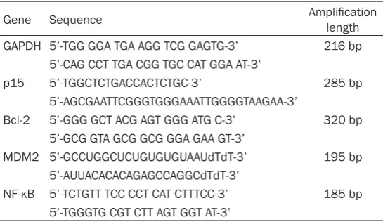
The muscle tissue at 1 cm under the knee was separated after euthanizing the rabbit at 0, 1, 2, 4, 6 weeks after seeding. Total RNA was extracted from the tumor tissue. The cDNA was synthesized using the RNA. Each real-time RT-PCR reaction (in 20 µL) contained 2.5×SYBR Green Real-time PCR Master Mix (TIANGEN), 0.5 µM primers and 0.5 µL of template cDNA. The cycling conditions for real-time RT-PCR reaction consisted of an initial, single cycle of 5 min at 95°C, followed by 40 cycles of 30 s at 95°C, 30 s at 60°C, and 30 s at 70°C. Gene expression levels were quantified relative to the expression of GAPDH using an optimized com-parative Ct (ΔΔCt) value method [7]. All real time PCR reagents were bought from Shanghai Novland Co., LTD. The primers for p15, Bcl-2, MDM2, NF-kB and GADPH were listed in Table 1.
Immunohistochemistry
[image:2.629.99.372.91.249.2]Lung, stomach, kidney, and bladder were extracted and maintained in 10% formalin after euthanizing the rabbit at 0, 1, 2, 4, 6 weeks
Table 1. Primers used for PCR
Gene Sequence Amplification length GAPDH 5’-TGG GGA TGA AGG TCG GAGTG-3’ 216 bp
5’-CAG CCT TGA CGG TGC CAT GGA AT-3’
p15 5’-TGGCTCTGACCACTCTGC-3’ 285 bp 5’-AGCGAATTCGGGTGGGAAATTGGGGTAAGAA-3’
Bcl-2 5’-GGG GCT ACG AGT GGG ATG C-3’ 320 bp 5’-GCG GTA GCG GCG GGA GAA GT-3’
MDM2 5’-GCCUGGCUCUGUGUGUAAUdTdT-3’ 195 bp 5’-AUUACACACAGAGCCAGGCdTdT-3’
NF-κB 5’-TCTGTT TCC CCT CAT CTTTCC-3’ 185 bp 5’-TGGGTG CGT CTT AGT GGT AT-3’
dle through proximal tibial articu-lar surface for about 2.0 cm in depth. The wound was sealed by bone wax after injecting 0.25 ml tumor cells suspension. The left tibia received the same opera-tion and was injected 0.3 ml saline as control.
Bone tumor pathological feature
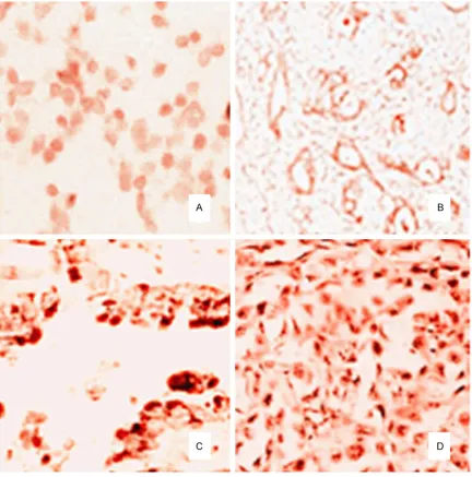
after seeding. Pathological tissue section was prepared for SP immunohistochemical stain-ing. Positive cells appeared brown granules. Improved Shimzu method was applied as 10 visions cells were counted and proliferation index was calculated by the number of positive staining cells percentage [8]. Expression rate <50% was considered negative, while 50% or higher was positive. All steps were in strict accordance with the immunohistochemical kit manual. Morphological changes were observed and apoptosis related gene P15, MDM2, NF-κB, and Bcl-2 expressions were measured to
spec-ulate their relevance with bone tumor metasta-sis. All immunohistochemical kits were provid-ed by Bioleaf co., LTD. SP immunohistochemi-cal reagent were provided by Beijing Xin Xing Tang biological technology co., LTD. DAB solu-tion were provided by Beijing Hapten and Protein Biomedical Institute.
Statistical analysis
[image:3.629.101.529.79.514.2]P15, MDM2, NF-κB, and Bcl-2 in bone tumor
deviation (± SD). Differences between multiple groups were analyzed by one-way ANOVA.
P<0.05 was considered as significant differ-ence. Spearman correlation and chi-square tests were used to evaluate the relationship between immunohistochemical detected gene and cancer distant metastasis.
Results
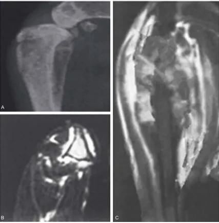
Tibia bone tumor pathology characteristics de-tected by X-ray
No significant tissue pathological change was observed within the first week after inoculation. In the second week, X-ray detection found that local bone destruction began to appear. Tibial periosteal thickening and skeletal muscle sar-comatoid structure were clearly observed at the 4th and 6th week, and skeletal muscle fibrosis
became worse (Figure 1).
P15, NF-κB, MDM2 and Bcl-2 gene expression in bone tumor tissue at different times
P15, NF-κB, MDM2 and Bcl-2 relative expres-sion levels showed no obvious differences in
Its expression increased obviously in the above-mentioned organs at the 2nd, 4th, and 6th week
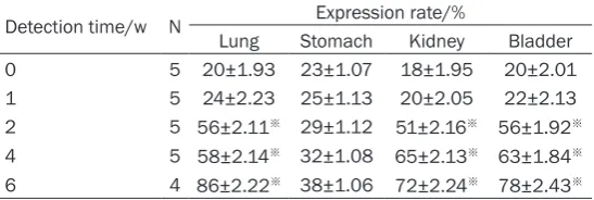
compared with control (P<0.05). However, Bcl-2 expression was negative in stomach and showed no significant difference (P>0.05) (Table 3 and Figure 2).
Immunohistochemical detection of MDM2 ex-pression in different organs
MDM2 expression was negative at the first week after inoculation. It presented positive expression in the lung and bladder at the 2nd,
4th, and 6th week and showed an escalating
trend. MDM2 expression elevated significantly compared with control (P<0.05), while its expression was negative in stomach and kidney with no significant difference (P>0.05) (Table 4).
Immunohistochemical detection of P15 ex-pression in different organs
[image:4.629.98.375.106.237.2]P15 was highly expressed both in the experi-mental groups and control with no significant
Table 2. P15, NF-κB, MDM2 and Bcl-2 gene expression and cor-relation analysis in bone tumor tissue
Index Group Detection time/w
0 1 2 4 6
P15/% Control 0.02 0.04 0.08 0.12 0.10 Experimental group 0.05 0.13 0.85※ 1.13※ 1.32※
NF-κB/% Control 0.07 0.09 0.11 0.10 0.12 Experimental group 0.03 0.22# 0.76※,# 1.14※,# 1.34※,#
MDM2/% Control 0.05 0.04 0.08 0.13 0.12 Experimental group 0.02 0.36# 0.93#,※ 1.23#,※ 1.42#,※
Bcl-2/% Control 0.02 0.06 0.09 0.14 0.11 Experimental group 0.04 0.24# 0.83#,※ 1.22#,※ 1.35#,※
※P<0.05, compared with control; #P<0.05, compared with experimental groups
at different time points.
control at different time points (P>0.05). Following the inocula-tion time prolonged, P15 gene expression level declined obvi-ously in the experimental group, while NF-κB, MDM2 and Bcl-2 levels elevated. They showed sig-nificant differences compared with control at the 2nd, 4th, and 6th
week (P<0.05). Spearman corre-lation revealed that P15 present-ed no significant correlation with the rest three genes (r=0.22, P= 0.22), while NF-κB, MDM2 and Bcl-2 were markedly positively correlated with each other be- tween the expression level of (r=0.85, P=0.03; r=0.72, P=0.04; r=0.91, P=0.02) (Table 2).
Immunohistochemical detection of Bcl-2 expression in different organs
At one week after inoculation, Bcl-2 expression was negative in viscera tissues. It began to express in the lung, kidney, and bladder from the second week and showed an elevation trend.
Table 3. Bcl-2 expression in different organs of rabbit bonetu-mor model
Detection time/w N Expression rate/%
Lung Stomach Kidney Bladder 0 5 20±1.93 23±1.07 18±1.95 20±2.01 1 5 24±2.23 25±1.13 20±2.05 22±2.13 2 5 56±2.11※ 29±1.12 51±2.16※ 56±1.92※
4 5 58±2.14※ 32±1.08 65±2.13※ 63±1.84※
6 4 86±2.22※ 38±1.06 72±2.24※ 78±2.43※
[image:4.629.100.374.308.400.2]difference (P>0.05), suggesting that P15 was not correlated with tumor metastasis (Table 5).
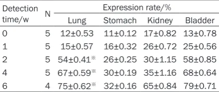
Immunohistochemical detection of NF-κB ex-pression in different organs
NF-κB expressed negatively in all organs at the first week after inoculation. It exhibited positive expression in an escalating trend in the lung from the second week and. NF-κB expression elevated markedly compared with control (P< 0.05), while its expression was negative in stomach, bladder and kidney with no significant difference (P>0.05) (Table 6).
Discussion
[image:5.629.99.532.79.517.2]P15, MDM2, NF-κB, and Bcl-2 in bone tumor
tumor is of great significance to better assess bone tumor progression for timely diagnosis and treatment.
A large number of molecular biological studies found that many apoptotic genes play a critical role in malignant tumor cells proliferation and metastasis [10-13]. Once activated, transcript-ed and amplificattranscript-ed, they can act on cell prolif-eration and differentiation in different periods, thus control tumor cells abnormal growth and metastasis. Studies showed that P15 gene abnormally expressed in a variety of malignant tumors tissues, such as renal cancer, bladder cancer, tongue squamous cell carcinoma, naso-pharyngeal carcinoma, and multiple myeloma, etc. [14]. P15 gene positive expression is
nega-In this paper, we established rabbit tibia malig-nant bone tumor model, and detected the apoptotic gene MDM2, Bcl-2, P15 and NF-κB expression in the tissue surrounding the tibia bone tumor. We further measured gene expres-sion in multiple organs to determine their cor-relation with bone tumor formation and metas-tasis. The results showed that no within no sig-nificant pathological changes in the tissues surrounding the tibia in the first week after inoculation; X-ray examination presented bone destruction from the second week after inocu-lation; tibial periosteal thickening and skeletal muscle sarcomatoid structure can be observed at the 4th week, and skeletal muscle fibrosis
[image:6.629.96.322.106.198.2]became worse. Real time-PCR detection reve- aled that P15 gene expression significantly
Table 4. MDM2 expression in different organs of rabbit bonetumor model
Detection time/w N
Expression rate/%
Lung Stomach Kidney Bladder 0 5 16±0.21 17±0.17 20±1.02 19±0.16 1 5 18±0.23 19±0.14 22±1.12 29±0.18 2 5 54±0.21※ 23±0.12 25±1.05 52±1.12※
4 5 63±0.19※ 29±0.09 31±1.26 59±1.21※
6 4 85±0.22※ 32±0.16 34±1.14 82±1.32※
[image:6.629.100.321.258.351.2]※P<0.05, positive expression compared with control.
Table 5. P15 expression in different organs of rab-bit bone tumormodel
Detection time/w N
Expression rate/%
Lung Stomach Kidney Bladder 0 5 85±0.23 89±0.14 86±1.05 89±0.58 1 5 80±0.33 83±0.18 82±1.12 85±0.16 2 5 74±0.21 76±0.12 72±1.26 77±0.62 4 5 70±0.19 65±0.09 69±1.09 72±0.24 6 4 64±0.22 62±0.16 53±1.32 56±0.82
Table 6. NF-κB expression in different organs of rabbit bone tumor model
Detection time/w N
Expression rate/%
Lung Stomach Kidney Bladder 0 5 12±0.53 11±0.12 17±0.82 13±0.78 1 5 15±0.57 16±0.32 26±0.72 25±0.56 2 5 54±0.41※ 26±0.25 30±1.15 58±0.85
4 5 67±0.59※ 30±0.19 35±1.16 68±0.64
6 4 75±0.62※ 32±0.16 65±0.84 79±0.71
※P<0.05, positive expression compared with control.
[image:6.629.99.320.397.490.2]reduced, while MDM2, Bcl-2 and NF-κB overex-pressed obviously in the tibia compared with control (P<0.05), indicating that these four genes were involved in bone tumor develop-ment and regulated tumor cell proliferation. It may be caused by P15 gene methylation, which reduced its expression rate and made it lose the function of inhibiting tumor cell proliferation and differentiation. MDM2 and Bcl-2 overex-pressed significantly and showed obvious posi-tive correlation (P<0.05), suggesting MDM2 and Bcl-2 may act on cell cycle and apoptosis indirectly through regulating P53 at the same time. NF-κB up-regulation may lead to it trans-fers from the cytoplasm into the nucleus, pro-moting tumor cell proliferation and transcrip-tion. Skeletal muscle sarcomatoid structure began to appear in the lung, kidney, and blad-der tissue from the second week. Among them, MDM2, Bcl-2, and NF-κB expression level ele-vated obviously in the lung (P<0.05); Bcl-2 up-regulated in the kidney (P<0.05); MDM2 and Bcl-2 expression increased in the bladder (P<0.05); while no obvious gene expression changes in stomach (P>0.05). It suggested that P15 gene only participated in the formation process of bone tumor, but not in bone tumor distant metastasis. MDM2, Bcl-2 and NF-κB involved in both bone tumor formation and metastasis to lung. Bcl-2 gene overexpression is associated with bone tumors metastasis to kidney and bladder, while MDM2 gene is par-ticipated in bladder metastasis. More tests about their roles and mechanism in bone tumor formation and metastasis were needed. To sum up, MDM2, Bcl-2, P15 and NF-κB all participate in primary bone tumor formation and development. Of them, P15 gene only relat-ed to bone tumor proliferation but not distant metastasis. MDM2, Bcl-2 and NF-κB were relat-ed to lung metastasis, Bcl-2 was associatrelat-ed with renal metastasis, and MDM2 and Bcl-2 were involved in bladder metastasis. It provides reference bases for bone tumor early diagnosis and treatment after metastasis.
Disclosure of conflict of interest
None.
Address correspondence to: Dr. Qinggang Meng, Department of Orthopaedics, The First Hospital of Harbin, 37 Yiyuan Street, Harbin 150000, Heilong- jiang, China. Tel: 451-84883003; Fax: +86-451-84883003; E-mail: [email protected]
References
[1] Ren BX, Ji Y, Tang JC, Sun DP, Hui X, Yang DQ, Zhu XL. Effect of Tanshinone IIA intrathecal in-jections on pain and spinal inflammation in mice with bone tumors. Genet Mol Res 2015; 14: 2133-2138.
[2] Liu S, Wang X, Zhao Q, Liu S, Zhang H, Shi J, Li N, Lei X, Zhao H, Deng Z, Cao Y, Ning L, Xia G, Duan E. Senescence of human skin-derived precursors regulated by Akt-FOXO3-p27/p15 signaling. Cell Mol Life Sci 2015; 72: 2949-60. [3] Liu L, Bernard D, Wang S. Case Study: Discov-ery of Inhibitors of the MDM2-p53 Protein-Pro-tein Interaction. Methods Mol Biol 2015; 1278: 567-585.
[4] Wang M, Lu X, Dong X, Hao F, Liu Z, Ni G, Chen D. pERK1/2 silencing sensitizes pancreatic cancer BXPC-3 cell to gemcitabine-induced apoptosis via regulating Bax and Bcl-2 expres-sion. World J Surg Oncol 2015; 13: 66. [5] Yao K, Xing HC, Wu B, Li Y, Liao AJ, Yang W, Liu
ZG. Effect of TIEG1 on apoptosis and expres-sion of Bcl-2/Bax and Pten in leukemic cell lines. Genet Mol Res 2015; 14: 1968-1974. [6] Trotter TN, Li M, Pan Q, Peker D, Rowan PD, Li
J, Zhan F, Suva LJ, Javed A, Yang Y. Myeloma cell-derived Runx2 promotes myeloma pro-gression in bone. Blood 2015; 125: 3598-608.
[7] Molaee N, Abtahi H, Ghannadzadeh MJ, Karimi M, Ghaznavi-Rad E. Application of Reverse Transcriptase -PCR (RT-PCR) for rapid detec-tion of viable Escherichia coli in drinking water samples. J Environ Health Sci Eng 2015; 13: 24.
[8] Ma D, Fang Q, Wang P, Gao R, Sun J, Li Y, Hu XY, Wang JS. Downregulation of HO-1 promot-ed apoptosis inducpromot-ed by decitabine via in-creasing p15(INK4B) promoter demethylation in myelodysplastic syndrome. Gene Ther 2015; 22: 287-296.
[9] Piccione M, Salzano E, Vecchio D, Ferrara D, Malacarne M, Pierluigi M, Ferrara I, Corsello G. 4p16.1-p15.31 duplication and 4p terminal deletion in a 3-years old Chinese girl: Array-CGH, genotype-phenotype and neurological characterization. Eur J Paediatr Neurol 2015; 19: 477-83.
[10] Hajiahmadi S, Panjehpour M, Aghaei M, Sha-bani M. Activation of A2b adenosine receptor regulates ovarian cancer cell growth: involve-ment of Bax/Bcl-2 and caspase-3. Biochem Cell Biol 2015: 93: 321-9.
P15, MDM2, NF-κB, and Bcl-2 in bone tumor
clamp sliding during DNA replication and re-pair. Nat Commun 2015; 6: 6439.
[12] Bailly A, Perrin A, Bou Malhab LJ, Pion E, Larance M, Nagala M, Smith P, O’Donohue MF, Gleizes PE, Zomerdijk J, Lamond AI, Xirodimas DP. The NEDD8 inhibitor MLN4924 increases the size of the nucleolus and activates p53 through the ribosomal-Mdm2 pathway. Onco- gene 2015; [Epub ahead of print].
[13] Arismendi M, Giraud M, Ruzehaji N, Dieude P, Koumakis E, Ruiz B, Airo P, Cusi D, Matucci-Cerinic M, Salvi E, Cuomo G, Hachulla E, Diot E, Caramaschi P, Riccieri V, Avouac J, Kayser C, Allanore Y. Identification of NF-κB and PLCL2 as new susceptibility genes and highlights on a potential role of IRF8 through interferon signa-ture modulation in systemic sclerosis. Arthritis Res Ther 2015; 17: 71.
[14] Han X, Wang XL, Li Q, Dong XX, Zhang JS, Yan QC. HIF-1alpha SUMOylation affects the stabil-ity and transcriptional activstabil-ity of HIF-1alpha in human lens epithelial cells. Graefes Arch Clin Exp Ophthalmol 2015; 253: 1279-90. [15] Sayama C, Willsey M, Chintagumpala M,
Bray-ton A, Briceno V, Ryan SL, Luerssen TG, Hwang SW, Jea A. Routine use of recombinant human bone morphogenetic protein-2 in posterior fu-sions of the pediatric spine and incidence of cancer. J Neurosurg Pediatr 2015; 16: 4-13. [16] Sarveswaran S, Ghosh R, Morisetty S, Ghosh J.
MK591, a Second Generation Leukotriene Bio-synthesis Inhibitor, Prevents Invasion and In-duces Apoptosis in the Bone-Invading C4-2B Human Prostate Cancer Cells: Implications for the Treatment of Castration-Resistant, Bone-Metastatic Prostate Cancer. PLoS One 2015; 10: e0122805.
[17] Pan H, Chen J, Shen K, Wang X, Wang P, Fu G, Meng H, Wang Y, Jin B. Mitochondrial Modula-tion by Epigallocatechin 3-Gallate Ameliorates Cisplatin Induced Renal Injury through De-creasing Oxidative/Nitrative Stress, Inflamma-tion and NF-kB in Mice. PLoS One 2015; 10: e0124775.
[18] Zhang LL, Mu GG, Ding QS, Li YX, Shi YB, Dai JF, Yu HG. PTEN represses colon cancer pro-gression through inhibiting paxillin transcrip-tion via PI3K/AKT/NF-kB pathway. J Biol Chem 2015; 290: 15018-29.
[19] Baravalle C, Silvestrini P, Cadoche MC, Becca-ria C, Andreotti CS, Renna MS, Pereyra EA, Ortega HH, Calvinho LF, Dallard BE. Intramam-mary infusion of Panax ginseng extract in bo-vine mammary gland at cessation of milking induces changes in the expression of toll-like receptors, MyD88 and NF-kB during early invo-lution. Res Vet Sci 2015; 100: 52-60.
[20] Hunniger T, Felbinger C, Wessels H, Mast S, Hoffmann A, Schefer A, Martlbauer E, Paschke-Kratzin A, Fischer M. Food Targeting: A Real-Time PCR Assay Targeting 16S rDNA for Direct Quantification of Alicyclobacillus spp. Spores after Aptamer-Based Enrichment. J Agric Food Chem 2015; 63: 4291-4296.
[21] Huang H, Li S, Sun L, Zhou G. Digital Detection of Multiple Minority Mutants and Expression Levels of Multiple Colorectal Cancer-Related Genes Using Digital-PCR Coupled with Bead-Array. PLoS One 2015; 10: e0123420. [22] Chen L, Rousseau RF, Middleton SA, Nichols
GL, Newell DR, Lunec J, Tweddle DA. Pre-clini-cal evaluation of the MDM2-p53 antagonist RG7388 alone and in combination with che-motherapy in neuroblastoma. Oncotarget 2015; 6: 10207-21.
[23] Sen A, Terzioglu G, Atmaca P, Celik G, Ozgun O, Arslan S. Modulatory actions of o-coumaric acid on carcinogen-activating cytochrome P450 isozymes and the potential for drug inter-actions in human hepatocarcinoma cells. Pharm Biol 2015; 53: 1391-8.
[24] Abliz G, Mijit F, Hua L, Abdixkur G, Ablimit T, Amat N, Upur H. Anti-carcinogenic effects of the phenolic-rich extract from abnormal Savda Munziq in association with its cytotoxicity, apoptosis-inducing properties and telomerase activity in human cervical cancer cells (SiHa). BMC Complement Altern Med 2015; 15: 23. [25] Cingeetham A, Vuree S, Jiwatani S, Kagita S,