used for specific immunotherapy
Tong Sun1*, Kang Yin2*, Lu-Yi Wu2, Wen-Jie Jin2, Yang Li2, Bin Sheng2, Yu-Xin Jiang3
1Department of Endocrinology, Xuanwu Hospital of Capital Medical University, 45 Changchun Street, Xicheng
District, Beijing 100053, China; 2Department of Clinical Medicine, School of Clinical Medicine, Wannan Medical
College, 22 Wenchang Road, Wuhu 241002, China; 3Department of Physiology, School of Basic Medicine,
Wannan Medical College, 22 Wenchang Road, Wuhu 241002, China. *Equal contributors.
Received July 21, 2014; Accepted August 23, 2014; Epub August 15, 2014; Published September 1, 2014
Abstract: Immunization with DNA-based constructs has been shown to be against the antigen and the response is skewed in such a way as to ameliorate the symptoms of allergic disease. This approach is particularly useful in the treatment of allergic inflammatory diseases, such as asthma. The major group 1 allergen from house dust mites is one of the triggers of allergic asthma. This study explores whether a chimeric gene R8, derived from the major group 1 allergen of house dust mite species (Dermatophagoides farinae and Dermatophagoides pteronyssinus), can be expressed in Human Embryonic Kidney 293 cells (HEK 293T) and whether such a construct can be used as a DNA vaccine in asthma therapy. The eukaryotic expression vector pcDNA3.1 was used to express the R8 molecule in HEK 293T cells and successful expression of R8 was confirmed using a fluorescence microscope and western blot analysis. The efficacy of R8 as DNA vaccine was also assessed in a mouse asthma model. The in vivo data showed that R8 rectified the TH1/TH2 imbalance typical of allergic inflammation and stimulated the proliferation of regulatory T (Treg) cells. Immunization with the R8 construct also decreased serum allergen-specific IgE production in this mouse asthma model. Our findings suggest that R8 may be a feasible potential DNA vaccine for specific im -munotherapy (SIT) in the treatment of allergic asthma.
Keywords: Dermatophagoides allergen 1 group, chimeric gene, DNA vaccine, asthma
Introduction
Allergic asthma is one of the most common
type I allergic inflammatory diseases and the
prevalence of asthma continues to rise in most industrialized countries [1-3]. The pathogenesis of asthma remains unclear. Asthma is
charac-terized by chronic airway inflammation, goblet cell hyperplasia, and variable airflow obstruc -tion with airway hyperresponsiveness (AHR) [4]. Another well-recognized characteristic of asth-ma is an imbalance in the activation of T helper type 1 (TH1) and type 2 (TH2) [5], which is accompanied by the predominance of type 2 cytokines secreted by the increased number of TH2 cells. Moreover, regulatory T (Treg) cells,
important in regulating the inflammatory
response and maintaining the TH1 and TH2 balance, also play a central role in the patho-genesis of allergic asthma [6].
Specific immunotherapy (SIT) is a conventional
treatment for allergic asthma [7, 8] and involves the repetitive application of an allergen by sub-cutaneous injection or sublingual application. This treatment approach is designed to modify the natural course of the asthmatic response and alleviate the severity of a patient’s asthma symptoms when they are re-exposed to the inhaled allergens [9].
Rodent models of allergic inflammation have
shown that immunization with DNA encoding an allergen protein is a feasible form of SIT [9-12]. DNA vaccination has been shown to induce anti-allergic immune responses through the
recruitment of allergen-specific TH1 cells and
the establishment of a TH1 cytokine milieu,
opti-Construction of recombinant vectors for the DNA vaccine
The allergen gene, ProDer f1 (GenBank No. AB034946.1) served as template for the poly-merase chain reaction (PCR). Target DNA was
amplified using specific primers as follows: 5’-
TAT GGA TCC CGT CCA GCT TCA ATC AAA ACT -3’ (BamH I) and 5’- GGC CTC GAG TCA CAT GAT TAC AAC ATA TGG -3’ (Xho I) for ProDer f1 and R8. The length of the ProDer f1 and R8 genes was 963 bp. To create the reporter vector, enhanced
green fluorescent protein (eGFP) (GenBank No. KJ667592.1) was amplified using PCR (sense
strand primer: 5’- ATG GTG AGC AAG GGC GAG GAG CTG T -3’; antisense strand primer: 5’- TTA CTT GTA CAG CTC GTC CAT GCC G -3’). Pac I and
Not I restriction sites were inserted at the 5’ and 3’ ends, respectively. The length of the
eGFP gene was 720 bp.
The PCR amplifications were carried out in a total reaction volume of 50 μl, containing 10 ng
cDNA, 1× PCR buffer (50 mM KCl, 1.5 mM MgCl2, 10 mM Tris-HCl pH 9.0), 0.2 mM dNTP,
0.4 μM of primer (sense and antisense primers
as described above), and 2.5 units of Taq DNA polymerase (Sangon Biotech, Shanghai, China).
For PCR analysis, denaturation was performed
at 94°C for 4 min, followed by 35 cycles, and carried out as follows: 94°C for 30 s, 58°C for
30 s, and 72°C for 1 min, with a final elongation
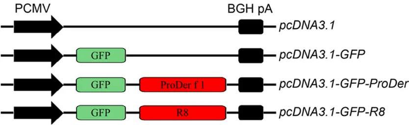
at 72°C for 10 min. Subsequently, the PCR products were cloned into the pcDNA3.1 vector to create recombinant plasmids pcDNA3.1-GFP, pcDNA3.1-GFP-ProDer f1 and pcDNA3.1-GFP-R8 recombinant plasmids (Figure 1).
Nucleotide sequences were confirmed by DNA
sequencing. mize DNA vaccines that are able to restore the
balance to the TH1/TH2 allergic inflammatory
response typical in allergic asthma.
Previously, the R8 gene that encodes the chi-meric R8 allergic protein was discovered from a screen of the major group 1 allergens of house dust mite (ProDer f1 and ProDer p1). The R8 protein is hypoallergenic and hyperimmunogen-ic [14] and exhibits properties similar to that of
the parental allergen ProDer f1. Specifically, R8
has similar IgE immunoreactivity to ProDer f1 and restores the imbalance of TH1/TH2 cells
during an allergic inflammatory response [15].
Although the R8 gene is an attractive therapeu-tic tool for immunotherapy, DNA vaccination using the chimeric R8 gene has not been previ-ously explored. In the present study, the expres-sion of R8 in HEK 293T cells was characterized and the use of the chimeric R8 gene as a DNA
vaccine for the alleviation of allergic inflamma -tion was explored using a mouse asthma model.
Materials and methods
Animals
Female BALB/c mice (6-8 week-old) were pur -chased from the Center for Comparative Medicine, Yangzhou University (License No: SCXK 2007-0001). Mice were housed in the animal facility of Wannan Medical College and were provided with food and water ad libitum under specific-pathogens free conditions. All
[image:2.612.90.507.74.202.2]procedures were approved by the Animal Research Ethics Board of Wannan Medical College.
proteins) of each treatment were separated using 10% SDS-PAGE according to Laemmli’s method [16] in a Mini-PROTEAN 3 system (Bio-Rad), and then transferred onto an Immobilon-P membrane (EMD Millipore, Billerica, MA, USA). Membranes were incubated in blocking buffer (10% bovine serum, 3% BSA, in PBS, pH 7.2) for
2 h at 37°C. GFP-specific rabbit polyclonal anti -serum (1:500, Sangon Biotech) diluted in PBS was added to the blocking buffer and the mem-branes were incubated overnight at 4°C. The following day, the membranes were washed three times with blocking buffer for 10 min each and a horseradish peroxidase-conjugated goat anti-rabbit IgG (1:5000, ZSGB-BIO, Beijing, China) diluted in PBS was added as a second-ary antibody, followed by three washes in
block-ing buffer (10 min each). Antibodies specific to β-actin (Sangon Biotech) served as an internal
control. The target proteins were visualized using enhanced chemiluminescence (ECL) reagent (Sangon Biotech) according to the man-ufacturer’s instructions.
Immunization protocols
Sixty mice were randomly assigned to seven groups (n = 10 for each) as follows: (i) PBS group (PBS): mice treated, sensitized and chal-lenged with PBS; (ii) asthma group (asthma): mice sensitized and challenged with the ProDer f1 allergen; (iii) pcDNA3.1 group (pcDNA3.1): mice treated with the pcDNA3.1 plasmid (100 µg/mouse), sensitized and challenged with the ProDer f1 allergen; (iv) pcDNA3.1-GFP group
(GFP): mice treated with the pcDNA3.1-GFP
plasmid (100 µg/mouse), sensitized and chal-lenged with the ProDer f1 allergen; (v)
pcDNA3.1-GFP-ProDer f1 group (ProDer f1): mice treated with the pcDNA3.1-GFP-ProDer f1
plasmid (100 µg/mouse), sensitized and chal-lenged with the ProDer f1 allergen; (vi)
pcDNA3.1-GFP-R8 group (R8): mice treated
Transfections of recombinant DNA vectors in vitro
HEK 293T cells (Sangon Biotech) were cultured in RPMI-1640 media (Gibco®, Invitrogen, Shanghai, China) supplemented with 10% fetal
bovine serum (FBS) in a 5% CO2 incubator at 37ºC, overnight. Then, cells were transfected with the pcDNA3.1, pcDNA3.1-GFP, pcDNA3.1-GFP-ProDer f1 or pcDNA3.1-GFP-R8 plasmids. The transfection of the plasmids into the cells was performed using DNAfect Transfection Reagent (CWBiotech, Beijing, China) according to the manufacturer’s instructions. HEK 293T
cells grew to 80%-90% confluence and were transfected with 10 μl (about 4 μg) of pcDNA3.1,
pcDNA3.1-GFP, pcDNA3.1-GFP-ProDer f1 or
pcDNA3.1-GFP-R8 plasmid, dissolved in 100 μl RPMI-1640 without FBS. After a 6 h transfec -tion, the culture media was replenished with
fresh RPMI-1640 containing 10% FBS. After 46
h in culture, the cells were observed under a
fluorescent microscope (Ix17, Olympus, Tokyo, Japan) using a 488 nm wavelength filter. Western blot analysis
The HEK 293T cells were treated with trypsin (0.25%) and rinsed with 1× phosphate-buffered saline (PBS, 11.9 mM phosphate, pH 7.4, 137 mM NaCl, 2.7 mM KCl, pH 7.2). The cells were collected and lysed using lysis buffer (50 mM Tris, pH 7.4, 150 mM NaCl, 1% NP-40, 1% SDS)
containing 1 μl protein inhibitor and 1 μl phenyl
-methanesulfonyl fluoride (PMSF), at 4°C for 10
min. The lysates were centrifuged at 12000 ×g for 10 min at 4°C, and the supernatants were collected. The protein concentration of each sample was measured using the Quick Start™ Bradford protein assay (Bio-Rad, Berkeley, CA, USA), with bovine serum albumin (BSA) to
[image:3.612.91.529.81.150.2]gen-erate a concentration curve for quantification. Equal volumes (about 80 μg of total soluble
TGF-β in the BALF according to the manufac -turer’s protocol (R&D Systems, Minneapolis, MN, USA).
Determination of allergen-specific IgE, IgG1
and IgG2a antibodies in the sera
Blood sample from each mouse was collected and centrifuged at 4000 ×g for 30 min. The sera was separated and stored at -80°C for fur-ther analysis. ELISA plate wells were coated overnight with ProDer f1 or R8 (500 ng/well) in coating buffer (15 mM Na2CO3 and 35 mM NaHCO3, pH 9.6) at 4°C. The plates were then washed 5 times with 0.1% Tween-20 in PBS (PBST), and blocked for 1 h at 37°C with 150 µl of PBST supplemented with 0.5% bovine serum albumin (BSA; Sigma-Aldrich® Co. LLC., St. Louis, MO, USA). Sera samples were diluted in PBST to appropriate concentrations and then incubated for 1 h at 37ºC. After washing 5 times
with PBST, the amount of specific IgE, IgG1 and IgG2a was detected after incubation with HRP-goat anti-mouse IgE, IgG1 and IgG2a (1:1000 in PBST; Sigma-Aldrich® Co. LLC.) at 37ºC for 2 h. The plates were then washed 5 times with PBST, and 3, 3’, 5, 5’- Tetramethylbenzidine (TMB) substrate solution (Sigma-Aldrich® Co. LLC.) was added, allowing it to react 20 min at 37ºC. Stop buffer of 50 µl for each well was immediately used to terminate the reaction. A450 nm was measured in a microplate reader (BioTek, Winooski, VT, USA).
Statistical analysis
The statistical data for each group was expressed as the mean ± SD and analyzed using SPSS for Windows, version 16.0 (SPSS, Chicago, IL, USA), to determine the one-factor analysis of variance. The group comparisons
were performed using the least significant dif -ference-t (LSD-t or Thamhane’s T2) method. A
P-value of less than 0.05 was accepted as
significant.
Results
Expression of R8 in vitro in HEK 293T cell line
To determine whether the R8 protein could be successfully expressed in the HEK 293T cell
line, the sequences encoding for GFP and R8
were inserted into the pcDNA3.1 vector and the resulting plasmid (pcDNA3.1-GFP-R8) was
used to transfect HEK 293T cells. Forty-six
with the pcDNA3.1-GFP-R8 plasmid (100 µg/ mouse), sensitized and challenged with the ProDer f1 allergen.
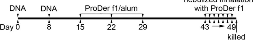
In this study, the asthma model and vaccination protocols were the same as described by
Maecker et al. [17], with some modifications
(Figure 2). On day 0, the mice were injected with 100 µg of plasmid DNA (dissolved in 100 µl of PBS) intramuscularly (i.m.) in the tibialis anterior muscles. On day 8, the mice were once again injected i.m. with the same amount of plasmid DNA. The mice in the PBS and asthma groups were injected with PBS or alum only, respectively. On days 15, 22, and 29, the asth-ma, pcDNA3.1, GFP, ProDer f1, and R8 groups
were injected intraperitoneally (i.p.) with 10 µg of the ProDer f1 allergen. The allergen mix was dissolved in 200 µl of PBS containing 2% (W/V) Al(OH)3 suspension. The PBS group was inject-ed with only PBS. On day 43, the PBS group was challenged with nebulized inhalation of PBS and the remaining groups were challenged with a nebulized inhalation of the ProDer f1 allergen (10 µg/ml). All groups were challenged for 30 min with PBS or the ProDer f1 allergen for next 7 successive days (days 43-49).
Histopathologic evaluation of the pulmonary tissues
Twenty-four hours after the last aerosol
chal-lenge, the lungs were harvested, fixed in 10% formalin overnight, and embedded in paraffin. The pulmonary sections (5 μm) were placed
onto poly-L-lysine-coated slides and stained with hematoxylin and eosin (HE). The histologi-cal changes in the tissues were microscopihistologi-cally assessed according to the scoring protocol described by Underwood [18] which is based
on the extent of eosinophil infiltration, epithelial
damage, and edema in the lung.
Detection of IFN-γ, IL-5, IL-10, and TGF-β cyto
-kines in the BALF
Twenty four hours after the final inhalation chal -lenge, the mice were anesthetized with an i.p. injection of 100 µl of 0.5% pentobarbital sodi-um. The trachea of each mouse was
cannulat-ed and bronchoalveolar lavage fluid (BALF) was
harvested and handled as previously described [15]. Enzyme-linked immunosorbent assay (ELISA) was performed on each sample to
hours later, the cells were collected and further
analyzed under a fluorescent microscope. The pcDNA3.1-GFP, pcDNA3.1-GFP-ProDer f1 and
pcDNA3.1-GFP-R8 plasmids were successfully expressed in the HEK 293T cells (Figure 3). To evaluate the expression of recombination proteins in the transfected cells, we examined
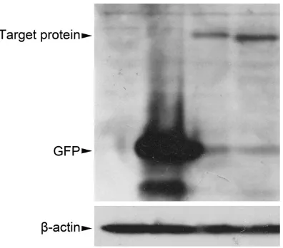
the expression of R8 protein by western blot analysis. Distinctive bands were observed in the lysates from the cells transfected with the
pcDNA3.1-GFP-ProDer f1 and pcDNA3.1-GFP-R8 plasmids (lanes 3 and 4 in Figure 4) and these bands corresponded to the predicted
molecular weight of ProDer f1 and
GFP-R8. These bands were not detected in the cell lysates from the control cells that had been transfected with the pcDNA3.1 or pcDNA3.1-GFP plasmids (lanes 1 and 2 in Figure 4) and
only GFP was found in the cells that had been
transfected with the pcDNA3.1-GFP plasmid (lane 2 in Figure 4).
Pathological changes in the pulmonary tissue
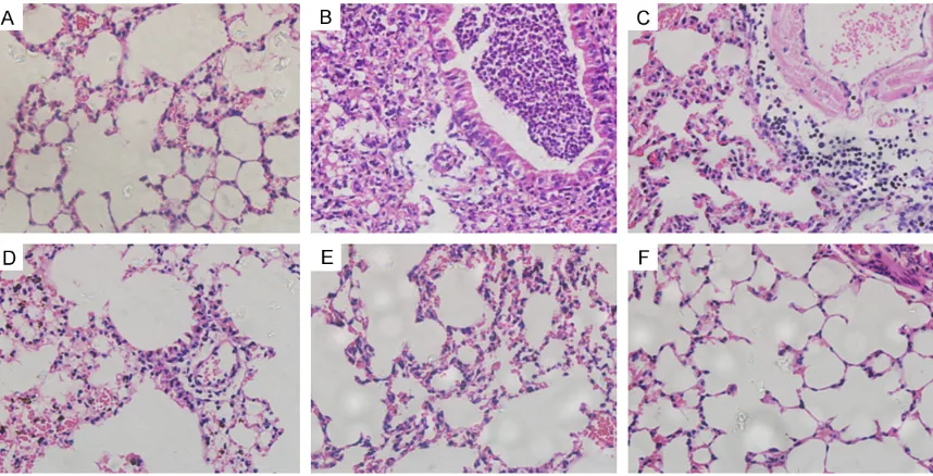
Histological examination of the lung tissues from mice in the asthma group (Figure 5B)
showed intensive peribronchial inflammatory infiltration and submucosal airway wall thicken -ing. The bronchial epithelial cells were overtly hypertrophied. Epithelial shedding and goblet cells hyperplasia were also noted. The same conditions were also present in the pcDNA3.1
and GFP groups (Figure 5C, 5D). Conversely, the negative controls exhibited minimal
peri-bronchial cellular infiltration and airway wall
thickening (Figure 5A). ProDer f1- and R8- immunized mice that had received DNA
[image:5.612.92.522.71.306.2]vac-Figure 3. Detection of R8 gene expression in HEK 293T cells by fluoresent microscopy, 46 hours after treatment with the DNA-liposome complex (×200).
[image:5.612.92.290.356.530.2]cines also showed a general alleviation of these
inflammatory markers (Figure 5E, 5F), com-pared with the asthma group. Moreover, fewer pathological changes were observed in the R8
group compared to the ProDer f1 groups (Figure 5F).
Evaluation of the prophylactic potential of R8 in immunized mouse models
An imbalance in the activation of TH1/TH2 cells is considered an important pathogenic mecha-nism of asthma in most animal asthma models. To determine whether pcDNA3.1-GFP-R8
immunization could alter the TH1/TH2 cytokine
profile characteristic of allergic inflammation in
our mouse model, ELISA was performed to
measure the amount of IFN-γ and IL-5 in the BALF. In the R8 group, IFN-γ production (409.54 ± 26.33 pg/ml) was significantly increased
compared with the PBS group (146.77 ± 43.68 pg/ml, P < 0.001), the asthma group (55.31 ± 19.76 pg/ml, P < 0.001), the pcDNA3.1 group (58.96 ± 14.55 pg/ml, P < 0.001), and the GFP
group (74.27 ± 12.5 pg/ml, P < 0.001), as shown in Figure 6A. Mice immunized with R8 also produced a higher amount of IFN-γ than
mice in the ProDer f1 group (294.13 ± 71.76 pg/ml, P < 0.05). Conversely, animals vaccinat-ed with R8 produced a lower amount of IL-5 (118.65 ± 24.23 pg/ml) than the mice in the asthma group (319.10 ± 57.36 pg/ml, P < 0.001), the pcDNA3.1 group (305.11 ± 48.87
pg/ml, P < 0.001), the GFP group (345.11 ±
59.53 pg/ml, P < 0.001), and the ProDer f1 group (209.58 ± 21.67 pg/ml, P < 0.001), as shown in Figure 6B. The amount of IL-5 in the ProDer f1, and R8 groups were still higher than in the PBS group (48.11 ± 15.76 pg/ml, P < 0.001).
To evaluate whether R8 could stimulate Treg
cell proliferation, the amount of IL-10 and TGF-β in the BALF was also measured by ELISA. The
data showed that IL-10 production (709.47 ±
120.08 pg/ml) in the R8 group was significantly
higher compared with the PBS group (77.18 ± 25.10 pg/ml, P < 0.001), the asthma group (413.42 ± 93.10 pg/ml, P < 0.001), the pcDNA3.1 group (301.18 ± 46.75 pg/ml, P <
0.001), and the GFP group (380.18 ± 59.28
pg/ml, P < 0.001). However, there was no
sta-tistically significant difference between the
ProDer f1 group (744.03 ± 66.14 pg/ml), and the R8 group (P > 0.05) in terms of IL-10 in the
BALF, as shown in Figure 6C. A similar trend
was observed with the amount of TGF-β in the BALF of the immunized mice (Figure 6D).
In addition, there was no statistically significant difference in IFN-γ, IL-5, IL-10, and TGF-β in the BALF of mice in the PBS, pcDNA3.1, and GFP
treatment groups.
[image:6.612.93.522.71.290.2]We examined the levels of specific IgE, IgG1, and IgG2a to determine possible mechanisms
Figure 6. Detecting cytokines in the BALF of DNA vaccinated mice. Cytokines in the BALF from all animals were de -tected by quantitative ELISA. Data are shown as the mean ± SD of ten mice from each group. Note: a, compared with the PBS group, P < 0.001; b, compared with the asthma group, the pcDNA3.1 group, and the GFP group, P < 0.001; c, compared with the PBS group, P < 0.05; d, compared with the asthma group, the pcDNA3.1 group, and the GFP group, P < 0.05; e, compared with the ProDer f1 group, P < 0.05; f, compared with the ProDer f1 group, P < 0.001.
[image:7.612.92.520.520.646.2]promising immunotherapeutic strategies for the treatment of allergic diseases [26, 27], because they have fewer side-effects and are easier to use [28]. DNA vaccination has been shown to induce anti-allergic immune respons-es [29, 30] that are induced through the
recruit-ment of allergen-specific TH1 cells and estab -lishment of a TH1 cytokine milieu, most notably
IFN-γ [13].
In this study, we began by establishing the expression of R8 recombinant protein in HEK 293T cells through plasmid transfection. This proof-of-concept experiment showed stable expression of R8 in HEK 293T cells was possi-ble with this system. Next, we used introduced this transfection system into a mouse asthma
model and measured several allergic inflamma -tory biomarkers in vivo. As expected, the
amount of IFN-γ, a cytokine predominantly secreted by TH1, in the BALF of mice that had
been treated with the R8 gene was higher com-pared to mice that had been treated with a plasmid that carried the gene that encoded the ProDer f1 allergen. The amount of IL-5,
primari-ly produced by TH2 cells, found in the BALF of these mice was significantly lower, when com
-pared to the BALF of mice that had been treat -ed with plasmid carrying the gene encoding the parental allergen. The shift in the TH1/TH2
cytokine profile from predominantly TH2
towards TH1 implies that the chimeric DNA vac-cine R8 can activate TH1 cell differentiation, while downregulating TH2 cell growth. This data
agrees with previous studies. For example,
Jarman et al. reported that a Der p1 DNA
vac-cine could reverse airway inflammation caused
by the Der p1 allergen, as indicated by lower
levels of IL-5 in BALF [31]. Pulsawat et al. also found that a Der p1 DNA vaccine candidate
could induce an allergen-specific TH1 response
[12].
Treg cells, mainly CD4+ CD25+ Treg, play an important regulatory role in the pathogenesis of allergic diseases and asthma [32-34] via the
secretion of IL-10 and TGF-β [33, 35, 36]. Some
studies have reported an association between
the elevated production of IL-10 and TGF-β by
Treg cells and the suppression of allergic responses after DNA immunization [32, 37, 38]. Our results support these observations because they show an increased production of
IL-10 and TGF-β after treatment with the R8
DNA vaccine in our mouse model. behind the effects of DNA-based vaccines. The
resulting serum IgE, IgG1, and IgG2a antibody levels suggested that the IgE level in the R8
group (48.80 ± 5.94 ng/ml) was significantly
lower than that of the PBS group (5.71 ± 2.29 ng/ml, P < 0.001), the asthma group (78.02 ± 8.63 ng/ml, P < 0.001), the pcDNA3.1 group (80.21 ± 9.00 ng/ml, P < 0.001), and the GFP
group (82.70 ± 10.40 ng/ml, P < 0.001; Figure 7A). The IgG1 levels were also significantly dif -ferent between the R8 group (46.67 ± 7.89 ng/ ml) and the PBS group (7.44 ± 4.36 ng/ml, P < 0.001), the asthma group (85.11 ± 11.48 ng/ ml, P < 0.001), the pcDNA3.1 group (93.44 ± 11.37 ng/ml, P < 0.001), and the GFP group
(84.54 ± 10.37 ng/ml, P < 0.001; Figure 7B).
Finally, the level of IgG2a in the R8 group (70.21
± 16.22 ng/ml) was significantly higher com -pared to the PBS group (15.68 ± 7.81 ng/ml, P
< 0.001), the asthma group (35.31 ± 7.31 ng/ ml, P = 0.003), the pcDNA3.1 group (37.48 ± 9.92 ng/ml, P < 0.001) and the GFP group
(34.28 ± 8.98 ng/ml, P < 0.001; Figure 7C).
However, there was no statistically significant difference in the amount of specific IgE, IgG1, and IgG2a in the serum of mice in the ProDer f1 and the R8 groups (P > 0.05; Figure 7A-C).
Discussion
Asthma is one of the most serious health prob-lems worldwide. It has long been recognized that there exists an imbalance in the number of activated TH1 and TH2 cells during the allergic
inflammatory response that characterizes asth -ma [6]. In addition, Treg cells play an important role in the development of asthma [19], and
patients with asthma have a significant
decrease in the number of Treg cells [20]. SIT is the only etiologic treatment available to patients with allergies [21]. Successful SIT is accompanied by a reduction in allergen-specif-ic IgE production, a shift in the balance of TH1/
TH2, as well as induction of allergen-specific
CD4+ CD25+ Treg cells [22]. Several recombi-nant allergens such as Der p1 or Der p2 from D. pteronyssinus, Bet v 1a from birch pollen, and Api m 1 and Api m 2 from bee venom have been introduced as possible immunotherapy aller-gens for use in pre-clinical studies [23-25]. However, traditional SIT has frequent
side-effects, making it difficult to achieve the desired
myeloid DCs drives airway hyperresponsive-ness in asthma-susceptible mice. PLoS One 2008; 3: e3879.
[4] Galli SJ, Tsai M and Piliponsky AM. The devel-opment of allergic inflammation. Nature 2008; 454: 445-454.
[5] Prescott SL. Maternal allergen exposure as a risk factor for childhood asthma. Curr Allergy Asthma Rep 2006; 6: 75-80.
[6] Ying L, Fu Z, Luo J, Zhou C, Chen Y, Wang L and Liu E. Cytotoxic T lymphocyte antigen 4 immu-noglobulin modified dendritic cells attenuate allergic airway inflammation and hyperrespon -siveness by regulating the development of T helper type 1 (Th1)/Th2 and Th2/regulatory T cell subsets in a murine model of asthma. Clin Exp Immunol 2011; 165: 130-139.
[7] Jacobsen L. Preventive aspects of immuno-therapy: prevention for children at risk of de-veloping asthma. Ann Allergy Asthma Immunol 2001; 87: 43-46.
[8] Rolland JM, Douglass J and O’Hehir RE. Allergen immunotherapy: current and new therapeutic strategies. Expert Opin Investig Drugs 2000; 9: 515-527.
[9] Zhang F, Huang G, Hu B, Song Y and Shi Y. Induction of immune tolerance in asthmatic mice by vaccination with DNA encoding an al-lergen-cytotoxic T lymphocyte-associated anti-gen 4 combination. Clin Vaccine Immunol 2011; 18: 807-814.
[10] Zhou B, Ensell M, Zhou Y, Nair U, Glickstein J, Kermany MH, Cai Q, Cai C, Liu W, Deng YP, Kakigi A, Barbieri M, Mora M, Kanangat S and Yoo TJ. Prevention and treatment of DNA vac-cine encoding cockroach allergen Bla g 1 in a mouse model of allergic airway inflammation. Allergy 2012; 67: 166-174.
[11] Wang X, Yang Q, Wang P, Luo L, Chen Z, Liao B and Li G. Derp2-mutant gene vaccine inhibits airway inflammation and up-regulates Toll-like receptor 9 in an allergic asthmatic mouse model. Asian Pac J Allergy Immunol 2010; 28: 287-293.
[12] Pulsawat P, Piboonpocanun S, Sirivichayakul S, Buranapraditkun S, Jacquet A, Shimada M, Okuda K and Ruxrungtham K. Production and immunogenicity of hypoallergenic codon-opti-mized DNA vaccine encoding mature Der p1 allergen. J Investig Allergol Clin Immunol 2010; 20: 582-590.
immune response that is associated with
asth-ma. Furthermore, the treatment of sensitized
and challenged mice with the R8 DNA vaccine resulted in a predominately TH1-biased allergic response. The results agree with our previous study which demonstrated that using R8 aller-gen as a vaccine for SIT can rectify the imbal-ance in TH1/TH2 cells [15].
In conclusion, this study demonstrates that the chimeric gene R8 was more effective than its parental allergens in reversing an allergen-induced Th2-skewed immune response. Likewise, R8 DNA vaccine was also more
effec-tive in preventing allergic pulmonary inflamma
-tion through a decrease in specific IgE levels.
Therefore, it can be considered an extremely promising candidate vaccine for clinical SIT tri-als in patients with house dust mite allergy in future.
Acknowledgements
This work was supported by National Natural
Science Foundation of China (No. 81172790),
National Training Programs of Innovation and Entrepreneurship for College Students (201310368022, 201310368031), Anhui Provincial Training Programs of Innovation and Entrepreneurship for College Students (AH201310368022, AH201310368031).
Disclosure of conflict of interest
None.
Address correspondence to: Dr. Yuxin Jiang, Department of Physiology, School of Basic Medicine, Wannan Medical College, Wuhu 241002, China. E-mail: [email protected]
References
[1] Beasley R. The burden of asthma with specific reference to the United States. J Allergy Clin Immunol 2002; 109: S482-489.
[25] Chen KW, Blatt K, Thomas WR, Swoboda I, Valent P, Valenta R and Vrtala S. Hypoallergenic Der p1/Der p2 combination vaccines for im-munotherapy of house dust mite allergy. J Allergy Clin Immunol 2012; 130: 435-43, e4. [26] Valenta R and Niederberger V. Recombinant
allergens for immunotherapy. J Allergy Clin Immunol 2007; 119: 826-830.
[27] Velickovic TC and Jankov RM. Design and mod-ifications of allergens for improving specific im -munotherapy. Inflamm Allergy Drug Targets 2008; 7: 270-278.
[28] Passalacqua G, Compalati E and Canonica GW. Advances in allergen-specific immuno -therapy. Curr Drug Targets 2009; 10: 1255-1262.
[29] HuangFu T, Lim LH and Chua KY. Efficacy eval -uation of Der p1 DNA vaccine for allergic asth-ma in an experimental mouse model. Vaccine 2006; 24: 4576-4581.
[30] Lombardi V, Singh AK and Akbari O. The role of costimulatory molecules in allergic disease and asthma. Int Arch Allergy Immunol 2010; 151: 179-189.
[31] Jarman ER and Lamb JR. Reversal of estab-lished CD4+ type 2 T helper-mediated allergic airway inflammation and eosinophilia by thera -peutic treatment with DNA vaccines limits pro-gression towards chronic inflammation and remodelling. Immunology 2004; 112: 631-642.
[32] Xia ZW, Zhong WW, Xu LQ, Sun JL, Shen QX, Wang JG, Shao J, Li YZ and Yu SC. Heme oxy-genase-1-mediated CD4+CD25high regulatory T cells suppress allergic airway inflammation. J Immunol 2006; 177: 5936-5945.
[33] Shevach EM. CD4+ CD25+ suppressor T cells: more questions than answers. Nat Rev Immunol 2002; 2: 389-400.
[34] Prescott SL and Dunstan JA. Immune dysregu-lation in allergic respiratory disease: the role of T regulatory cells. Pulm Pharmacol Ther 2005; 18: 217-228.
[35] van Scott MR, Justice JP, Bradfield JF, Enright E, Sigounas A and Sur S. IL-10 reduces Th2 cy-tokine production and eosinophilia but aug-ments airway reactivity in allergic mice. Am J Physiol Lung Cell Mol Physiol 2000; 278: L667-674.
[36] Umetsu DT, Akbari O and Dekruyff RH. Regulatory T cells control the development of allergic disease and asthma. J Allergy Clin Immunol 2003; 112: 480-487; quiz 488. [37] Fonseca DM, Wowk PF, Paula MO, Campos LW,
Gembre AF, Turato WM, Ramos SG, Dias-Baruffi M, Barboza R, Gomes E, Silva CL, Russo M and Bonato VL. Recombinant DNA immuno-therapy ameliorate established airway allergy [13] Klinman DM. Use of CpG
oligodeoxynucleo-tides as immunoprotective agents. Expert Opin Biol Ther 2004; 4: 937-946.
[14] Guo W, Jiang YX and Li CP. [Prokaryotic expres-sion of chimeric gene derived from the group 1 allergens of dust mites and bioactivity identifi -cation]. Zhongguo Ji Sheng Chong Xue Yu Ji Sheng Chong Bing Za Zhi 2012; 30: 274-278. [15] Li C, Jiang Y, Guo W and Liu Z. Production of a
chimeric allergen derived from the major aller-gen group 1 of house dust mite species in Nicotiana benthamiana. Hum Immunol 2013; 74: 531-537.
[16] Laemmli UK. Cleavage of structural proteins during the assembly of the head of bacterio-phage T4. Nature 1970; 227: 680-685. [17] Maecker HT, Hansen G, Walter DM, DeKruyff
RH, Levy S and Umetsu DT. Vaccination with allergen-IL-18 fusion DNA protects against, and reverses established, airway hyperreactiv-ity in a murine asthma model. J Immunol 2001; 166: 959-965.
[18] Underwood S, Foster M, Raeburn D, Bottoms S and Karlsson JA. Time-course of antigen-in-duced airway inflammation in the guinea-pig and its relationship to airway hyperresponsive-ness. Eur Respir J 1995; 8: 2104-2113. [19] Cassis L, Aiello S and Noris M. Natural versus
adaptive regulatory T cells. Contrib Nephrol 2005; 146: 121-131.
[20] Hartl D, Koller B, Mehlhorn AT, Reinhardt D, Nicolai T, Schendel DJ, Griese M and Krauss-Etschmann S. Quantitative and functional im-pairment of pulmonary CD4+CD25hi regulato-ry T cells in pediatric asthma. J Allergy Clin Immunol 2007; 119: 1258-1266.
[21] La Rosa M, Ranno C, Andre C, Carat F, Tosca MA and Canonica GW. Double-blind placebo-controlled evaluation of sublingual-swallow im-munotherapy with standardized Parietaria ju-daica extract in children with allergic rhinocon-junctivitis. J Allergy Clin Immunol 1999; 104: 425-432.
[22] Frew AJ. Allergen immunotherapy. J Allergy Clin Immunol 2010; 125: S306-313.
[23] Hartl A, Hochreiter R, Stepanoska T, Ferreira F and Thalhamer J. Characterization of the pro-tective and therapeutic efficiency of a DNA vaccine encoding the major birch pollen aller-gen Bet v 1a. Allergy 2004; 59: 65-73. [24] Kussebi F, Karamloo F, Rhyner C,