Case Report
Multifocal canalicular adenoma of the
minor labial salivary glands
María Elena Samar1, Rodolfo Esteban Avila2, Ismael Bernardo Fonseca2, William Anderson3, Gabriel M Fonseca1,4, Mario Cantín4,5,6
1Faculty of Dentistry, National University of Cordoba, Cordoba, Argentina; 2Faculty of Medical Sciences, National
University of Cordoba, Cordoba, Argentina; 3Department of Pathology, New Cross Hospital, Wolverhampton, United
Kingdom; 4CIMA Research Group, Faculty of Dentistry, Universidad de La Frontera, Temuco, Chile; 5Doctoral Program
in Morphological Sciences, CEMyQ, Faculty of Medicine, Universidad de La Frontera, Temuco, Chile; 6Center of
Research in Biomedical Sciences, Universidad Autónoma de Chile, Temuco, Chile
Received September 5, 2014; Accepted October 31, 2014; Epub October 15, 2014; Published November 1, 2014
Abstract:Canalicular adenoma (CA) is an uncommon benign neoplasia of salivary glands which is clinically difficult to recognise. Despite having an excellent prognosis, the histological diagnosis and clinical management of this entity can be troublesome. While the main differential diagnosis to consider is basal cell adenoma (BCA), similar histological patterns and multifocality have been observed in adenoid cystic carcinoma (ACC) and polymorphous low-grade adenocarcinoma (PLGA), both locally-aggressive malignancies which require radically different treatment to CA. An emphasis has been placed on the value of immunohistochemistry in avoiding diagnostic and surgical er-rors. CA is positive for AE1/AE3, CD117 and S-100 protein, and negative for p63, α-SMA, Ki 67 and vimentin. Here we discuss the case of a 61-year-old female with CA in her right upper lip, showing multifocal growth histologically. The differential diagnosis with other adenomas is discussed in addition to the role of immunohistochemical studies that can confirm the clinical and surgical findings.
Keywords: Lip, minor salivary glands, benign neoplasia, canalicular adenoma
Introduction
Canalicular adenoma (CA) is a benign neoplasia of salivary glands, which although representing less than 1% of salivary gland tumours, is the second or third most common benign tumour
of minor salivary glands [1, 2]. Historically con -sidered a variant of basal cell adenoma (BCA),
it wasn’t until 1991 that the World Health Organisation (WHO) recognised this entity as
being different to other salivary gland adeno-mas, due to its distinct clinical, morphological, and immunohistochemical characteristics [3- 5]. After this the older term “monomorphic ade-noma”, used to differentiate this and other neo-plasms from pleomorphic adenoma fell into disuse [6].
Peak incidence of CA is in the seventh decade and shows a female predominance (1.8:1) [5, 7]. It occurs almost exclusively in the oral cavi-ty, principally involving the minor salivary glands
in the mucosa of the upper lip in around 80% of cases [5]. Some authors have reported rare cases in the palate (10%), parotid, oesophagus, and mandible [3, 5, 8-10].
Clinically, CA usually presents as a well-demar-cated, occasionally blue-tinged nodule measur-ing between 0.5-2 cm, which is otherwise as- ymptomatic. Prognosis is excellent, and recur-rence is extremely rare even if just locally excised [5].
The histological appearance of CA is uniform, with cells distributed in solid structures, trabec-ulae, tubules, and cribriform or membranous patterns, therefore presenting a diagnostic
challenge [7, 11]. There may be a fibrous cap -sule, and the presence of multiple adjacent foci
is a relatively frequent finding microscopically
[5]. Here we present an unusual case of CA
Case report
A 61-year-old female with a history of smoking presented with a 12-month history of a slow-growing nodule in the mucosa of her right upper lip. Examination of the oral cavity revealed a tender, solitary nodular lesion with a violaceous overlying mucosa without ulceration or bleed-ing. An informed consent was signed. Once the tumour was removed under local anaesthesia, macroscopic examination showed a brown, 2.3×1.6×1 cm lesion with an irregular surface. Areas of vascular congestion were seen on sec-tioning. The tissue was stained with
haematox-ylin and eosin (H & E), periodic acid Schiff (PAS),
and toluidine blue at pH 3.8 and immunohisto -chemical studies were done with AE1/AE3,
CD117, S-100 protein, vimentin, α-SMA, p63 and Ki 67.
Histopathological examination revealed a mu-cosal surface lined with stratified squamous
epithelium. Deep to this were mixed labial sali-vary glands, predominantly mucous acini, which were partially surrounded by skeletal muscle
and frequent nerve fibres. The main lesion was
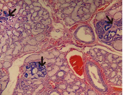
within the glandular tissue and was surrounded by smaller islands with similar histological char-acteristics (Figure 1). These lesions were com-posed of canalicular structures with a central lumen and were branching and anastomosing (Figure 2A). These foci were covered by one or two layers of cuboidal to columnar epithelial cells with vesicular nuclei and inconspicuous nucleoli. No cytological pleomorphism or
mitot-ic figures were observed.
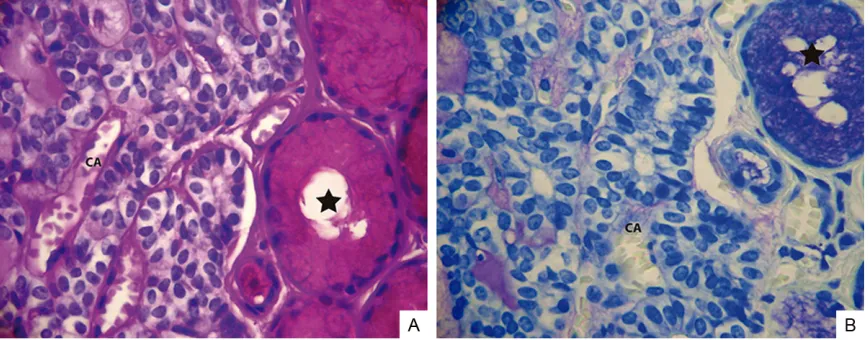
The tumour cells were arranged in a loose, oth-erwise paucicellular stroma with a prominent vascular pattern (Figure 2B). The stroma was PAS-positive and metachromatic with toluidine blue (Figure 3A and 3B). Secretory intralobular ducts were dilated.
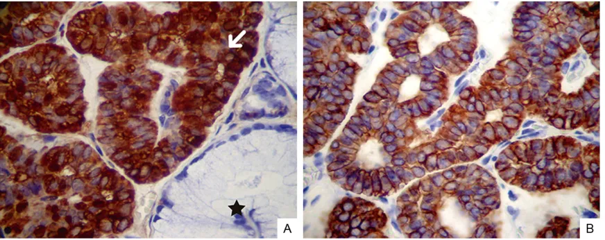
The tumour cells were positive for AE1/AE3, S-100 and CD117 (Figure 4A and 4B). Ex- pression of p63 (a selective marker of
myoepi-thelial and basal cells in stratified epithelia) was negative, as was α-SMA, vimentin, and the proliferation marker Ki 67. Given these micro
-scopic and immunohistochemical findings, a
di-agnosis of multifocal canalicular adenoma was made. At the time of writing, there has been no recurrence of the lesion.
Discussion
Salivary gland tumours are relatively uncom-mon in comparison with other tumours. Al- though they represent less than 2% of all plasias and 2-6.5% of all head and neck neo-plasias, these tumours are of particular inter-est, given their varied histological and clinical characteristics [12-16].
Despite originating from the same progenitor cells, tumours of the minor salivary glands (approximately 14-22% of all salivary gland tu- mours) have a tendency to develop in particular sites, and thereby exhibit different biological behaviour. Although the majority of tumours in- volve the palate (due to its numerous glands), in the case of CA, it is most commonly located in the upper lip (80% of cases) [5, 7, 12, 17]. The most common presentation of epithelial tumours of the minor salivary glands is as a
simple nodule. However, CA affecting the upper
lip may occasionally present multifocally [6]. Rousseau et al. [18] have proposed that
[image:2.612.90.289.72.226.2]multi-focal variants be classified into three catego -ries according to their growth pattern (clinical, microscopic and recurrent). These authors be- lieve that recurrent tumours most likely repre-sent growth of microscopic foci, which persist-ed following resection of larger, clinically-evi-dent tumours. In this case, although the multi-focal tumour was completely removed by exci-sion biopsy, conferring an excellent prognosis [5], we recognise that patients should receive long-term follow-up (up to 11 years) since multi-focal lesions have higher rates of local recur-rence, albeit without malignant transformation [2, 6].
Although the clinical details in this report cor-respond to those predicted by the literature as most frequent for CA (sex, age, and location), this tumour has been described as a rare enti-ty, relatively unknown to practising dental sur-geons, and its histological diagnosis and
clini-cal management can therefore be difficult [19]. Matsuzaka et al. [20] reported that from 18,093 oral biopsies carried out between 1996 and 2004 in Tokyo Dental College, only one
case of CA was identified. In addition, the clini
-cal characteristics of CA, its firm or slightly fluc -tuant consistency and occasionally blue-tinged mucosa may imitate a mucocele, a lesion which is much more common and (in contrast to CA) usually develops in the lower lip [18, 21].
Other differential diagnoses include benign and malignant neoplasias of glandular tissue, sialolithiasis with secondary sialadenitis, vas-cular anomalies and even lipomas [19]. It has been stressed that CA must be clearly differen-tiated from polymorphous low-grade adenocar-cinoma (PLGA) and adenoid cystic caradenocar-cinoma (ACC), since it’s similar histological features may lead to an overdiagnosis with excessive surgery and radiotherapy [2].
In agreement with Sivolella et al. [19], the
[image:3.612.91.526.71.243.2]histo-logical features identified in our case are typi -cal of CA; importantly however, these features have also been observed in some adenocarci-nomas, meaning that immunohistochemical
Figure 2. Multifocal canalicular adenoma of the upper lip. A. Interconnecting and branching canal-like structures (Ar-row). H/E. Original magnification ×200; B. Branching and interconnecting tubules lined in a nearly acellular stroma. H/E. Original magnification ×400.
[image:3.612.90.524.304.474.2]studies are necessary in order to confirm the
diagnosis. In our case, the immunological
pro-file of CA was confirmed by positivity for
pan-cytokeratin AE1/AE3, CD117, and S-100 pro-tein. This indicated a probable origin in the luminal epithelial cells of secretory ducts or the intercalated duct cells [3, 5, 11, 22]. The lack of
p63 and α-SMA reactivity excluded myoepithe -lial origin [22, 23], and negativity for vimentin precluded a diagnosis of PLGA according to the majority of reports [6, 24]. Negativity for Ki 67
was compatible with a benign neoplasia, as some authors have reported [2, 19].
CA is included in the group of salivary gland
adenomas by the WHO, and its most important
differential diagnoses are ACC and basal cell adenoma (BCA). In addition, its multifocal and cribriform patterns should not be interpreted as evidence of malignancy. BCA is a rare benign epithelial neoplasia characterised by a predom-inance of basaloid cells, without the chondro-myxoid component of pleomorphic adenomas [5]. Although the major salivary glands repre-sent its most frequent site (parotid: 75%, sub-mandibular gland: 5%), some reports suggest that when rarely seen in the minor salivary glands, it has a predilection for the upper lip [5, 15, 25]. Immunohistochemistry has now estab-lished that CA develops from luminal ductal cells whereas BCA is originated in the salivary gland parenchyma [6]. BCA is positive for p63,
α-SMA, cytokeratins and EMA, supporting its
ductal and myoepithelial differentiation, while CA shows pure luminal ductal cell
differentia-tion [11, 25]. Importantly, hybrid forms of CA
and BCA have also been reported by WHO.
Interestingly, Furuse et al. [24] report that al- though the differential diagnosis of CA most commonly includes BCA, distinguishing these
two entities has minimal therapeutic signifi -cance, when compared with differentiating PL- GA, a lesion of greater clinical importance. This malignant entity is locally aggressive and often has a histological pattern identical to CA, yet it requires a very different surgical approach. PLGA is the second most frequent malignant tumour of the minor salivary glands, and al- though its preferred location is the palate (60%), there are reports of cases involving the upper lip [7]. PLGA shares clinical similarities with CA, given its mean age of presentation (59 years) and female predominance (2:1). These underscore the need for reliable methods when considering the differential diagnosis [7, 11, 15].
PLGA develops from intercalated duct cells and microscopically it shows a varied architecture, uniform cells and nuclei, frequent perineural invasion and a low metastatic potential. It is non-encapsulated, always invades adjacent tis-sues, and within the same tumour there may be different patterns of growth not always appreci-ated in an incisional biopsy [24]; the predomi-nance of the canalicular form can erroneously be diagnosed as CA. Matsuzaka et al. [20] point out that where CA is resected in a fragmented way or has a cystic structure, it may be misin-terpreted as PLGA. A diagnosis of PLGA is
[image:4.612.90.526.71.244.2]firmed by immunohistochemistry showing posi
-tivity for p63, α-SMA, and vimentin. Furuse et al. [24] claim that in isolated cases CA may overlap with areas of BCA. This would justify the presence of occasional small foci of vimentin-positive cells, clearing this great diagnostic
dif-ficulty. The same authors clarify that PLGA is always negative for CK13 while CA may express
it, and that S-100, a test normally requested by pathologists to identify salivary gland tumours, is not that useful in differentiating CA from PLGA, given that both would be positive.
Despite no critical differences in management, the characteristics of PLGA should to be com-pared with the cribriform pattern of ACC. Al- though ACC is a malignant glandular neoplasia which shows a more aggressive behaviour (us- ually with perineural invasion) and frequently involves the sinonasal cavity, its involvement of minor salivary glands and appearance in the upper lip (more common in the lower) have been reported [5, 7]. It is distinguished from CA by its cribriform architecture with characteristic
microcystic spaces filled with a mucoid sub -stance and a poorly-vascularised stroma. The neoplastic cells are epithelial and myoepithelial in nature, clearly differentiated by immunohis-tochemical stains [11].
Other diagnostic dilemmas have been reported regarding this tumour. It has been reported that CA may be confused with papillary cystadeno-carcinoma [26], given its tendency to show a large single cyst or multiple small cysts, and its consequent papillary projections. These
how-ever, usually locally infiltrate the glandular
pa-renchyma and surrounding connective tissue. The presence of fragmented cysts with haem-orrhagic foci and granulation tissue is a
com-mon finding [5, 20].
The importance of an adequate differential diagnosis of nodular lesions in the oral mucosa has been emphasised; we agree with Stra- mandinoli-Zanicotti et al. [6]. In that although clinical details, aspiration biopsies and routine histopathology may provide valuable
informa-tion, the definitive diagnosis of CA should only
be established through immunohistochemical studies. In this way, the clinical and surgical
findings may be confirmed and potential diag -nostic errors avoided.
Disclosure of conflict of interest None.
Address correspondence to: Dr. Mario Cantín, CIMA Research Group, Faculty of Dentistry, Universidad de La Frontera, Avenue Manuel Montt 112, Temuco, Chile. Tel: 056-45-2325574; E-mail: mario.cantin@ ufrontera.cl
References
[1] Siqueira CS, Fernandes KS, Vivas AP, Pinto Ddos S Jr, de Sousa SC. Clinical and histologi-cal features of multifohistologi-cal canalicular adeno-mas of the upper lip. Braz Dent J 2013; 24: 542-546.
[2] Tyralik D, Dzierwa-Gawron A, Ryś J. Canalicular adenoma of the upper lip. Metachronous (mul-tifocal) canalicular adenoma of the upper lip: a case report of an unusual finding. Pol J Pathol 2013; 64: 71-74.
[3] Huebner TA, Almubarak H, Drachenberg CB, Papadimitriou JC. Canalicular adenoma--sear- ch for the cell of origin: ultrastructural and im-munohistochemical analysis of 7 cases and review of the literature. Ultrastruct Pathol 2014; 38: 74-82.
[4] Smullin SE, Fielding AF, Susarla SM, Pringle G, Eichstaedt R. Canalicular adenoma of the pal-ate: case report and literature review. Oral Surg Oral Med Oral Pathol Oral Radiol Endod 2004; 98: 32-36.
[5] World Health Organization: Classification of Tumours. Pathology and Genetics of Head and Neck Tumours. In: Barnes L, Everson JW, Re- ichart P, editors. Lyon: IARC Press; 2005. [6] Stramandinoli-Zanicotti RT, Cesa TS, Giustina
JD, Bahr JA, Schussel JL, Sassi LM. Canalicular adenoma of the minor salivary gland in the up-per lip: case report. J Oral Diag 2012; 1:4-6. [7] Yih WY, Kratochvil FJ, Stewart JC. Intraoral mi
-nor salivary gland neoplasms: review of 213 cases. J Oral Maxillofac Surg 2005; 63: 805-810.
[8] Dayisoylu EH, Pampu AA, Mungan S, Taskesen F. Intra-mandibular canalicular adenoma: re-port of a rare case. J Pak Med Assoc 2012; 62: 1239-1241.
[9] Grimm EE, Rulyak SJ, Sekijima JH, Yeh MM. Canalicular adenoma arising in the esopha-gus. Arch Pathol Lab Med 2007; 131: 1595-1597.
[10] Maamouri F, Bellil K, Bellil S, Chelly I, Mekni A, Kchir N, Haouet S, Zitouna M. Canalicular ad -enoma of buccal mucosa. Pathologica 2007; 99: 69-70.
[11] Ellis GL, Auclair PL. Tumors of the salivary glands: AFIP Atlas of tumor pathology, series 4. Washington: Armed Forces Institute of Pa-thology (AFIP); 2008.
salivary gland tumors. Dent Res J (Isfahan) 2012; 9: S9-S19.
[13] Avila RE, Samar ME, Ferraris L, Ferraris RV, Fonseca I, Corball A, Asís OG, Olmedo L. Non-Hyalinizing Clear Cell Carcinoma of the Parotid: Report of Two Cases with Different Grade of Differentiation. Int J Morphol 2013; 31: 1056-1061.
[14] Laishram RS, Kumar KA, Pukhrambam GD, Laishram S, Debnath K. Pattern of salivary gla-nd tumors in Manipur, Igla-ndia: A 10 year study. South Asian J Cancer 2013; 2: 250-253. [15] Samar ME, Avila RE. Tumores epiteliales de
glándulas salivales. Saarbrücken: Editorial Ac- adémica Española; 2013.
[16] Sunil S, Gopakumar D. Pleomorphic Adenoma: A Case Report and Review of Literature. Int J Odontostomat 2013; 7: 171-174.
[17] Pereira MC, Pereira AA, Hanemann JA. Immu-nohistochemical profile of canalicular adeno -ma of the upper lip: a case report. Med Oral Patol Oral Cir Bucal 2007; 12: E1-3.
[18] Rousseau A, Mock D, Dover DG, Jordan RC. Multiple canalicular adenomas: a case report and review of the literature. Oral Surg Oral Med Oral Pathol Oral Radiol Endod 1999; 87: 346-350.
[19] Sivolella S, Valente M, De Biagi M, Mazzoleni S, Stellini E. Canalicular adenoma immunopro-file: a case report. Gerodontology. 2013; [Epub ahead of print].
[20] Matsuzaka K, Murakami S, Shimono M, Inoue T. Canalicular adenoma arising in the upper lip: review of the pathological findings. Bull Tokyo Dent Coll 2004; 45: 229-233.
[21] Chen YK, Lin CC, Lai S, Chen CH, Wang WC, Lin YR, Hsue SS, Lin LM. Pleomorphic adenoma with extensive necrosis: report of two cases. Oral Dis 2004; 10: 54-59.
[22] Oliveira-Santos C, Freitas-Faria P, Damante JH, Consolaro A. Asymptomatic nodules of the up-per lip: report of a canalicular adenoma with immunoprofile presentation. Gerodontology 2012; 29: e1121-e1124.
[23] Edwards PC, Bhuiya T, Kelsch RD. Assessment of p63 expression in the salivary gland neo-plasms adenoid cystic carcinoma, polymor-phous low-grade adenocarcinoma, and basal cell and canalicular adenomas. Oral Surg Oral Med Oral Pathol Oral Radiol Endod 2004; 97: 613-619.
[24] Furuse C, Tucci R, Machado de Sousa SO, Ro- darte Carvalho Y, Cavalcanti de Araújo V. Co-mparative immunoprofile of polymorphous low-grade adenocarcinoma and canalicular adenoma. Ann Diagn Pathol 2003; 7: 278-280.
[25] Samar ME, Avila RE, Fonseca I, Ferraris RV, Ra-bino M. Adenoma de células basales de glán-dulas salivales, variedades sólido y tubular: estudio histopatológico e inmunohistoquímico. Patología (Rev Latinoamer Mx) 2008; 46: 276-283.