Temuco, Chile; Center for Biomedical Research, Universidad Autónoma de Chile, Chile; Dental School, Federal University of Ponta Grossa, Brazil; 5Univeersidad de Tarapacá, Chile; 6Piracicaba Dental School, State University of Campinas, Piracicaba, Brazil
Received June 18, 2013; Accepted July 7, 2013; Epub July 15, 2013; Published August 1, 2013
Abstract: Surgical procedures involving the rehabilitation of the maxillofacial region frequently require bone grafts; the aim of this research was to evaluate the interface between recipient and graft with cortical or cancellous con-tact. 6 adult beagle dogs with 15 kg weight were included in the study. Under general anesthesia, an 8 mm diameter block was obtained from parietal bone of each animal and was put on the frontal bone with a 12 mm 1.5 screws. Was used the lag screw technique from better contact between the recipient and graft. 3-week and 6-week eutha-nized period were chosen for histometric evaluation. Hematoxylin-eosin was used in a histologic routine technique and histomorphometry was realized with IMAGEJ software. T test was used for data analyses with p<0.05 for sta-tistical significance. The result show some differences in descriptive histology but non stasta-tistical differences in the interface between cortical or cancellous bone at 3 or 6 week; as natural, after 6 week of surgery, bone integration was better and statistically superior to 3-week analyses. We conclude that integration of cortical or cancellous bone can be usefully without differences.
Keywords: Bone defects, bone grafting, animal study
Introduction
Surgical procedures involving the rehabilitation of the maxillofacial region frequently require bone grafts [1]. This graft incorporation hap-pens dynamically through reabsorption and apposition of the new bone tissue [2], and it
can be influenced by factors that are inherent
to the patient or external factors [3-5].
One factor that can interfere with the incorpo-ration process is the graft’s structural charac-teristic [6] that can be cancellous, cortical or a mixture of them [7]. Medullary grafts are typi-cally revascularized faster involving the entire region, whereas cortical grafts are slower and incomplete [7].
Thus, the bone incorporation is made harder because of the fewer blood vessels in the graft region. This reduces the number of precursor osteoblast cells [8]. As a result, the cortical
graft incorporation process is slower than the cancellous one, and at the end of the process there are still regions that did not undergo any bone tissue formation and they show remaining islands of grafted material [9]. Despite that,
several studies show the efficacy for both grafts
in bone reconstruction before implant insertion [10-12].
Since that cortical and cancellous bone has morphological and chemical differences [13], the object of this study is to assess autogenous graft block incorporation through a histometric analysis when grafts are of the cortical-medul-lary or cortical-cortical contact.
Material and method
Preoperative
Were selected 6 adult beagles dog (15 kg approximately) for the study. Thirty minutes before the procedure the animals received intramuscular benzathine benzylpenicillin (0.1 ml/kg) and dexamethasone (0.5 mg/kg). Before the surgical procedures, the animals were sedated with the anesthetic inductor ket-amine chlorohydrate (0.15 mL/kg) and under-went general anesthesia receiving a 3% pento-barbital sodium (30 mg/kg) intravenous.
Surgical procedure
The bone graft was obtained from de parietal bone. With a trephine of 8 mm diameter was realized the bone cut until the dura mater which preserved with a carefully dissection. With a hand piece (10.000 rpm), two cortical-cancel-lous grafts (from each side of parietal bone) measuring 5 mm high (2 mm cortical and 3 mm cancellous bone) and 8 mm diameter were removed. Bone fragments that were removed
were fixed on the frontal bone region without
gap between the graft and recipient area, using 12 mm (1.5 mm system) metallic screws with a compression (lag screw) technique. Recipient site was not decorticalized (Figure 1).
Blocks fixtures were randomly; one of them was fixed with the cortical region to recipient con
-tact (Group I) and another one was fixed with
the cancellous region to the recipient contact (Group II). After bone graft’s stabilization and
fixation, periosteum and temporal muscles
were approximated with sutures by using
absorbable stitch (polyglactin 910). Superficial
planes were sutured with monofilament 4-0
nylon stitch.
Euthanasia
Animals were randomly divided into two groups corresponding to the two euthanasia periods:
the first group consisted of 3 animals which
were euthanized three weeks after surgical pro-cedure; the second group consisted of 3 ani-mals which were euthanized six weeks after surgical procedure, which corresponds to 1 and 2 months in humans, i.e., (primary and second-ary repair according to Turner et al. [14]). Euthanasia took place with a 19.1% potassium chlorideintravenous overdose until cardiorespi-ratory arrest. Following this stage, access to the animal’s skull was created and the grafted region exposed.
[image:2.612.325.523.72.383.2]Bone blocks were obtained by cross and coro-nal sectioning of the bone with a 702 tapered
[image:2.612.90.288.72.201.2]Figure 1. Intraoperatory image of graft position being one with cortical-bone-contact recipient and another with cancellous-bone-contact recipient. All graft was fixed with one compression 12 mm screws.
drill in high-speed turbine under constant saline solution irrigation with a 5 mm to 10 mm safety margin for the previously operated areas.
Histological analysis
Specimens were immersed in 4% buffered for-malin. Posteriorly, the specimens were subse-quently dehydrated in an ascending series of
ethyl alcohols and infiltrated with methylmeth -acrylate. The hardened blocks were position-ed in a microtome (Microslice 2, Ultra Tec, Santa
Sony, Tokyo, Japan) coupled to a light micro-scope (BX50; Olympus, Tokyo, Japan). Each
area was quantified using the public domain
image-processing program IMAGEJ (National Institutes of Health, Bethesda, MD); the type of
tissue was identified manually, marked and
assigned to a color. In detail, the connective tis-sue, blood vessel and old bone were described and areas of new bone formation were calcu-lated per total bone defect area and expressed as a percentage [15, 16].
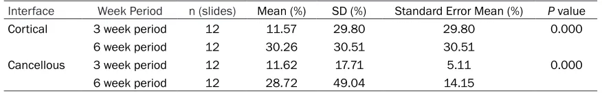
3 week Cortical 12 11.57 29.80 8.60 0.961
Cancellous 12 11.62 17.71 5.11
6 week Cortical 12 30.26 30.51 8.80 0.365
[image:3.612.89.526.88.151.2]Cancellous 12 28.72 49.04 14.15
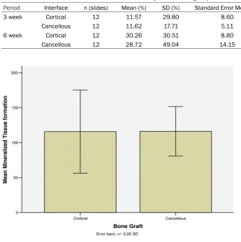
Figure 3. Cortical and cancellous interface standard error deviation values on 3 weeks period.
Ana, CA) and sec-tioned perpendic-ularly to in-terface recipient-graft to obtain sec- tions of about 30
μm; then were
stained using he- matoxylin-eosino-phile for light mi-croscopy analysis. Finally, were obta-ined 4 slices by each graft area. Histological analy-sis was done using a light microscope (DMLB, Leica, Ben- sheim, Germany), and for histomor-phometric analys-is the images were acquired in X4 ma-
gnification using a
[image:3.612.88.433.187.534.2] [image:3.612.90.522.191.257.2]The mean values for mineralized tissue forma-tion (MTF) on the interface graft and recipient area in the group I (cortical interface) was 11.57% (± 8.60% of standard error mean) and for group II (cancellous interface) was 11.62% (± 5.11% of standard error mean) as shows Table 2. It was not observed statistically signifi -cant difference (p=0.961). Mineralized tissue formation showed statistically the same results in each group (Figure 3).
Statistical analysis
Descriptive statistics were used for histological analysis referring the presence or not of blood vessel, connective tissue and mineralized tis-sue formation. For 48 examined slices with his-tomorphometric analysis, t test for paired anal-ysis was used (Biostat 5.0 software) with a 5%
[image:4.612.90.523.71.238.2]significance level.
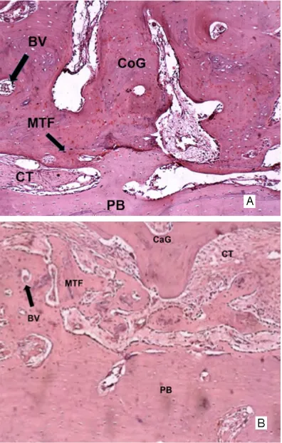
Figure 4. A, B: Histological analysis (10x) in a 6 week perior. A: Group I - Cortical interface (CoG). B: Group II - Can-cellous interface (CaG). Is observed more calcified material inside (MT) than 3 weeks period, blood vessel (BV) and connective tissue (CT).
Figure 5. Cortical and cancellous interface standard error deviation values on 6 weeks period.
Results
3 weeks
Descriptive analy-ses noted on the interface for both group the pres-ence of connective
tissue fulfilling the
[image:4.612.92.430.316.547.2]sia periods. In both cases there was graft inporation at the 6-week assessment, which cor-responds to 2 months in human beings as described by Turner et al. [14]. Gerressen et al.
[22], in a human sinus floor elevation surgery
research used iliac bone with pure cancellous in one sinus and a mixture of cortical and can-cellous in another sinus; the results demon-strate non differences between density or qual-ity in both maxillary sinus; also, don’t show differences between 4 or 7 month of evaluation.
Besides some authors noted that
decorticaliza-tion of receipt area have influence on bone
graft healing, improving their integration [4, 5, 7], we observed that both of interface were integrated without any preparation of recipient
area, suggesting that it does not influence on
bone graft integration.
Patient factors can influence the graft incorpo -ration process [5, 7]. External factors include the graft’s structural characteristics [6], the cancellous-type showing a better repair pro-cess when compared to cortical grafts because of a greater angiogenesis. This supposedly facilitates osteoblast precursor cell migration to the graft region, thus speeding up the incor-poration due to greater absorption and new mineralized connective tissue formation [7]. Despite that, this study could not observe any statistic difference between the two groups in mineralized tissue formation for both euthana-sia periods. This way, it may be suggest that, when analyze the probability to relapse, both grafts are similar, in agreed with other authors suggests [6, 7].
Based in our result, it may be concluded that both the cancellous or cortical graft interface in
autogenous block does not influence in bone
graft incorporation. both groups the presence of blood vessels and
less connective tissue fulfilling the interface
between the graft and recipient area, being this
region almost fulfilled by mineralized tissue for -mation. The recipient area and graft were not evidenced as shows Figure 4.
The mean values for MTF on the interface graft and recipient area for the group I was 30.26% (± 8.80% of standard error mean) and for the group II was 28.72% (± 14.15% of standard error mean) as shows Table 2. It was not
observed significant difference (p=0.365). MTF
showed statistically the same results in each group (Figure 5).
When analyzed the comparison between the 6 weeks and 3 weeks periods, both groups,
[image:5.612.87.525.87.155.2]corti-cal and cancellous interface, presented signifi -cant difference (p=0.000) for MTF as shows Table 3.
Discussion
The use of bone graft for treatment of
congeni-tal or acquired bone deficiencies has increased
in line with dental implant therapy [17, 18]. This study used autogenous bone, since this is the gold standard because of its properties of osteoconduction, osteoinduction, and osteo-genesis. Also, this material is the most used to treat these kinds of defects [19].
The lag screw technique reduces the space between the graft and recipient area [20]. This technique, used on the present research, reduced the gap on the interface graft-recipient area, improving the incorporation of the graft probably because it promotes primary reparation.
tis-[11] Silva FM, Cortez AL, Moreira RW, Mazzonetto R. Complications of intraoral donor site for bone grafting prior to implant placement. Im-plant Dent 2006; 15: 420-6.
[12] Sverzut AT, Stabile GA, de Moraes M, Maz-zonetto R, Moreira RW. The influence of tobac-co on early dental implant failure. J Oral Maxil-lofac Surg 2008; 66: 1004-9.
[13] Kuhn LT, Grynpas MD, Rey CC, Wu Y, Ackerman JL, Glimcher MJ. A comparison of the physical and chemical differences between cancellous and cortical bovine bone mineral at two ages. Calcif Tissue Int 2008; 83: 146-154.
[14] Turner AS. Animal models of osteoporosis – Necessity and limitations. Eur Cells Mater 2001; 1: 66-81.
[15] Parfitt AM, Drezner MK, Glorieux FH, Kanis JA, Malluche H, Meunier PJ, Ott SM, Recker RR. Bone histomorphometry: standardization of nomenclature, symbols, and units. Report of the ASBMR Histomorphometry Nomenclature Committee. J Bone Miner Res 1987; 2: 595-610.
[16] Recker RR. Bone Histomorphometry: Tech-niques and Interpretation. Boca Raton, FL: CRC; 1983.
[17] Callan DP, Rohrer MD. Use of bovine derived hydroxiapatite in the treatment of edentulous ridge deffects: a human clinical and histologic case report. J Periodontol 1993; 64: 575-82. [18] Pallesen L, Schou S, Aaboe M,
Hjørting-Han-sen E, Nattestad A, MelHjørting-Han-sen F. Influence of par-ticle size of autogenous bone grafts on the early stages of bone regeneration: a histologic and stereologic study in rabbit calvarium. Int J Oral Maxillofac Implants 2002; 17: 498-506. [19] Chaves Netto HDM, Olate S, Chaves MMGA,
Barbosa JR, Mazzonetto R. Análisis histológico del proceso de reparación en defectos óseos. Reconocimiento de defectos críticos. Int J Mor-phol 2009; 27: 1121-1127.
[20] Ellis E 3rd. Is lag screw fixation superior to plate fixation to treat fractures of the mandibu-lar Symphysis? J Oral Maxillofac Surg 2012; 70: 875-882.
[21] Franceschi C. Genes involved in immune re-sponse/inflammation, IGF1/insulin pathway and response to oxidative stress play a major role in the genetics of human longevity: the les-son of centenarians. Mech Ageing Dev 2005; 126: 351-61.
[22] Gerresen M, Hermanns-Sachweh B, Riediger D, Hilgers RD, Spiekermann H, Ghassemi A. Purely cancellous vs. corticocancellous bone in sinus floor augmentation with autogenous iliac crest: a prospective clinical trial. Clin Oral Impl Res 2009; 20: 109-115.
Disclosure of conflict of interest
None.
Address correspondence to: Dr. Sergio Olate, Division of Oral and Maxillofacial Surgery, School of Medicine, University of La Frontera, Claro Solar No
115, Temuco, Chile. Phone: 56-45-325000; E-mail: [email protected]
References
[1] Jensen J, Sindet-Pedersen S. Autogenous man-dibular bone grafts and osseointegrated im-plants for reconstruction of the severely atro-phied maxilla: a preliminary report. J Oral Maxillofac Surg 1991; 49: 1277-87.
[2] Schliephake H. Bone growth factors in maxil-lofacial skeletal reconstruction. Int J Oral Max-illofac Surg 2002; 31: 469-84.
[3] Issa MJP, Tiossi R, Pitol DL, Mello SAS. TGF-ß and new bone formation. Int J Morphol 2006; 24: 399-405.
[4] Jensen SS, Broggini N, Hjorting-Hansen E, Schenk R, Buser D. Bone healing and graft re-sorption of autograft, anorganic bovine bone ß-tricalcium phosphate. A histologic and histo-morphometric study in the mandible of minip-igs. Clin Oral Impl Res 2006; 17: 237-43. [5] Wang YN, Feng CC, Liu JL, Li F, Fu L, Wang Z.
Transforming Growth Factor beta Alleviates Acute Graft-versus-Host-Disease after Alloge-neic Bone Marrow Transplantation in Murine Model. Zhongguo Shi Yan Xue Ye Xue Za Zhi 2008; 16: 1135-9.
[6] Tong L, Buchman SR. Facial bone grafts: con-temporary science and thought. J Craniomaxil-lofac Trauma 2000; 6: 31-41.
[7] Nunamaker DM. Experimental models of frac-ture repair. Clin Orthop Relat Res 1998; 355 Suppl: S56-65.
[8] Bhatt KA, Chang EI, Warren SM, Lin SE, Basti-das N, Ghali S, Thibboneir A, Capla JM, McCar-thy JG, Gurtner GC.Uniaxial Mechanical Strain: An In Vitro Correlate to Distraction Osteogene-sis. J Surg Res 2007; 143: 329-36.
[9] Kübler A, Neugebauer J, Oh J, Scheer M, Zöller JE. Growth and proliferation of human osteo-blasts on different bone graft substitutes. An in vitro study. Implant Dent 2004; 13: 171-9. [10] Serra E Silva FM, Ricardo de