Original Article
In vivo
biocompatibility of the PLGA microparticles in
parotid gland
Mario Cantín1,2, Patricio Miranda3, Iván Suazo Galdames2, Daniela Zavando3, Patricia Arenas4, Luis Velásquez4,5, Cristian Vilos4,5
1CIMA, Department of Integral Dentistry, Faculty of Dentistry, Doctoral Program in Morphological Science,
Uni-versidad de La Frontera, Temuco, Chile; 2Center of Research in Biomedical Sciences, Universidad Autónoma de
Chile, Temuco, Chile; 3Private Practice, Chile; 4Center for the Development of Nanoscience and Nanothechnology
(CEDENNA), Santiago, Chile; 5Universidad Andres Bello, Facultad de Medicina, Center for Integrative Medicine and
Innovative Science, Santiago, Chile
Received August 13, 2013; Accepted September 3, 2013; Epub October 15, 2013; Published November 1, 2013
Abstract: Poly(lactic-co-glycolic acid) (PLGA) microparticles are used in various disorders for the controlled or sus-tained release of drugs, with the management of salivary gland pathologies possible using this technology. There is no record of the response to such microparticles in the glandular parenchyma. The purpose of this study was to assess the morphological changes in the parotid gland when injected with a single dose of PLGA microparticles. We used 12 adult female Sprague Dawley rats (Rattus norvegicus) that were injected into their right parotid gland with sterile vehicle solution (G1, n=4), 0.5 mg PLGA microparticles (G2, n=4), and 0.75 mg PLGA microparticles (G3, n=4); the microparticles were dissolved in a sterile vehicle solution. The intercalar and striated ducts lumen, the thickness of the acini and the histology aspect in terms of the parenchyma organization, cell morphology of acini and duct system, the presence of polymeric residues, and inflammatory response were determined at 14 days post-injection. The administration of the compound in a single dose modified some of the morphometric parameters of parenchyma (intercalar duct lumen and thickness of the glandular acini) but did not induce tissue inflammatory response, despite the visible presence of polymer waste. This suggests that PLGA microparticles are biocompatible with the parotid tissue, making it possible to use intraglandular controlled drug administration.
Keywords: Salivary glands, parotid gland, drug delivery, microparticles, PLGA
Introduction
Various polymeric macromolecules have been used in new drug delivery systems [1], and bio-degradable hydroxy-functional aliphatic polyes-ters such as poly(lactic-co-glycolic acid) (PLGA) have been particularly important in the formu-lation of microparticles for a sustained release device that prolongs pharmacological exposure over time. The Food and Drug Administration (FDA) have approved its use in humans, for use in medicines with many applications [2], includ-ing in labile tissues such as the brain [3] or in the perineurium [4]. The devices made from these polymers can be adapted so that the duration of the release of the encapsulated drug may be from hours to weeks and months, depending on the characteristics of the mic-roparticles and the route of administration.
This feature makes such microparticles ideal for managing multiple drugs; controlling their bioavailability becomes possible, and side ef- fects are reduced.
controlled release in the parenchyma may reduce the likelihood of tissue damage, control-ling the unwanted effects of these treatments; however, several authors have indicated that the microparticles are not biologically inert. For Kohane et al. [4], Colombo et al. [9] and Acuña
et al. [10] the polymeric microparticles are not innocuous, because they can cause inflamma-tion, which is associated with the presence of polymer waste. However, there is no record of the response to the microparticles in parenchy-ma. Thus, the purpose of this study was to assess the morphological changes in the parot-id gland when injected with a solution of PLGA microparticles.
Materials and methods
Were used 12 female Sprague Dawley (Rattus norvegicus) healthy rats, between 250 and 300 g, maintained in the animal facility of the Universidad de Talca. The rats were maintained in a light/dark cycle of 12/12 hours at room temperature of 22 ± 21°C and humidity of 60 ± 10%, and were fed with commercial pellets and water ad libitum throughout the experiment. The animals were treated in compliance with the protocol of bioethics adopted by the Universidad de Talca, in accordance with the “Guide for Care and Use of Laboratory Animals” published by the U.S. National Institutes of Health (NIH Publication no. 85-23, http:// grants.nih.gov/grants/olaw/olaw.htm).
PLGA microparticle formulation
Were used poly(D,L-lactide-co-glycolide) with a 50:50 monomer ratio, ester-terminated, with a viscosity of 0.26-0.54 dL/g, which was purchased from Lactel Absorbable Polymers (Pelham, AL, USA). The poly(vinyl-alcohol) (PVA) was 87% to 90% hydrolyzed, with average mol. Wt. of 30,000 to 70,000, from Sigma-Aldrich (St. Louis, MO, USA). Dichloromethane (DCM) was purchased from Sigma-Aldrich (St. Louis, MO, USA).
PLGA microparticles were prepared by double emulsion-evaporation method (w/o/w). In the first emulsion (w/o), 0.4 mL of double-distilled water solution was added into 1 mL of DCM containing 25 mg of PLGA, which was emulsified at 35,000 rpm using a high-speed homogenizer, the Ultra-Turrax system (Thermo Fisher Scienti- fic, Waltham, MA). The second emulsion (w/o/w)
was mixed with 2 mL of PVA 1% (1 mg/mL) and emulsified by the same system. The particles were stirred (orbital shaker at 100 rpm) at room temperature overnight for the solvent evapora-tion. The microparticles were then collected by filtration. Once sieved, the microparticles were lyophilized and stored at -20°C.
The diameters and morphology of PLGA mic-roparticles were determined by analysis of images obtained by phase-contrast on confo-cal microscopy and processed in the LSM Image Examiner software.
Experimental procedure
The animals were randomly assigned to form study groups as follows:
▪ Group 1 (G1): control injected with sterile vehi-cle solution (n=4).
▪ Group 2 (G2): experimental; injected with 0.5 mg of PLGA microparticles in a sterile vehicle solution (n=4).
▪ Group 3 (G3): experimental; injected with 0.75 mg of PLGA microparticles in a sterile vehicle solution (n=4).
The dilution and homogenization of microparti-cles in the vehicle solution for the G2 and G3 groups was made with a stirrer for 15 seconds prior to administration.
The rats were injected extraorally. They were positioned horizontally, directly exposing the parotid region and placing palp surface of the right parotid gland, and were injected with a single puncture on the parotid gland using a disposable 1 mL syringe graduated with a 20 G needle (day 0). The procedure was performed by a single operator who had been previously trained. The animals of all groups were then kept in care under the individual animal man-agement protocol for this research.
On day 14 post–intraglandular injection, the rats of all the groups were anesthetized with ether and were euthanized by cervical dislocation.
with four sheets for each piece. These were observed consistently through magnification at 400x using an Olympus CX21 microscope (Olympus ®) and analyzed by MicroImaging Software.
Morphometric parameters
Striated duct lumen: Duct diameter expressed in μm measured from the basal surface of an epithelial cell wall through the cell from the opposite surface. This dimension was deter-mined in all cut, transversely striated ducts, vis-ible on the plate analyzed.
Intercalar duct lumen: The major duct diameter expressed in μm measured from the basal sur-face of an epithelial cell wall to cell through the opposite surface. This dimension was deter-mined in all cross-sectioned intercalated ducts, visible on the plate analyzed.
Thickness of the glandular acini: Linear dimen-sion measured in μm from the acinar duct mar-gin at the periphery of the acinus, determined by all cross-cut acini seen in the plate analysis.
This corresponds to the thickness of acinar and myoepithelial cell.
Histology
Parenchyma cell morphology of acini and duct system was analyzed for morphological chang-es in organization. Also observed was the prchang-es- pres-ence of microscopic polymeric residues, which were defined as any mass at least partially, composed of microparticles and with the degree of inflammatory response according to the system established by Setzen and Williams [11].
Statistical analysis
Based on these parameters descriptive statis-tical differences were obtained and were evalu-ated by one-way ANOVA test and Bonferroni post-test. In all cases statistical significance was achieved when the p value is <0.05.
Results
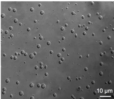
Our results showed that the size (diameter, µm) of PLGA microparticles was 2.1 ± 0.368 (µm), and normal particle size distribution with a range betwen 1.6-3.1 (µm) as shown the Figure 1. The phase-contrast on confocal microscopy images showed an uniform polymeric spherical microosized particles (Figure 2).
[image:3.612.94.286.67.369.2]The striated duct lumen was higher in G2, but the differences with the other groups were not significant (p=0.3113). An assessment of the Figure 1. Size distribution of PLGA microparticles
[image:3.612.323.522.70.243.2]ob-tained by confocal microscopy.
intercalar duct lumen parameter revealed sig-nificant differences between groups (p= 0.0154). Bonferroni post-test analysis showed significant differences (p<0.05) only when com-paring G1 with G3. An analysis of the glandular acinus thickness parameter revealed signifi-cant differences between groups (p=0.0244). Assessment by Bonferroni post-test showed only the differences between G1 and G2 to be significant. Table 1 shows the results.
With regard to histology, the glandular tissue in G1 was found to be disorganized; acini showed an irregular arrangement, with interacinary spaces apparently increased. Striated and intercalar ducts showed a typical organization, with easy viewing. There were no inflammatory cells. This is shown in Figure 3.
Histologically, G2 showed changes in both the organization and height of acinus-glandular excretory cells. The appearance of the striated and intercalar ducts was enlarged lumen. Polymeric residues were found on the periph-ery of the acinar cells, but no inflammatory cells were found (Figure 4).
The histologic appearance of G3 was a greater disorganization of the parenchyma, with large
amounts of polymer waste peri-acinar. Acinar cells and epithelium of striated and intercalar ducts showed greater separation between them, leading to increased centri-acinar lumen and ducts. Despite the abundant presence of polymer waste, no inflammatory response was observed (Figure 5).
Discussion
This study evaluated the hypothesis that PLGA microparticles can be safely applied directly to the parotid gland tissue. We found that applica-tion of these substances modifies some mor-phometric parameters of parenchyma (interca-lar duct lumen and thickness of the glandu(interca-lar acini), but does not induce tissue inflammatory response, despite the presence of polymer waste.
[image:4.612.91.525.97.163.2]Although at present both the research on and the use of microparticles as a means of trans-port are wide, its application in salivary gland tissue has not been reported. Authors such as Enders [12], using models of Macacus mulatta, have sought ways to inoculate the parotid gland with inactivated mumps virus to immunize the
Table 1. Descriptive statistic of histologycal parameters striated duct lumen, intercalar lumen duct and glandular acinus thickness. Values expressed in microns
Histologycal parameters MeanGroup 1 SD MeanGroup 2 SD MeanGroup 3 SD Striated duct lumen 293.72 94.99 350.47 120.90 338.11 115.51 Intercalar lumen duct 120.38 45.53 140.91 31.69 162.13 41.30 Glandular acinus thickness 220.53 33.57 196.41 15.93 214.50 23.95
Figure 3. Histological image of parotid gland tissue,
[image:4.612.324.521.183.338.2] [image:4.612.91.288.184.338.2]host, thus suggesting the use of means of transport that have protective properties to keep the virus and promote a humoral response.
In our study, we injected the parotid glands because of the easy identification of anatomi-cal landmarks and their potential use for the treatment of several diseases. However, a num-ber of intraparotid injection complications, such as facial nerve injury, have been reported. To avoid such complications, existing ultra-sound-guided techniques that minimize any complications of this type can be applied [13, 14].
The use of botulinum toxin (BT) by parotid gland injection in the treatment of sialorrhea in Parkinson’s disease has been shown to obtain very good results, reducing salivary flow with-out interfering with other processes [15]. PLGA microparticles used as a means of transporta-tion would be a good treatment for synergy of both. Bothwell et al. [16] reported the use of this BT in children with neurological problems and excessive salivation. Symptoms decreased significantly, with 55% of parents calling it a successful treatment, showing itself to be effective. In patients with cerebral palsy, treat-ment of sialorrhea with botulinum neurotoxin serotype A is effective and safe. The injection of the neurotoxin must be repeated to achieve effective treatment, as availability to the tis-sues is limited over time. Using micro-devices for transportation and protection, in addition to gauging the range of actions over time, is an
advantage compared with conventional thera-py. It reduces the adverse effects of intrapa-rotid injection and the dissemination of this toxin to muscle groups such as the pterygoid, masseter, or pharyngeal muscles, which alters their normal function. This effect could be low-ered by restricting the toxin through encapsula-tion, with slow and sustained release over time. Complications such as muscle paralysis, dys-phagia, and chewing problems have been reported. The poor dissemination of the mic-roparticles observed through the glandular tis-sues counteracts this problem.
For several treatments, the parotid gland is usually injected in two or three different sites. In general, the duration of action varies up to six weeks [17]. Based on this knowledge, the use of microparticles would result in a reduc-tion of doses, number of injecreduc-tion sites, and number of injections over time due to the pos-sibility of controlling the delivery of encapsulat-ed drug over time.
Modifying the function of the salivary glands produces a change of the acinar cell. Leal et al.
[image:5.612.91.288.73.220.2][18] subjected rats to periods of liquid diet in which the histological appearance was similar to the parotid glands in our control group. Glandular parenchyma, intercellular spaces, and the cell nucleus do not suffer any signifi-cant alteration. However, serous acini showed a mild atrophy and some degranulated acinar cells. Salivary ducts were considered normal. Vacuoles were observed in the cytoplasm of cells. Other studies have reported histological alterations of the salivary glands of rats fed a liquid diet, with vacuoles and a reduction in the size of the acinar and duct cells [19, 20]. In the interpretation of the polymeric residues on his-topathological sections there should also be taken into account possible lesions of acinar vacuolar degeneration. Here, there should be imposed the performance of a differential diag-nosis with alterations such as virus infection, intoxication, neoplasm or others pathologies. Several authors have noted that one of the most important histological parameters evalu-ated, using these micro-devices for drug deliv-ery, is the inflammatory response. Jang and Shea [21] encapsulated plasmids using PLGA microparticles, which were injected into striat-ed muscle. They observstriat-ed that at 2 days post-injection there was a marked inflammatory Figure 5. Histological image of parotid gland tissue,
cal applications of polylactic acid/polyglycolic acid copolymers. Biomaterials 1996; 17: 93-102.
[3] Menei P, Daniel V, Montero-Menei C, Brouillard M, Pouplard-Barthelaix A, Benoit JP. Biodegra-dation and brain tissue reaction to poly(D,L-lactide-co-glycolide) microspheres. Biomateri-als 1993; 14: 470-78.
[4] Kohane DS, Lipp M, Kinney RC, Anthony DC, Louis DN, Lotan N, Langer R. Biocompatibility of lipid-protein-sugar particles containing bupi-vacaine in the epineurium. J Biomed Mater Res 2002; 59: 450-9.
[5] Manrique D. Application of botulinum toxin to reduce the saliva in patients with amyotrophic lateral sclerosis. Braz J Otorhinolaryngol 2005; 71: 566-9.
[6] Fuster Torres MA, Berini Aytés L, Gay Escoda C. Salivary gland application of botulinum toxin for the treatment of sialorrhea. Med Oral Patol Oral Cir Bucal 2007; 12: E511-7.
[7] Nahlieli O, Shacham R, Shlesinger M, Eliav E. Juvenile recurrent parotitis: a new method of diagnosis and treatment. Pediatrics 2004; 114: 9-12.
[8] Crampsey DP, Cochrane L, Roebuck D, Hartley BE. Chronic facial pain following injection of sodium tetradecyl sulphate into an intraparot-id haemolymphangioma. J Laryngol Otol 2008; 122: 1002-4.
[9] Colombo G, Langer R, Kohane DS. Effect of ex-cipient composition on the biocompatibility of bupivacaine-containing microparticles at the sciatic nerve. J Biomed Mater Res A 2004; 68: 651-9.
[10] Acuña L, Suazo GI, Zavando D, Elgueta S, Ve-lásquez L, Vilos C, Cantín M. Morphometrics and histopathologic changes in skeletal mus-cle induced for injectable PLGA micropartimus-cles. Int J Morphol 2011; 29: 403-8.
[11] Setzen G, Williams EF 3rd. Tissue response to suture materials implanted subcutaneously in a rabbit model. Plast Reconstr Surg 1997; 100: 1788-95.
[12] Enders JF. Chemical, clinical and immunologi-cal studies on the products of human plasma fractionation. X. The concentrations of certain antibodies in globulin fractions derived from human blood plasma. J Clin Invest 1944; 23: 510-30.
[13] Lin YC, Shieh JY, Cheng ML, Yang PY. Botulinum toxin type A for control of drooling in Asian pa-tients with cerebral palsy. Neurology 2008; 70: 316-8.
[14] Banerjee KJ, Glasson C, O’Flaherty SJ. Parotid and submandibular botulinum toxin A injec-tions for sialorrhoea in children with cerebral palsy. Dev Med Child Neurol 2006; 48: 883-7. [15] Nóbrega AC, Rodrigues B, Melo A. Does botuli-num toxin injection in parotid glands interfere
response around the muscle fibers, but at 50 days inflammatory cells were located mainly adjacent to the microparticles. We did not observe inflammatory response at 14 days post-injection of PLGA microparticles. It is likely that the presence of the microencapsulated plasmids model of Jang and Shea is responsi-ble for part of this response. In a similar model Acuña et al. [10] reported a mild inflammatory response by applying PLGA microparticles on striated muscle. The absence of inflammatory response observed in this study suggests that PLGA microparticles originate a poor cellular and molecular response, slowing this reabsorp-tion. This is relevant because stability in the glandular tissue is essential for the develop-ment of microencapsulated drug delivery for treatment of chronic parenchymal diseases as Sjörgen’s syndrome, sialorrhea [5, 6], juvenile recurrent parotitis with the application of hydro-cortisone solutions [7], or the encapsulation of antiviral and antineoplastic agent for local therapies.
In conclusion PLGA microparticles can be safe-ly applied directsafe-ly to the parotid gland tissue in an animal model, because they modifies slight-ly morphometric parameters of parenchyma (intercalar duct lumen and thickness of the glandular acini), but does not induce tissue inflammatory response, despite the presence of polymer waste.
Acknowledgements
The support from CONICYT under Project BASAL FB0807 and Project Tesis en la Industria TPI06, are gratefully acknowledged.
Disclosure of conflict of interest
None.
Address correspondence to: Dr. Mario Cantín, CIMA, Department of Integral Dentistry, Faculty of Dentistry, Doctoral Program in Morphological Sci- ence, Universidad de La Frontera, Avenue Manuel Montt 112, Temuco, Chile. Tel: 056-045-2325574; E-mail: mario.cantin@ufrontera.cl
References
[1] Okada H, Toguchi H. Biodegradable micro-spheres in drug delivery. Crit Rev Ther Drug Carrier Syst 1995; 12: 1-99.
clini-[19] Hall HD, Merig JJ Jr, Schneyer CA. Metrecal-in-duced changes in human saliva. Proc Soc Exp Biol Med 1967; 124: 532-6.
[20] Sreebny LM, Johnson DA. Effect of food consis-tency and decreased food intake on rat parotid and pancreas. Am J Physiol 1968; 215: 455-60.
[21] Jang JH, Shea LD. Intramuscular delivery of DNA releasing microspheres: microsphere properties and transgene expression. J Control Release 2006; 112: 120-8.
with the swallowing dynamics of Parkinson’s disease patients? Clin Neurol Neurosurg 2009; 111: 430-2.
[16] Bothwell JE, Clarke K, Dooley JM, Gordon KE, Anderson R, Wood EP, Camfield CS, Camfield PR. Botulinum toxin A as a treatment for exces-sive drooling in children. Pediatr Neurol 2002; 27: 18-22.
[17] Winterholler MG, Erbguth FJ, Wolf S, Kat S. Botulinum toxin for the treatment of sialor-rhoea in ALS: serious side effects of a trans-ductal approach. J Neurol Neurosurg Psychia-try 2001; 70: 417-8.