Original Article
A preliminary study on the peripheral retinal
refractive status and the development of
myopia in RP patients with myopia
Shuang-Shuang Zhou1*, Zhou Zhou2*, Qiu-Xia Xie1, Yi Shao3, Zheng Yang4, An-Hua Wu1, Xue-Xiang Zou1, Ying Zhou1, Gang Tan1
1Department of Ophthalmology, The First Affiliated Hospital of University of South China, Hengyang 421001,
Hunan Province, China; 2Medical College, University of South China, Hengyang 421001, Hunan Province, China; 3Department of Ophthalmology, The First Affiliated Hospital of Nanchang University, Jiangxi Province Clinical
Oph-thalmology Institute, Nanchang 330006, Jiangxi Province, China; 4Department of Cardiology, The First Affiliated
Hospital of Sun Yat-sen University, Guangzhou 510080, Guangdong Province, China. *Equal contributors.
Received December 4, 2017; Accepted September 5, 2018; Epub November 15, 2018; Published November 30, 2018
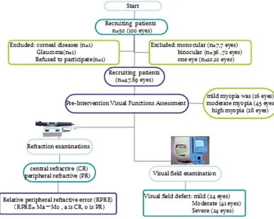
Abstract: Purpose: To study the relationship between peripheral retinal refraction and the development of myopia in retinitis pigmentosa patients with myopia. Methods: A total of 47 subjects (89 eyes) of RP patients with different degrees of myopia underwent peripheral refraction and visual field measurement. The visual field test results were grouped according to the AGIS scoring system, and classified into mild visual field defect group (24 eyes), moder-ate visual field defect group (41 eyes), and severe group (24 eyes). According to shadow moving, neutralization in the open view field condition, relative peripheral refractive errors (RPREs) were measured in the center and at the stare angles of 10°, 20° and 30° in the nasal and temporal fields, respectively. A complete set of data for each eye included one central diopter and six peripheral diopters. The RPREs of the nasal and temporal fields at 10°, 20° and 30° were determined by subtracting each peripheral diopter from central diopter, RPRE = Ma-M0, a is central diopter, o is peripheral diopter. Results: The relative refractive errors of temporal fields between mild and moderate group were significantly different at temporal field eccentricities of 20° and 30° (Independent-samples t-tests, P = 0.01, 0.004). The peripheral hyperopia state was positively correlated with the degree of visual field damage. In mild to moderate group, there was also a positive correlation between peripheral hyperopia state and visual field dam-age degree (P = 0.012, <0.05). Conclusion: For RP patients with myopia, there was a positive correlation between peripheral hyperopia state and retinal damage when the damage was mild. For the severe retinal damaged group, the central diopter did not correlate with the peripheral retinal damage.
Keywords: Retinitis pigmentosa (RP), myopia, peripheral refraction, visual field
Introduction
With the application of electronic products, refractive errors have become the most com-mon eye diseases. However, the pathogenesis of myopia is unclear [1]. Genetic studies dem-onstrate that, the genomic loci of high myopia causative genes are proximal to those of RP. Clinically, genetic RP is usually combined with high myopia [2]. A large number of animal and clinical studies demonstrate that peripheral refractive state have a substantial impact on emmetropization, resulting in the onset and progression of myopia [3-10]. Compared to
peripheral refraction state of sporadic RP patients leads to myopia is unclear and war-rants detailed characterization. Our study retro-spectively collected and analyzed data from sporadic RP patients associated with myopia, and investigated the relationship between the peripheral retinal refraction state and the development of myopia.
Materials and methods
Subjects
A total of 47 RP patients (89 eyes) with differ-ent degrees of myopia who visited the Eye Center of the First Affiliated Hospital of University of South China from March, 2014 to May, 2016 were recruited in the study. Patients with other known ocular diseases such as cata-ract, glaucoma, corneal diseases or blind and systemic disease or previous surgery history were excluded. Outline of study procedures shown in Figure 1. Among these patients, there were 36 patients (76.6%) with binocular RP and 7 patients (7.8%) with monocular RP. Patients ranged in age from 30 years old to 40 years old, with a mean age of 33.94±2.16 years; 61.7% of the patients were men (n = 29) and 38.2% were women (n = 18). Moreover, there were 58.5% patients with uncorrected visual acuity (UCVA) less than 0.1 and 2.2% greater than or equal to 0.3. The percentage of high myopia (≥-6.00 D) was 31.4%, moderate myopia (-3.00 D~-6.00
surements with the subjects turning their eyes and fixating on the object in the nasal and tem-poral fields at 10°, 20°, and 30° angles were recorded sequentially, under the condition of an open visual field. Each subject was instruct-ed to stay steady to avoid any head movement, open the pupil with diameter greater than 4 mm, and stare at a cross Maltese object at 2.5 m distance (the size of the cross Maltese object was 25 cm×20 cm, contrast was greater than 80%) at 10° intervals from nasal 30° to tempo-ral 30° visual fields. According to the neuttempo-ral condition of shadow moving, peripheral refrac-tive errors were measured at the visual angle of 10°, 20°, and 30°, respectively.
Each measurement was calculated as the arith-metic mean of three replicates. For each visual angle, a complete set of data for each eye included one CR and six PR. The relative periph-eral refractive error (RPRE) was determined by subtracting each peripheral refractive (PR) with central refractive (CR) measurement (RPRE = Ma-M0, a is CR, o is PR) [13].
Visual field examination
[image:2.612.94.367.74.292.2]Visual field tests were conducted with a Humphrey720i perimeter using the central 30-2 threshold test with SITA standard thresh-old strategy. Subjects stayed in a darkroom for 5~10 min before examination to ensure the pupil diameter greater than 4 mm. Tests were Figure 1. Outline of study procedures.
D) was 50.6% and mild myo-pia was 17.9% (≤-3.00 D). Refraction examinations
adjusted with appropriate correction lens based on the patient’s refractive status and age. There was a 30 minute break between examinations. All patients were examined three or more times. Excluding two test sites above and below the physiologic blind spot, there were a total of 74 test sites for analysis. The visual field test results were grouped according to the Advanced Glaucoma Treatment Study (AGIS) scoring system [14]. AGIS system was scored by the defect depth and quantity of nor-mal threshold in the overall deviation figure from the nasal visual field, the upper half visu-al-field and the bottom half visuvisu-al-field visual field tests. The scores ranged from 0 (no defect) to 20 (worst). The highest score was 20 that included two points from nasal and 9 points from each half visual-field. AGIS were classified into five stages: first stage: 0 points, normal visual field; second stage: 1-5 points, mild al field defect; third stage: 6-11, moderate
visu-Results
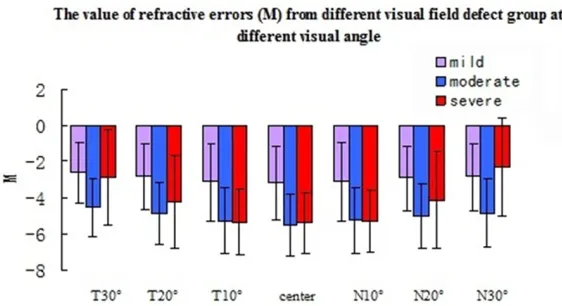
[image:3.612.92.373.91.212.2]As illustrated in Figure 2, in the mild visual field defect group, mild myopia accounted for about 50%, moderate myopia was 37.5%, and high myopia was 12.5%. In the moderate group, mild myopia was 7.3%, moderate myopia was 53.6%, and high myopia was 39%. In severe group, mild myopia accounted for 4.2%, moder-ate myopia and high myopia was 58.3% and 37.5%, respectively. In the mild visual field defect group, mild myopic was the major form. And moderate to severe visual field defect group mainly showed moderate myopia. As shown in Figures 3 and 4 the relative refrac-tive errors of temporal fields between mild and moderate group were significantly different at temporal field eccentricities of 20° and 30° (Independent-samples t-tests, P-value = 0.01, 0.004). The peripheral hyperopia sta- Figure 2. The ratio of different myopic degree in mild, moderate and severe
visual field defect group visual field defect groups.
Figure 3. The value of refractive errors (M) from mild, moderate and severe visual field defect group at temporal field eccentricities of 10° (T10°), 20° (T20°) and 30° (T30°), at the 10° (N10°), 20° (N20°), and 30° (N30°) nasal field eccentricities.
al field defect; 4th stage: 12-17, severe visual field defect; 5th stage: 18-20 points: absolute visual field defect. According to inclusion and exclusion criteria, 24 eyes (26.97%) were mild visual field defect, 41 eyes (46.06%) were moderate visual field defect, and 24 eyes (26.97%) were severe visual field defect. Statistical analysis
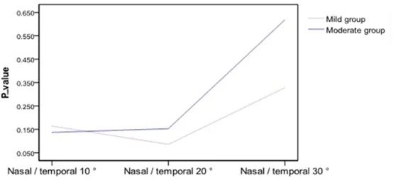
[image:3.612.91.372.260.413.2]te was positively correlated with the degree of visual field damage. In mild to moderate group, there was also a positive correlation between peri- pheral hyperopia state and visual field damage deg- ree (P-value = 0.012, <0.05). The peripheral state was hyperopic at the 10° temporal field eccentricities, but there was no significant cor-relation between the peripheral retinal degen-eration and the peripheral hyperopia state near the macula (Independent-samples t-tests, P-value = 0.125, >0.05). In addition, RP pat- ients showed a peripheral hyperopia state at the 10°, 20°, and 30° nasal field eccentricities, but no significant differences were obser- ved in the degree of pigmentary degeneration (Indepen- dent-samples t-tests, P-value = 0.190, 0.217, 0.101, >0.05).
Figures 3 and 4 indicated that RP patients in the moderate to severe group showed relative peripheral hyperopic errors. However, about half of RP patients preserved only a narrowing
gree of pigmentary degeneration and the devel-opment of myopia. The results of the peripheral refraction degree also showed asymmetry between nasal and temporal retina, which is consistent with the conclusions from Millodot.
Discussion
[image:4.612.95.372.276.403.2]Retinitis pigmentosa and myopia are common eye diseases. Myopia could result in peripheral retinal degenerations that may lead to retinal holes or retinal detachment, causing constrict-ed visual fields and eventually visual loss [15]. Similarly, retinal degeneration in the majority of RP patients starts from the peripheral retina, leading to tunnel vision and blindness gradual-ly. RP patients are often myopic and have cylin-drical refractive errors. However, the exact pathogenesis and progresses are different. Paula (8) and Bokd’ [16] showed that most spo-radic RP patients with high incidence of myopia were mild to moderate myopia. Moreover, high myopia can be caused by genetic factors [17]. Figure 4. The RPRE from mild, moderate and severe visual field defect group
at temporal field eccentricities of 10° (T10°), 20° (T20°) and 30° (T30°), at the 10° (N10°), 20° (N20°), and 30° (N30°) nasal field eccentricities.
Figure 5. Paired t-tests results about RPRE from nasal and temporal retinal fields between each two visual field defect groups.
central visual acuity in the severe group, and the periph-eral refraction could not be measured by retinoscopy at nasal field eccentricities of 20° and 30°, and we were not able to evaluate the RPRE effectively. Meanwhile, there was no significantly correla-tion of RPRE between nasal field eccentricities of 20° and 30° (P-value = 0.930, 0.787, >0.05). In AGIS scores, there was no statistical significance in the central refractive state in moderate and severe group, suggesting that for severe pig-mentary degeneration patien- ts, the central refractive state is not associated with the peripheral retinal degenera- tion.
Studies have indicated that high myopia might be polygenic inheritance [16]. The genomic loci of high myopia causative genes are proximal to those of RP. As a consequence, genetic RP is usually combined with high myopia. However, according to the clinical manifestation, high myopia is different from sporadic retinitis pig-mentosa. Sporadic RP patients lose night vision first, followed by the loss of peripheral vision, which causes tunnel vision. Eventually the vision of the macula, a central region of the retina, is lost. It is well known that rod photore-ceptor cells are active in dim-light environment and allow for night vision. Cone photoreceptor cells are responsible for sensing different light wavelengths and allow for daytime and color vision[18].As mentioned above, the progres-sive atrophy of the rod photoreceptor cells leads to a secondary death of the cone cells. On the contrary, most high myopia starts with poor light vision due to the degeneration of peripheral retina, followed by poor night vision. Furthermore, ocular fundus changes are differ-ent. The main retinal lesions of sporadic RP patients are that bone spicule-shaped pigment deposits in the retina along with RPE atrophy. As a result, the visual impairment or the ocular fundus changes in RP and high myopia are dis-tinct from each other. In order to exclude the hereditary factor of RP patients, our study col-lected and analyzed data from sporadic RP patients to investigate the relation between myopic errors and non-hereditary RP patients. Over the past decade, as shown in Table 1, there are large amount of experiments confirm-ing that relative hyperopia in the periphery could influence the development of myopia. Through animal studies, Wiesel [19] and Smith [3] claimed that hyperopia in the periphery caused an elongation of the eye globe, while myopia in the periphery could prevent this
[image:5.612.91.524.84.190.2]progress. In contrast, Earl [8] discovered that unrestricted central vision was not sufficient to ensure normal refractive development, and the fovea was not essential for emmetropizing responses and ametropias production. How- ever, the peripheral retina is essential for this process. Mutti’s [10] study suggested that the optical state in the periphery retina was closely associated with the development of axial myo-pia. The main pathological changes of myopia are comus and leopard fundus. In addition to the pathologic changes of posterior pole fun-dus, the peripheral retina is often the patho-logic site as well. In myopia, the peripheral reti-nal lesions are mainly diffusive choroidal thinning, solitary choroidal lesion and cystoid degeneration of retina. According to the statis-tic analysis of the axial length and the periph-eral retinal lesions from 513 consecutive patients, Pierro [20] confirmed that the per-centages of eyes with lesions in each axial length varied. It has been reported that eyes of greater axial length have a higher incidence of peripheral retinal lesions. In other words, there is a positive correlation between myopic errors and peripheral retinal degenerations. However, all findings mentioned above need more long-term trials or a larger number of samples. Yet, the typical pathological changes of sporadic RP patients are retina degeneration. The retina degeneration starts from the peripheral retina to the macula in general. At the same time, bone spicule-shaped pigment deposits accu-mulate at the peripheral retina. This manifests as the loss of peripheral vision at the begin-ning, followed by tunnel vision and then total blindness [21]. Thus, visual field defects in the peripheral retina from RP patients cause hyper-opia state in the periphery. We collected a large amount of data from RP patients with visual field defects, to investigate the relation between refraction hyperopic state and the development
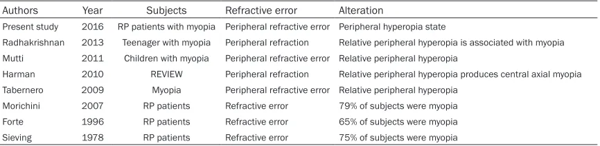
Table 1. Peripheral retinal refractive status in RP patients with myopia Authors Year Subjects Refractive error Alteration
Present study 2016 RP patients with myopia Peripheral refractive error Peripheral hyperopia state
Radhakrishnan 2013 Teenager with myopia Peripheral refraction Relative peripheral hyperopia is associated with myopia Mutti 2011 Children with myopia Peripheral refractive error Relative peripheral hyperopia
Harman 2010 REVIEW Peripheral refraction Relative peripheral hyperopia produces central axial myopia
Tabernero 2009 Myopia Peripheral refractive error Relative peripheral hyperopia
Morichini 2007 RP patients Refractive error 79% of subjects were myopia
Forte 1996 RP patients Refractive error 65% of subjects were myopia
of myopia. Clinically, RP patients can be diag-nosed with the clinical presentations of night blindness, pigmentary degeneration, constrict-ed visual fields, abnormal ERG or specific molecular genetic defect. Generally, the chang-es of constricted visual fields could be regard-ed as indicators of the disease severity. Therefore, this experiment regarded visual fields examination as an assessment to evalu-ate the RP condition. And based on the degree of visual field damage, we explored the relation-ship between the peripheral retinal hyperopia state and the development of myopia.
In this investigation, peripheral refractive errors were measured by retinoscopy. In recent years, retinoscopy method is often applied to examine the peripheral refractive errors of rhesus mon-keys. Hung [12] measured peripheral refractive errors of anesthetized infant rhesus monkeys by retinoscopy. And the peripheral refractive errors (RPE) were measured at the 15°, 30°, and 45° field eccentricities in the nasal, tempo-ral and the centtempo-ral field. They also found that, in the longitudinal infants group, the relative peripheral refractive errors (RPRE) of the rela-tive peripheral myopia underwent emme-tropization in nature and increased as vision-dependent growth. Based on that, Huang [22] used retinoscopy to investigated the effects of form deprivation on refractive development in infant rhesus monkeys. It is reported that, like humans with myopia, monkeys with form-depri-vation myopia exhibit relative peripheral hyper-opia, indicating that abnormal visual experi-ence can alter the shape of the posterior globe and the pattern of relative peripheral refractive errors in infant primates. These results showed that retinoscopy could be used to examine the spherical-equivalent refractive errors in the periphery, the regular astigmatism, the refrac-tive status, and ocular axial dimensions. Although this process uses subjective shadow moving as the readout, and examination is dis-continuous, slow and time-consuming, mea-surements in the study were obtained at a 1 m working distance, and the visual angle was set for 10°, 20°, and 30°. It is reasonable to use the retinoscopy method to measure the periph-eral refractive errors. Various methods have been used to define the peripheral refractive errors of human eyes, including subjective refraction, retinoscopy, autorefraction, wave-front technology, double-pass technique, and
so forth. Previous studies have validated these methods. Among these, retinoscopy is the most accessible method for current study.
Our results showed that among different visual field damage extent, RP population with mild to moderate myopia was the majority, and the myopic degree increased with the increase of visual field damage degree (the degree of pig-mentary degeneration). In addition, statistical analysis showed that RP patients exhibited rel-ative peripheral hyperopia at temporal field eccentricities of 20 and 30 degrees. Fur- thermore, there were significant differences in peripheral hyperopia degree between low and moderate group, and the peripheral hyperopia state was positively associated with the visual field damage degree. In the moderate group, peripheral hyperopia state was similar to the visual field damage degree. The peripheral state exhibited hyperopic at the 10° temporal field eccentricities. However, there was no sig-nificant correlation between the peripheral reti-nal degeneration and the peripheral hyperopia state near the macula. In addition, retinitis pig-mentosa patients showed a peripheral hypero-pia state at the 10°, 20°, and 30° nasal field eccentricities, and no significant differences were observed in the degree of pigmentary degeneration. The above results were similar to the Smith [23] and Atchison [24] that there was a positive correlation between the peripheral hyperopia degree and myopic degree. At the temporal field, there was a correlation between the degree of pigmentary degeneration and the myopia degree [25]. However, that was not exactly symmetrical at the nasal field. It is con-sistent with the conclusions from Millodot that the results of the peripheral refraction degree were asymmetric between nasal and temporal retina [26].
Our findings suggest that there were significant differences in peripheral hyperopia degree between low and moderate group, and the peripheral hyperopia state was positively asso-ciated with the visual field damage degree, which may help us to understand the patho-physiology between RP patients and the devel-opment of myopia, and could provide a research platform for clinical treatment of myopia. It has some drawbacks in this research. Because the peripheral refractive errors of severe pigmen-tary degeneration were measured by retinos-copy, a certain number of RP patients only remained tunnel vision so that we were unable to examine the peripheral refractive state. To some extent, once the peripheral retinal lesions affect vision, the central refractive state is not associated with the peripheral retinal degener-ation. Considering the limited number of patients in this study, common and stable clini-cal examination were applied to the current research. Our results need to be validated on a larger number of subjects.
Acknowledgements
This study was supported by the National Nat- ural Science Foundation of China (81100648, 81160118 and 81400372); Hunan Province Education Department Outstanding Youth Science Foundation (15B210).
Disclosure of conflict of interest
None.
Address correspondence to: Gang Tan, Department of Ophthalmology, The First Affiliated Hospital of University of South China, Hengyang 421001, Hunan Province, China. E-mail: tangang99@hotmail. com
References
[1] Morgan I, Rose K. How genetic is school myo-pia. Prog Retin Eye Res 2005; 24: 1-38. [2] Costa KA, Salles MV, Whitebirch C, Chiang J,
Sallum JMF. Gene panel sequencing in Brazil-ian patients with retinitis pigmentosa. Int J Retina Vitreous 2017; 3: 33.
[3] Smith EL, Hung LF, Huang J. Relative peripher-al hyperopic defocus peripher-alters centrperipher-al refractive development in infant monkeys. Vis Res 2009; 49: 2386-2392.
[4] Hartwig A, Charman WN, Radhakrishnan H. Baseline peripheral refractive error and
chang-es in axial refraction during one year in a young adult population. J Optom 2016; 9: 32-39. [5] Atchison DA, Rosén R. The possible role of
pe-ripheral refraction in development of myopia. Optom Vis Sci 2016; 93: 1042-1044.
[6] Kee CS. The role of peripheral vision in the re-fractive-error development of infant monkeys. Invest Ophthalmol Vis Sci 2004; 45: 1157. [7] Seidemann A, Schaeffel F, Guirao A, Lopezgil
N, Artal P. Peripheral refractive errors in myo-pic, metropic and hyperopic young subjects. J Opt Soc Am A Opt Image Sci Vis 2002; 19: 2363-2373.
[8] Smith EL 3rd, Kee CS, Ramamirtham R, Qiao-Grider Y, Hung LF. Peripheral vision can influ-ence eye growth and refractive development in infant monkeys. Invest Ophthalmol Vis Sci 2005; 46: 3965-3972.
[9] Earl L, Smith, Ramkumar R, Hung LF, Huang J, Kee CS. Effects offoveal ablation on emmetro-plzation and deprivation myopia. Invest Oph-thalmol Vis Sci 2007; 48: 3914- 3922. [10] Mutti DO, Hayes JR, Mitchell GL, Jones LA,
Moeschberger ML. Refractive error, axial length, and relative peripheral refractive error before and after the on set of myopia. Invest Ophthalmol Vis Sci 2007; 48: 2510-2519.
[11] Sieving PA, Fishman GA. Refractive error of retinitis pigmenlosa patients. Br J Ophthalmol 1978; 62: 164-167.
[12] Hung LF, Ramamirtham R, Huang J, Qiao-Grid-er Y, Smith EL 3rd. PQiao-Grid-eriphQiao-Grid-eral refraction in nor-mal infant rhesus monkeys. Invest Ophthalmol Vis Sci 2008; 49: 3747-3757.
[13] Lee TT, Cho P. Repeatability of relative periph-eral refraction in untreated and orthokeratolo-gy-treated eyes. Optom Vis Sci 2012; 89: 1477-1486.
[14] The advanced glaucoma intervention study (AGIS): 7. The relationship between control of intraocular pressure and visual field deteriora-tion.The AGIS investigators. Am J Ophthalmol 2000; 130: 429-440.
[15] Bok D. Contributions of genetics to our under-standing of jaherited monogenic retinadiseas-es and age related macujar degeration. Arch Ophthation 2007; 125: 160-164.
[16] Hammond CJ, Sneider H, Gilbert CE, Spector TD. Genes and environment in refractive error: the twin eye study. Invest Ophthalmol Vis Sci 2001; 42: 1232-1236.
[17] Anasagasti A, Irigoyen C, Barandika O, López de Munain A, Ruiz-Ederra J. Current mutation discovery approaches in retinitis pigmentosa. Vision Res 2012; 75: 117-119.
[19] Wiesel TN, Raviola E. Myopia and eye enlarge-ment after neonatal lid fusion in monkeys. Na-ture 1977; 266: 66-68.
[20] Pierro L, Carnesasca FI, Mischi M, Brancato R. Peripheral retinal changes and axial myopia. Retina 1992; 12: 12-17.
[21] Allard RE. Retinitis pigmentosa-an overview. J Am Optom Assoc 1983; 54: 793-800.
[22] Huang J, Hung LF, Rarnamirtham R, Blasdel TL, Humbird TL. Effects of form deprivation on peripheral refractions and ocular shape in in-fant rhesus monkeys (macaca mulatta). Invest Ophthalmol Vis Sci 2009; 50: 4033-4044. [23] Smith EL 3rd, Ramamirtham R, Qiao-Grider Y,
Hung LF, Huang J, Kee CS, Coats D, Paysse E. Effects of fovealablation on emmetropization and form-deprivation myopia. Invest Ophthal-mol Vis Sci 2007; 48: 3914-3922.
[24] Atchison DA, Pritchard N, White SD. Influence of age on peripheral refraction. Vision Res 2005; 45: 715-720.
[25] Verkicharla PK, Suheimat M, Schmid KL, Atchi-son DA. Peripheral refraction, peripheral eye length, and retinal shape in myopia. Optom Vis Sci 2016; 93.