Original Article
Role and clinical significance
of miRNA-381 in prostate cancer
Yi He1, Qimei Zhang2, Rui Jiang1, Yingchuan Li1
1Department of Urology, The Affiliated Hospital of Sichuan Medical University, Luzhou, P. R. China; 2Department of Oral Medicine, The Affiliated Hospital of Sichuan Medical University, Luzhou, P. R. China
Received December 30, 2015; Accepted May 18, 2016; Epub February 15, 2017; Published February 28, 2017
Abstract: Objective: This study was to investigate the role and mechanism of miRNA-381 in prostate cancer. Methods: Totally 51 prostate cancer patients diagnosed and operated in our hospital from February 2013 to August 2015 were enrolled. And, 39 healthy volunteers were set as controls. Cancer tissues, the corresponding peritumoral tis-sues and blood specimens were collected. Quantitative RT-PCR was applied to detect the changes of COX-2 mRNA and miRNA-381 expression. ELISA was applied to detect COX-2 protein expression in blood. Dual luciferase reporter assay was applied to verify whether miRNA-381 could direct bind to COX-2 mRNA. MTT assay was applied to detect the cell proliferation of agomiR-381 and siRNA transfected LNCaP human prostate cells. Western Blot was applied to detect the changes of COX-2 protein expression in agomiR-381 transfected LNCaP human prostate cells. Results: The expressions of COX-2 mRNA and protein were significantly up-regulated in cancer tissues and blood in prostate cancer patients, while the relative expression of miRNA-381 was significantly down-regulated. Up-regulated expres
-sion of miRNA-381 resulted in significantly down-regulated expres-sion of COX-2 mRNA and protein and decreased cell proliferation rate of LNCaP cells. Dual luciferase reporter showed that miRNA-381 regulated the expression of COX-2 by binding to the 3’-UTR. The cell proliferation rate of LNCaP cells were also decreased after siRNA silencing
COX-2 expression. Conclusion: The expression of COX-2 was significantly up-regulated in cancer tissues and blood
in prostate cancer patients and this increase may be related to the down-regulated expression of miRNA-381. Our data suggests that miRNA-381 may regulate cell proliferation and expression of related proteins in prostate cancer cells through COX-2.
Keywords: mir-381, COX-2, prostate cancer
Introduction
Prostate cancer (PCa) is a common cancer of the male urinary system and the incidence rate
ranks as the highest in USA [1]. The mortality
ranks as the second among male cancers in
USA [2]. About 5% of prostate cancer cases
were detected by pathological sections of
pros-tatic hyperplasia [3].
Digital rectal examination combined with
post-ate specific antigen (PSA) testing is widely rec -ognized as the best screening method for the
early detection of prostate cancer [4-7].
Bio-markers play an important role in the diagnosis process, and many biological markers
associ-ated with prostate cancer have been confirmed by previous investigators [8, 9]. Previous stud -ies found that the up-regulated expression of
cyclooxygenase-2 (COX-2) plays an important regulatory role in the occurrence and develop-ment of prostate cancer, and the pathogenesis of most prostate cancer are accompanied by a progressive increase in COX-2 [10, 11]. It is
found that up-regulated expression of miRNAs can inhibit the expression of COX-2 [12] and
say kit was purchased from JRDUN
Biotech-nology (Shanghai, China). Rabbit anti-human COX-2 primary antibody (ab15191), rabbit anti-
human β-actin primary antibody (ab129348)
and goat anti-rabbit secondary antibody were
all purchased from Abcam Inc. (MA, USA). Trizol
was purchased from Yeasen Co, Ltd (Shang- hai, China). The iQ5 real-time PCR detection systems were purchased from Bio-Rad Cor-
poration (Hercules, CA, USA). Dual-Luciferase®
Reporter Assay System was purchased from
Promega Corporation (Madison, WI, USA).
Ima-ge lab3.0 software was purchased from
Bio-Rad Corporation (Hercules, CA, USA).
Quantitative RT-PCR
Total RNA was extracted by Trizol according to the manufacturer’s protocol. RNA was re- versely transcribed into cDNA, which was used as a template for PCR to detect the expre- ssion of COX-2 and miRNA-381. The expre-
ssion of β-Actin and U6 were set as internal
controls to COX-2 and miRNA-381, respec- tively. The primer sequences were as follows: COX-2: Sense: 5’-CAGCCATACAGCAAATCCTTG- 3’; Anti-sense: 5’-CAAATGTGATCTGGATGTCAAC-
3’, β-actin: Sense:
5’-CACCAGGGCGTGATGGT-3’; Anti-sense: 5’-CTCAAACATGATCTGGGTCAT- 3’, miRNA-381: Sense: 5’-ACACTCCAGCTGGG- TATACAAGGGCAAGCT-3’; Anti-sense: 5’-TGGTG-
TCGTGGAGTCG-3’, U6: Sense:
5’-CTCGCTTCG-GCAGCACA-3’; Anti-sense: 5’-AACGCTTCACGA- ATTTGCGT-3’. Quantitative PCR for COX-2 and
β-actin were performed with the following pro -cedure: 95°C for 30 s, followed by 40 cycles of 95°C for 5 s and 60°C for 34 s. And quan-
titative PCR for miRNA-381 and U6 were per -formed with the following procedure: 95°C for 5 min, followed by 40 cycles of 95°C for 15 s, 60°C for 30 s and 72°C for 30 s. The relative expression was calculated by 2-ΔΔCT method.
ELISA assay
Serum was separated from blood sample by centrifugation at 3000 rpm for 10 min. Then, 50 µL samples (1:4 dilution) or standard refer-ence solutions were added to the correspond-ing wells. HRP-conjugated detection antibody (100 µL) was added to each well, sealed and incubated in constant temperature incubator for 1 h. After washing for 5 times, 50 µL sub-strate A and B was added to each well. After incubation at 37°C for 15 min, 50 µL termina-tion solutermina-tions were added, and OD value of the mechanism of miRNA-381 in the
develop-ment of prostate cancer.
Materials and methods
Subjects
Totally 51 prostate cancer patients diagnosed and operated in our hospital from February 2013 to August 2015 were enrolled. And, 39 healthy volunteers were set as controls. Cancer tissues and the corresponding peritumoral tis-sues were collected from prostate cancer patients. Blood specimens were collected from prostate cancer patients and healthy controls. The age of prostate cancer patients was from 49 to 86 years, with a median age of 65.6 years. The age of control group was from 45 to 81 years, with a median age of 62.5 years. Among the 51 cases prostate cancer patients, 37 cases were hospitalized due to progressive
difficulty in urinating, 4 cases were due to uri -nary frequency and urgency, 4 cases were due to painless gross hematuria, 3 cases were due
to progressive urination difficulties associated
with painless gross hematuria, 2 cases were due to urinary frequency, urgency, dysuria and painless gross hematuria, and 1 case was due to urinary frequency associated with painless gross hematuria. All of the 51 prostate cancer
patients were the first time onset prostate can -cer patients, without any treatment history by hormone, traditional Chinese medicine, radio-therapy and chemoradio-therapy, etc, and they were all pathologically diagnosed. Prior written and informed consent was obtained from every patient and the study was approved by the
eth-ics review board of the Affiliated Hospital of Sichuan Medical University.
Reagents and instruments
each well at 450 nm wavelength were deter-mined within 15 minutes.
Bioinformatics prediction
The miRanda, TargetSean, PieTar, MiRanda, and BibiServ were applied for predicting COX-2 upstream regulatory miRNAs, and the possible regulatory sites were also predicted.
Dual luciferase reporter assay
Wild type and mutated type of the predicted
miRNA-381 binding site in the 3’-UTR of COX-2 gene were chemically synthesized in vitro, with adding Spe-1 and Hind III cleavage sites at both ends. The two DNA fragments were cloned into pMIR-REPORT luciferase reporter plasmid, and
the mutated 3’-UTR seed region containing
plasmid was set as a control. The 0.8 µg
plas-mids with 3’-UTR or mutated 3’-UTR were trans -fected into 293T cells by liposome method, and then transfected with agomiR-381 (100 nM).
After cultured for 24 h, the fluorescence value
was measured by GloMax 20/20 luminometer.
Renilla fluorescent activity was set as internal
control, and the procedures were strictly per-formed according to the instructions for dual luciferase report system kit.
Human prostate cancer cell line LNCaP trans-fection
The 3×10⁵ LNCaP cells in logarithmic grow-
th phase were seeded in 24-well plates,
cul-tured in antibiotic-free 10% FBS F12/DMEM
medium at one day before transfection. Cell transfection was performed when the cell
den-sity reached about 70%. Plasmids, siRNA, or
agomiR was added into 50 µL OptiMemi me- dium and incubated for 5 min. At the same time, 1 µL lipo2000 was also added to 50 µL OptiMemi medium and incubated for 5 min. Then the two mixtures were combined toge- ther and were added to cells. After incubation for 20 min, culture medium was added and co-cultured for 6 h. Then, the culture medium
was replaced with fresh 10% FBS F12/DMEM
medium and continued to culture. Cells were collected at 48 h after transfection, and the expression levels of targeted COX-2 mRNA and protein were detected.
Western blot
The total protein was extracted and the protein concentration was determined by BCA protein assay kit. The 20 µg samples were subjected
to 10% SDS-PAGE for Western Blot analysis.
The primary antibodies, including anti-COX-2
antibody (1:1000) and anti-β-actin antibody
(1:5000) were added and incubated overnight at 4°C. Then, secondary goat rabbit anti-bodies (1:3000) was added and incubated at room temperature for 1 h. Membrane was placed in ECL solution for color development. And, image was obtained by gel imaging system and analyzed by image lab3.0 software. The relative content of COX-2 protein was calculat-ed as the ratio of COX-2 gray value to β-actin
gray value. MTT assay
Cells were seeded in a 96-well plate at the den-sity of 2×10³/well, and each sample was
pro-vided with 3 parallel wells. The 20 µL of 5 g/L MTT reaction solution was added to each well at 24 h, 48 h, and 72 h. Then, 150 µL DMSO was added to each well at the last day, the absorbance value of each well was read at a wavelength of 490 nm after 4 h incubation. The cell proliferation curves were drawn.
Statistical analysis
All statistical analyses were performed by using the Statistical Package for Social Sciences software (SPSS, Windows version release 18.0;
SPSS Inc.; Chicago, IL, USA). Data were pre -sented as mean ± standard deviation. All data were analyzed with normality test. One-way ANOVA was applied for multiple sets of mea-surement data analysis. LSD and SNK method were applied when there was homogeneity of variance, and Tamhane’s T2 or Dunnett’s T3 method was applied when there was not homo-geneity of variance. A P value < 0.05 was
con-sidered statistically significant. Results
Changes of COX-2 mRNA expression in cancer tissues and blood samples
The qRT-PCR was applied to detect the expres-sion of COX-2 mRNA in cancer tissues, peritu-moral tissues, and blood samples. As shown in
Figure 1A, compared with that in the peritu-moral tissues, COX-2 mRNA expressions in
can-cer tissues were significantly increased in pros -tate cancer patients (P < 0.05). As shown in
Figure 1B, compared with that in control patients, COX-2 mRNA expressions in blood
indicat-ed that COX-2 may play a certain role in the regulation of prostate cancer.
Changes of COX-2 protein expression in cancer tissues and blood samples
Western Blot and ELISA were applied to de- tect the expression of COX-2 protein in cancer tissues, peritumoral tissues and blood sam-ples. As shown in Figure 2A, compared with
decreased in prostate cancer patients (P < 0.05). And, miRNA-381 expressions in blood samples of prostate cancer patients were
sig-nificantly lower than those of control patients (P
[image:4.612.91.375.74.191.2]< 0.05) (Figure 4B). These results indicated that miRNA-381 may play a certain role in the regulation of prostate cancer, and this regula-tory function may be acted through negatively regulating the transcription level of targeted COX-2 gene.
[image:4.612.91.376.252.574.2]Figure 1. Relative expression levels of COX-2 mRNA in cancer tissues, peritu-moral tissues (A) and blood (B). *P < 0.05 and **P < 0.01, compared with control group.
Figure 2. Relative expression levels of COX-2 protein in cancer tissues, peritumoral tissues (A) and blood (B). *P < 0.05 and **P < 0.01, compared with con-trol group.
that in peritumoral tissues, COX-2 protein expressions in cancer tissues were remark-ably increased in prostate cancer patients (P < 0.05). Similarly, COX-2 protein ex- pressions in blood samples of prostate cancer patients
were significantly higher than
those of control patients (P < 0.05) (Figure 2B). This result indicated that the expression of COX-2 protein was also up-regulated in cancer tissues and blood samples of pros-tate cancer patients, which was consistent with the trend of mRNA.
COX-2 is one of the target genes of miRNA-381
The COX-2 upstream regu- latory miRNAs were predict- ed with target gene predict- ion softwares. Results show- ed that there may be target- ed regulatory relationship be- tween miRNA-381 and COX-
2, and the specific regulatory
binding sequences as shown in Figure 3.
Changes of miRNA-381 ex-pression in cancer tissues and blood samples
The qRT-PCR was applied to detect the expression of mi- RNA-381 in cancer tissues, peritumoral tissues and blood samples. As shown in Figure 4A, compared with that in the peritumoral tissues, mi- RNA-381 expressions in
can-cer tissues were significantly
Figure 3. The predicted specific regulatory binding sequences of miRNA-381
Dual luciferase reporter assay
Dual luciferase reporter assay was applied to verify the relationship between miRNA-381 and COX-2. As shown in Figure 5, the fluores
-cence value was significantly decreased after
co-transfecting wild type COX-2 and
miRNA-381 (P < 0.05), while there was no significantly
difference in the mutant COX-2 transfected group (P > 0.05). This result indicated that miRNA-381 may regulate the expression of COX-2 through binding to the 3’-UTR of COX-2. Effect of agomiR-381 transfection on LNCaP cells
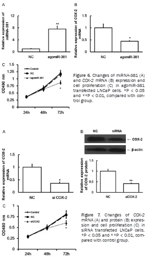
As shown in Figure 6, after transfected with agomiR-381, the expressions of miRNA-381 and COX-2 mRNA were significantly increased
(Figure 6A) and decreased (Figure 6B) in LNCaP cells, respectively. And, the cell
prolifer-ation was also slowed down significantly (Figure 6C). This result indicated that up-regulated expression of miRNA-381 may inhibit cell
prolif-that down-regulated expression of COX-2 may inhibit cell proliferation of LNCaP cells.
Discussion
In this study, we detected the expression levels of COX-2 mRNA and protein and miRNA-381 in cancer tissues and blood, preliminary explored the biological functions of miRNA-381 and COX-2, and studied the molecular mechanisms of miRNA-381 on prostate cancer.
Chronic inflammation is generally considered
as one of the physiological causes of tumors
[12]. The COX, especially the COX-2, plays an important role in the development of chronic
inflammation. COX-2 was induced by physical, chemical, biological and other external stimuli, which works as the necessary key rate-limiting enzyme to catalyze the synthesis of
prostaglan-dins (PGs) to involve in inflammation reaction [13]. Previous studies show that expression
change of COX-2 is closely related to the devel-opment of many kinds of tumors. The COX-2 inhibitors are developed for antipyretic analge-sic treatments, and also often for adjuvant
anti-tumor therapy [14-16]. In this study, a signifi -cant increase of COX-2 mRNA and protein expression was observed in prostate cancer tumor tissue, which is consistent with previous studies, indicating that the change of COX-2 expression is related to the development of prostate cancer, and the abnormal expression of COX-2 may be a key to the pathogenesis of
prostate cancer [17, 18]. Another obvious fea
-ture of tumor is invasion and metastasis [19, 20]. In theory, it is possible to determine the
COX-2 mRNA and protein in the blood depends
on blood or tissue fluid, which may indirectly reflect the malignant degree and metastasis of
[image:5.612.92.377.72.195.2]prostate cancer cells. Figure 4. Relative expression levels of miRNA-381 in cancer tissues (A) and
blood (B). *P < 0.05 and **P < 0.01, compared with control group.
Figure 5. Comparation of fluorescence values in dual
luciferase reporter assay. *P < 0.05 and **P < 0.01,
compared with cells transfected with wild type COX2.
eration of LNCaP cells through regulating COX-2 expression. Effect of siRNA transfection on LNCaP cells
As shown in Figure 7, after transfected with siRNA, the expression of COX-2 mRNA (Figure 7A) and protein (Fig- ure 7B) were significantly
de-creased in LNCaP cells. And, the cell proliferation was also
[image:5.612.94.287.254.386.2]After prediction, we found that miRNA-381 was closely related to COX-2. It is likely that miRNA-381 is an upstream regulation miRNA of COX-2. Endogenous, small, non-encoding miRNAs may splice and inhibit the translation of COX-2
mRNA [21]. miRNAs regulate the activities of
protein encoding genes, which play an
impor-tant role in the development of diseases [22, 23]. The expression of miRNA-381 was signifi -cantly downregulated in colon cancer and the down-regulated expression of miRNA-381 led
resulted in the decreased expression of COX-2 and cell proliferation rates of LNCaP cells. Dual luciferase reporter assay was applied to verify
the directly binding and the specific binding
sites of miRNA-381 to COX-2 mRNA. The results showed that miRNA-381 can directly bind to
the targeted seed region in 3’-UTR of COX-2 mRNA and regulate the expression of COX-2.
In summary, our findings suggest that
[image:6.612.90.379.70.592.2]miRNA-381 may play a role in the pathogenesis of Figure 6. Changes of miRNA-381 (A)
and COX-2 mRNA (B) expression and cell proliferation (C) in agomiR-381 transfected LNCaP cells. *P < 0.05 and **P < 0.01, compared with con-trol group.
to up-regulated expression of liver receptor homologue-1 (LRH-1), and resulted in prolif-eration and invasion of colon
cancer cells [24]. miRNA-381
could inhibit pituitary tumor
growth [25]. miRNA-381 and
multi-drug resistance gene 1 (MDR1) genes were closely related, and played an impor-tant role in multi-drug
resis-tance [26]. miRNA-381 could
inhibit the activity of renal cancer cell Cdc2, by working together with miRNA-424 to target on WEE1 gene [27].
And, miRNA-381 was also closely related to the patho-genesis of lung
adenocarci-noma [28]. These findings
su-ggest that miRNA-381 may have a very close relationship with the pathogenesis of tu- mors. Our results showed that the expressions of mi-
RNA-381 were significantly
decreased in cancer tissues and blood in prostate cancer patients. Considered that the expressions of COX-2 were abnormally increased in can-cer tissues and blood, we hypothesize that down-regu-lated expression of miRNA-381 may be one of reasons for COX-2 up-regulation, which may further affect the bio- logical characteristics of pro- state cancer cells. To fur- ther study the molecular me- chanism, we analyzed prolif-eration of LNCaP cells after agomiR-381 transfection. Re- sults showed that up-regulat-ed expression of miRNA-381 Figure 7. Changes of COX-2
prostate cancer through regulating the expres-sion of COX-2.
Acknowledgements
This work was supported by grants from Luzhou Municipal Science and Technology Bureau (2013-S-48 (28/30)) and The National Natural Science Fund (81070486).
Disclosure of conflict of interest
None.
Address correspondence to: Dr. Yingchuan Li, De-
partment of Urology, The Affiliated Hospital of Sichuan Medical University, 319 Zhongshan Road
Third Section, Jiangyang District, Luzhou 646000, P. R. China. Tel: +86-830-3115602; E-mail: gvm- 333@126.com
References
[1] Johnson LG, Madeleine MM, Newcomer LM, Schwartz SM and Daling JR. Anal cancer inci-dence and survival: the surveillance, epidemi-ology, and end results experience, 1973-2000. Cancer 2004; 101: 281-288.
[2] Kakehi Y. Watchful waiting as a treatment op-tion for localized prostate cancer in the PSA era. Jpn J Clin Oncol 2003; 33: 1-5.
[3] Guo Y. Prostate hyperplasia and prostate cancer. Beijing: People’s Medical Publishing House; 1998.
[4] Kattan MW, Cuzick J, Fisher G, Berney DM, Oliver T, Foster CS, Moller H, Reuter V, Fearn P, Eastham J and Scardino PT. Nomogram in-corporating PSA level to predict cancer-speci-
fic survival for men with clinically localized
prostate cancer managed without curative intent. Cancer 2008; 112: 69-74.
[5] Cuzick J, Fisher G, Kattan MW, Berney D, Oliver T, Foster CS, Moller H, Reuter V, Fearn P, Eastham J and Scardino P. Long-term outcome among men with conservatively treated local-ised prostate cancer. Br J Cancer 2006; 95: 1186-1194.
[6] Graham J, Baker M, Macbeth F and Titshall V. Diagnosis and treatment of prostate cancer: summary of NICE guidance. BMJ 2008; 336: 610-612.
[7] Catalona WJ, Richie JP, Ahmann FR, Hudson MA, Scardino PT, Flanigan RC, deKernion JB, Ratliff TL, Kavoussi LR, Dalkin BL, et al. Comparison of digital rectal examination and
serum prostate specific antigen in the early de -tection of prostate cancer: results of a
multi-center clinical trial of 6,630 men. J Urol 1994;
151: 1283-1290.
[8] McGrath SE, Michael A, Morgan R and Pandha H. EN2 in Prostate Cancer. Adv Clin Chem 2015; 71: 47-76.
[9] Jokerst JV, Chen Z, Xu L, Nolley R, Chang E,
Mitchell B, Brooks JD and Gambhir SS. A Magnetic Bead-Based Sensor for the Quan-
tification of Multiple Prostate Cancer
Bio-markers. PLoS One 2015; 10: e0139484.
[10] Yerokun T and Winfield LL. LLW-3-6 and cele -coxib impacts growth in prostate cancer cells
and subcellular localization of COX-2.
Anti-cancer Res 2014; 34: 4755-4759.
[11] Ceylan Y, Lekili M, Muezzinoglu T, Nese N and Isisag A. Predictive value of cyclooxygenase-2 over expression for identifying prostate cancer from benign prostatic hyperplasia in prostate
biopsy specimens. Minerva Urol Nefrol 2016;
68: 255-62.
[12] Eiro N and Vizoso FJ. Inflammation and cancer.
World J Gastrointest Surg 2012; 4: 62-72.
[13] Alhouayek M and Muccioli GG. COX-2-derived
endocannabinoid metabolites as novel in-
flammatory mediators. Trends Pharmacol Sci
2014; 35: 284-292.
[14] de Souza do Nascimento J, Carlos R, Delgado-Azañero W, Mosqueda Taylor A, de Almeida OP, Romañach MJ, de Andrade BA. Immunohis- tochemical expression of cyclooxygenase-2
(COX-2) in oral nevi and melanoma. J Oral
Pathol Med 2016; 45: 440-3.
[15] Bocaneti F, Altamura G, Corteggio A, Solcan C, Velescu E and Borzacchiello G. Expression of Cyclooxygenase-2 in naturally occurring bovine
cutaneous fibropapillomas. Pol J Vet Sci 2015;
18: 655-658.
[16] Kim HJ, Yim GW, Nam EJ and Kim YT. Sy-
nergistic Effect of COX-2 Inhibitor on
Pacli-taxel-Induced Apoptosis in the Human Ovarian Cancer Cell Line OVCAR-3. Cancer Res Treat 2014; 46: 81-92.
[17] Wang W, Bergh A and Damber JE. Cyclooxy- genase-2 expression correlates with local
chronic inflammation and tumor neovascular -ization in human prostate cancer. Clin Cancer Res 2005; 11: 3250-3256.
[18] Aparicio Gallego G, Diaz Prado S, Jimenez Fonseca P, Garcia Campelo R, Cassinello Espi- nosa J and Anton Aparicio LM. Cyclooxyge-
nase-2 (COX-2): a molecular target in prostate
cancer. Clin Transl Oncol 2007; 9: 694-702.
[19] Link MP, Gebhardt MC and Meyers PA. Principles and Practice of Podiatric Oncolngy Baltimore: Lippincutt Williams and Wilkins; 2002.
[21] Liu D, Wang D, Xu Z, Gao J, Liu M, Liu Y, Jiang
M and Zheng D. Dysregulated expression of miR-101b and miR-26b lead to age-associated
increase in LPS-induced COX-2 expression in
murine macrophage. Age (Dordr) 2015; 37: 97.
[22] Lewis BP, Burge CB and Bartel DP. Conserved
seed pairing, often flanked by adenosines, in -dicates that thousands of human genes are microRNA targets. Cell 2005; 120: 15-20.
[23] Chen K and Rajewsky N. The evolution of gene regulation by transcription factors and microR-NAs. Nat Rev Genet 2007; 8: 93-103.
[24] Liang Y, Zhao Q, Fan L, Zhang Z, Tan B, Liu Y and Li Y. Down-regulation of MicroRNA-381 promotes cell proliferation and invasion in co-lon cancer through up-regulation of LRH-1. Biomed Pharmacother 2015; 75: 137-141.
[25] Liang HQ, Wang RJ, Diao CF, Li JW, Su JL and Zhang S. The PTTG1-targeting miRNAs miR-329, miR-300, miR-381, and miR-655 inhibit pituitary tumor cell tumorigenesis and are in-volved in a p53/PTTG1 regulation feedback loop. Oncotarget 2015; 6: 29413-29427.
[26] Xu Y, Ohms SJ, Li Z, Wang Q, Gong G, Hu Y, Mao
Z, Shannon MF and Fan JY. Changes in the ex-pression of miR-381 and miR-495 are inverse-ly associated with the expression of the MDR1 gene and development of multi-drug resis-tance. PLoS One 2013; 8: e82062.
[27] Chen B, Duan L, Yin G, Tan J and Jiang X.
Simultaneously expressed 424 and miR-381 synergistically suppress the proliferation and survival of renal cancer cells---Cdc2 activ-ity is up-regulated by targeting WEE1. Clinics (Sao Paulo) 2013; 68: 825-833.