Original Article
All-trans retinoic acid in combination
with collagen IV induces the differentiation of
mouse embryonic stem cells into smooth muscle cells
Danan Liu1,2*, Xiaoping Shao1,2*, Zhixiong Zhong1,2, Hongming Zhang3
1Department of Cardiology, The Affiliated Hospital of Guizhou Medical University, Guiyang, P. R. China; 2Institute of
Medical Sciences, Guizhou Medical University, Guiyang, P. R. China; 3Department of Cardiology, General Hospital
of Jinan Military Region, Jinan, P. R. China. *Equal contributors.
Received March 21, 2017; Accepted November 4, 2017; Epub January 15, 2018; Published January 30, 2018
Abstract: All-trans retinoic acid (atRA) and collagen IV (ColIV) have been reported to play key roles in embryogenesis. Differentiation of smooth muscle cells (SMC) plays an important role in human diseases including atherosclerosis, hypertension and neoplasms. Here, atRA and ColIV were applied separately and in combination to investigate the differentiation of mouse embryonic stem cells (ESC) into SMC. The totipotency of ESC was examined by ALP (alka-line phosphatase) staining, SSEA-1 (stage-specific embyronic antigen-1) staining and teratoma formation test in vivo. Real-time Quantitative PCR (qRT-PCR), Western blot and immunofluorescence staining were used to detect α-smooth muscle actin (α-SMA) and smooth muscle myosin heavy chain (SM-MHC) expression in differentiation of ESC. We observed that expression of ALP and SSEA-1 was positively localized in ESC. The histochemical staining (HE) showed that ESC had the capacities of totipotency and could differentiate embryoid body. Interesting, we found that pretreatment of ESC with either atRA or ColIV could increase mRNA and protein expression levels of α-SMA and SM-MHC in differentiation of ESC at 7 and 14 days. More important, combined treatment with atRA and ColIV resulted in a significant promotion of α-SMA and SM-MHC expression compared with atRA or ColIV treated ESC at 7 and 14 days. In conclusion, ESC has the potential to differentiate to SMC. AtRA in combination with ColIV can ef-ficaciously induce the differentiation of mouse ESC into SMC.
Keywords: AtRA, ColIV, ESC, SMC, differentiation
Introduction
Embryonic stem cells (ESC) derived from the inner cell mass (ICM), which have unlimited self-renewal potential in vitro and can give rise to all three germ layers of embryo [1, 2].Cellular differentiation involves initial commitment of ESC to a specific cellular lineage and subse-quently differentiation of committed cells. Cell- ular differentiation is characterized by coordi-nate induction of a repertoire of cell-specific proteins necessary for specialized functions [3].
Differentiation and phenotypic plasticity of smooth muscle cells (SMC) act important roles in vasculogenesis and many human diseases [4, 5]. SMC are heterogeneous cells with a wi- de range of different phenotypes at different developmental stages [6, 7]. In the last few
signifi-cance and exact function of atRA and ColIV in SMC differentiation remain to be elucidated. In this study, we demonstrated that both atRA and ColIV could enhance mouse ESC differen-tiation from ESC. More important, AtRA in com-bination with ColIV could efficaciously induce the differentiation of mouse ESC into SMC.
Materials and methods
ESC culture
Mouse ESC was obtained from the American Type Culture Collection (ATCC, Rockville, Mary- land, USA). The ESC was cultured on γ-irradiated mouse embryonic fibroblasts (MEFs) at 37°C and 5% CO2 with daily change of the medium. The medium was composed of Dulbecco’s modified Eagle’s medium (DMEM, Invitrogen, Carlsbad, CA, USA), 15% fetal bovine serum (FBS, Invitrogen, Carlsbad, CA, USA), 1% of 100 U/ml penicillin, 1% of 100 mg/ml streptomycin sulfates, 0.1 mM mercaptoethanol, 2 mM glu-tamine, 0.1 mM nonessential amino acids, and 1,000 U/ml recombinant leukemia inhibitory factor.
SMC differentiation
The resuspended ESC was cultivated on 12-well cell culture plates at a density of approximately 3×10⁵/well at 37°C with 5% CO
2 in 2 ml of dif-ferentiation medium with the presence of 20 μM atRA or 10 μM ColIV or 10 μM atRA and 5 μM ColIV. The differentiation medium was made of DMEM, 15% fetal bovine serum, 2 mM L-glutamine, 1 mM MTG, 0.1 mM nonessential amino acids, 1% of 100 U/ml penicillin and 1% of 100 mg/ml streptomycin sulfates. The cul-ture was continued for 10 days with daily change of fresh media.
Starting from the 11th day, the differentiation medium was replaced by the serum-free cul-ture medium, which was composed of DMEM, 15% knock-out serum replacement, 2 mM L-glutamine, 1 mM MTG, 0.1 mM nonessential amino acids, 1% of 100 U/ml penicillin and 1%
Pluripotency in vivo was assessed by terato- ma formation in immunodeficient nude mice (BALB/cAJcl-nu/nu; CLEA Japan Inc. Tokyo, Japan). A 60 mm plate of undifferentiated ESC was washed with phosphate-buffered saline (PBS) and the cells were harvested with a cell scraper. The cell suspension was collected into a 15-ml conical tube and spun down at 1000 rpm for 4 min. The cell pellet was resuspended by addition of a 1:1 mixture of ESC culture medium and Matrigel (BD Biosciences, NJ, USA) to a final total volume of 400 μl. Approximately 2~5×10⁶ cells in 200 μl/injec-tion site were injected in the dorsolateral area into the subcutaneous space on both sides. Tumors were excised surgically. All animals were treated according to the Guide for the Care and Use of Laboratory Animals prepared by the Institute of Laboratory Animal Resources for the National Research Council. The study was approved by The Ethics Committee of the Affiliated Hospital of Guizhou Medical Univer- sity. All experiments with mice were subject to the 3 R consideration (refine, reduce replace) and all efforts were made to minimize animal suffering, and to reduce the number of animals used.
Total RNA isolation and real-time quantitative PCR (qRT-PCR)
Total RNAs were isolated from tissues and cells by TRIzol (Invitrogen, Carlsbad, CA, USA), according to the manufacturer’s protocols. The RNA was immediately frozen stored at -80°C until further experiments.
[image:2.612.90.325.84.139.2]For detect α-smooth muscle actin (α-SMA) and smooth muscle myosin heavy chain (SM-MHC) expression, reverse transcription was per-formed by using a Reverse Transcriptase M- MLV (Takara, Dalian, China). qRT-PCR reaction was performed by using a SYBR® Premix EX TaqTM ⅡPCR Kit (Takara, Japan) on ABI PRISM 7900 Sequence Detection System (Applied Biosystems, Foster City, CA, USA). β-actin was used as internal control. The primers sequenc-es were shown in Table 1. The α-SMA and
Table 1. The information of primers for qRT-PCR Gene Forward (5’-3’) Reverse (5’-3’)
α-SMA ACTGCCGAGCGTGAGATT TCCAGGGAGGAAGAGGAG SM-MHC AAGTCTAGGGCTATTCG ATGGCTTCCAGTGTCTCC β-Actin AATCGTGCGTGACATCAA AGAAGGAAGGCTGGAA
of 100 mg/ml streptomycin sulfates. The cultures were continued for another 10 days with daily change of the serum-free medium.
SM-MHC expression levels were normalized to those of β-Actin by using the 2-ΔΔCt method [15].
Western blot
Total protein was isolated by ice-cold RIPA buf-fer (Invitrogen, Carlsbad, CA, USA). Protein was quantitated by a BCA kit (Millipore, MA, USA) according to the manufacturer’s protocols. The protein samples were separated in 10% SDS-PAGE and transferred onto a polyvinylidene flu-oride membrane (PVDF; Millipore, Billerica, MA, USA). After that, the membrane was blocked in 5% non-fat milk overnight and incubated with the mouse monoclonal against α-SMA antibody (#ab7817; 1:500; Abcam, Cambridge, USA), SM-MHC antibody (#ab683; 1:1000; Abcam, Cambridge, USA) and β-actin antibody (#ab- 8226; 1:1000; Abcam, Cambridge, USA) at 37°C for 2 h. After washing with TBST buffer, the membrane was incubated with the HRP-conjugated goat mouse secondary anti-body (#ab6789; 1:2000; Abcam, Cambridge, USA) for 1 h at 37°C. The protein bands were detected by using an enhanced chemilumines-cence (ECL)-based detection system (Millipore, MA, USA).
Histological analysis
The tumor-containing tissues were fixed in 4% paraformaldehyde (PFA), embedded in paraffin and serially sectioned into 5 micron sections. Various parts of the tumor were stained with hematoxylin and eosin (HE), and subjected to histological analysis by certified pathologists.
ALP (alkaline phosphatase) staining
ESC was subjected to alkaline phosphatase staining on day 5 of culture. In detail, ESC was freed from culture media and fixed in 4% para-formaldehyde for 10 min, fixed cells were washed with DPBS and incubated in AP stain-ing solution containstain-ing of 25 mM Tris-HCl, 150 mM NaCl, 8 mM MgCl2, 0.4 mg/ml Naphthol AS-MX Phosphate and 1 mg/ml FastRed TR salt for 30 min at 37°C.
Immunofluorescence staining
[image:3.612.87.389.68.289.2]Prior to immunofluorescence staining, all cells were washed with 1×PBS, fixed for 10 minutes in 4% paraformaldehyde (Sigma), and rinsed twice in PBS. Cells were then permeabilized using 1% Triton X-100 (Sigma, USA) for 30 min-utes and subsequently incubated in 10% goat serum (Sigma, USA). After rinsing with 0.05% Tween 20, cells were incubated with the SSEA-1 (stage-specific embyronic antigen-SSEA-1) anti-α-SMA and SM-MHC primary antibody overnight at 4°C. For secondary antibody detection, the appropriate Alexa Fluor-conjugated antibodies were incubated at a 1:250 dilution in PBS for 30 minutes at room temperature in the dark. After secondary antibody incubation, the cells were washed with PBS and incubated in a DAPI solution (Invitrogen, Carlsbad, CA, USA) fol-lowed by washes with PBS. In a final step, the cell culture chambers were removed from the slides and slides and coverslips were mounted using ProLong Gold antifade mounting medium (Invitrogen, Carlsbad, CA, USA). The slides were
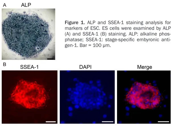
Figure 1. ALP and SSEA-1 staining analysis for markers of ESC. ES cells were examined by ALP (A) and SSEA-1 (B) staining. ALP: alkaline phos-phatase; SSEA-1: stage-specific embyronic anti -gen-1. Bar = 100 μm.
then stored in the dark overnight at 4°C prior to imaging. Images were acquired using a Laser Scanning Confocal Microscopy (Zeiss, Ger- many).
Statistical analysis
The SPSS 18.0 software (SPSS Inc; Chicago, IL, USA) was used to perform statistical analysis. All data were shown as the mean ± standard
[image:4.612.91.525.73.218.2]To further investigate the predisposition of ESC to differentiate in vivo, we measured the areas of histological components including ectoderm, mescderm and endoderm. The results showed that cartilage, epidermis, muscle and intestinal epithelium were successful generated by ESC, suggesting ESC has the capacities of totipoten-cy and can differentiate embryoid body (Figure 2).
Figure 2. Morphometric analysis of teratomas. The totipotency of ESC in vivo teratoma formation was detected by hematoxylin and eosin (HE) staining. Bar = 100 μm.
Figure 3. The mRNA expression of α-SMA and SM-MHC in differentiation of ESC at different times. Real-time Quantitative PCR (qRT-PCR) was performed to de-tect α-SMA (A and B) and SM-MHC (C and D) expression in differentiation of ESC at 7 and 14 days. β-Actin was used as internal control. α-smooth muscle actin: α-SMA; smooth muscle myosin heavy chain: SM-MHC. *P<0.05.
deviation (SD) from at least three times independently experiments. Student’s t- test or ANOVA was used to determine statistical signi- ficance. A value of P<0.05 was considered to be sta-tistically significant.
Results
ALP and SSEA-1 staining analysis for markers of ESC
Expression of pluripotency related marker genes like ALP and SSEA-1 were posi-tively localized in ESC at 5 days of culture. As shown in Figure 1, localization of these markers was cyto-plasmic and distributed ho- mogeneously all over the ESC.
[image:4.612.92.385.271.531.2]All-trans retinoic acid (atRA) and collagen IV (ColIV) enhance mouse ESC differentiation from ESC
ESC is characterized by unlimited self-renewal and the potential to differentiate into SMCs in vitro SMC [16, 17]. SMC can express specific cell markers such as α-SMA and SM-MHC [18, 19]. After pretreatment with atRA or ColIV, the mRNA expression of α-SMA and SM-MHC in dif-ferentiation of ESC at different times was deter-mined by qRT-PCR. As shown in Figure 3, the expression levels of α-SMA and SM-MHC in atRA or ColIV or atRA and ColIV treated ESC at 7 and 14 days were significantly increased compared with negative control group (P<0.05). The expression levels of α-SMA and SM-MHC in atRA and ColIV treated ESC have not statisti-cally significant.
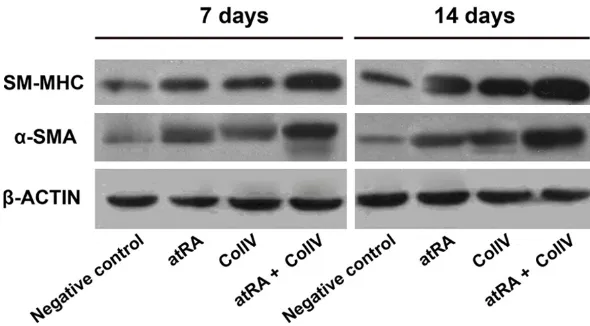
In addition, Western blot and immunofluores-cence staining was also applied to detected protein levels of α-SMA and SM-MHC in ESC. Interesting, as shown in Figures 4 and 5, we found that α-SMA and SM-MHC protein expres-sion in atRA or ColIV treated ESC at 7 and 14 days was also up-regulated compared with negative control group. The protein levels of α-SMA and SM-MHC in atRA and ColIV treated ESC have not statistically significant.
AtRA in combination with ColIV efficaciously induce the differentiation of mouse ESC into SMC
Pretreatment with atRA and ColIV in ESC, α-SMA and SM-MHC mRNA expression was
Discussion
[image:5.612.90.385.71.234.2]SMC is a key component of healthy and tissue engineered vessels and play a crucial role in vascular development and the pathogenic events of vascular remodeling [4, 5]. The over-all process of SMC differentiation is complex and involves the co-operative interaction of numerous factors. Myocardin, the transcrip-tional co-factor of serum response factor (SRF), is found to be required for the expression of many SMC differentiation markers [20]. It is crucial in the initial differentiation of SMC dur-ing development [21, 22]. Over-expression of myocardin induces ESC to express multiple SMC genes including α-SMA and SM-MHC [23]. ESC has the remarkable capability to differenti-ate into vascular SMC in response to specific stimuli, which provides a useful model for studying SMC differentiation [24, 25]. AtRA, a vitamin A derivative, exerts a wide range of bio-logical effects. Previous studies suggested that AtRA is involved in the control of cellular differ-entiation and proliferation [13]. Collagen IV (ColIV) has been reported to direct ESC differ-entiation to mesodermal lineages in both mouse and human [14]. However, the signifi-cance and exact function of atRA and ColIV on SMC differentiation remain to be elucidated. In this study, we found expression of pluripo-tency related marker genes like ALP and SSEA-1 were positively localized in ESC at 5 days of culture. The cartilage, epidermis, muscle and intestinal epithelium were successful generat-ed by ESC. These results suggestgenerat-ed that ESC
Figure 4. The protein expression of α-SMA and SM-MHC in differentiation of ESC at different times. Western blot was performed to detect α-SMA and SM-MHC expression in differentiation of ESC at 7 and 14 days.
had the capacities of totipotency and could dif-ferentiate embryoid body. Next, we detected the effects of AtRA and ColIV on SMC differen-tiation. Our results revealed that the expression levels of α-SMA and SM-MHC in atRA or ColIV treated ESC at 7 and 14 days were significantly increased compared with negative control group, while α-SMA and SM-MHC expression in atRA or ColIV treated ESC have not statistically significant. More important, Pretreatment with atRA and ColIV in ESC, α-SMA and SM-MHC expression was higher than atRA or ColIV treat-ed group. All of these data demonstrattreat-ed that both atRA and ColIV could enhance mouse ESC differentiation from ESC. More important, atRA in combination with ColIV could efficaciously induce the differentiation of mouse ESC into SMC.
In summary, we report that ESC has the poten-tial to differentiate to SMC. AtRA in combination with ColIV can efficaciously induce the differen-tiation of mouse ESC into SMC. These findings significantly increase our understanding of the molecular mechanisms in SMC differentiation and will benefit future applications in regenera-tive medicine.
Acknowledgements
This work was supported by the Project of Social Development of Guizhou Province [SY (2013)3017], the Project of Cultivation of High-Level Innovative Health Talents in Guizhou Province [(2015)4026], the Guizhou-Provincial
Outstanding Youth Science and Technology Talent Cultivation Object Special Funds [(2011) 26] and the Foreign Cooperation Project of Guizhou Province [(2011)7001]. This work was supported by the National Natural Science Foundation of China (no. 81660083).
Disclosure of conflict of interest
None.
Address correspondence to: Hongming Zhang, De- partment of Cardiology, The General Hospital of Jinan Military Region, Jinan 250031, P. R. China. Tel: (86) 531-51666666; E-mail: 13295416075@163. com
References
[1] Itskovitz-Eldor J, Schuldiner M, Karsenti D, Eden A, Yanuka O, Amit M, Soreq H and Benvenisty N. Differentiation of human embry-onic stem cells into embryoid bodies compro-mising the three embryonic germ layers. Mol Med 2000; 6: 88-95.
[2] Wobus AM and Boheler KR. Embryonic stem cells: prospects for developmental biology and cell therapy. Physiol Rev 2005; 85: 635-678. [3] Niwa H, Miyazaki J and Smith AG. Quantitative
expression of Oct-3/4 defines differentiation, dedifferentiation or self-renewal of ES cells. Nat Genet 2000; 24: 372-376.
[4] Rudijanto A. The role of vascular smooth mus-cle cells on the pathogenesis of atherosmus-clero- atherosclero-sis. Acta Med Indones 2007; 39: 86-93. [5] Tang Z, Wang A, Yuan F, Yan Z, Liu B, Chu JS,
Helms JA and Li S. Differentiation of
[image:6.612.91.527.72.264.2]tent vascular stem cells contributes to vascu-lar diseases. Nat Commun 2012; 3: 875. [6] Alexander MR and Owens GK. Epigenetic
con-trol of smooth muscle cell differentiation and phenotypic switching in vascular development and disease. Annu Rev Physiol 2012; 74: 13-40.
[7] Gittenberger-de Groot AC, DeRuiter MC, Bergwerff M and Poelmann RE. Smooth mus-cle cell origin and its relation to heterogeneity in development and disease. Arterioscler Thromb Vasc Biol 1999; 19: 1589-1594. [8] Xie CQ, Zhang J, Villacorta L, Cui T, Huang H
and Chen YE. A highly efficient method to dif -ferentiate smooth muscle cells from human embryonic stem cells. Arterioscler Thromb Vasc Biol 2007; 27: e311-312.
[9] Xiao Q, Pepe AE, Wang G, Luo Z, Zhang L, Zeng L, Zhang Z, Hu Y, Ye S and Xu Q. Nrf3-Pla2g7 interaction plays an essential role in smooth muscle differentiation from stem cells. Arter- ioscler Thromb Vasc Biol 2012; 32: 730-744. [10] Yamaguchi S, Yamahara K, Homma K, Suzuki
S, Fujii S, Morizane R, Monkawa T, Matsuzaki Y, Kangawa K and Itoh H. The role of microR-NA-145 in human embryonic stem cell differ-entiation into vascular cells. Atherosclerosis 2011; 219: 468-474.
[11] Xiao Q, Wang G, Yin X, Luo Z, Margariti A, Zeng L, Mayr M, Ye S and Xu Q. Chromobox protein homolog 3 is essential for stem cell differentia-tion to smooth muscles in vitro and in embry-onic arteriogenesis. Arterioscler Thromb Vasc Biol 2011; 31: 1842-1852.
[12] Simpson RM, Hong X, Wong MM, Karamariti E, Bhaloo SI, Warren D, Kong W, Hu Y and Xu Q. Hyaluronan is crucial for stem cell differentia-tion into smooth muscle lineage. Stem Cells 2016; 34: 1225-1238.
[13] Axel DI, Frigge A, Dittmann J, Runge H, Spyridopoulos I, Riessen R, Viebahn R and Karsch KR. All-trans retinoic acid regulates proliferation, migration, differentiation, and ex-tracellular matrix turnover of human arterial smooth muscle cells. Cardiovasc Res 2001; 49: 851-862.
[14] Schenke-Layland K, Angelis E, Rhodes KE, Heydarkhan-Hagvall S, Mikkola HK and Macl- ellan WR. Collagen IV induces trophoectoderm differentiation of mouse embryonic stem cells. Stem Cells 2007; 25: 1529-1538.
[15] Schmittgen TD and Livak KJ. Analyzing real-time PCR data by the comparative C(T) meth-od. Nat Protoc 2008; 3: 1101-1108.
[16] Gerecht-Nir S, Ziskind A, Cohen S and Itskovitz-Eldor J. Human embryonic stem cells as an in vitro model for human vascular development and the induction of vascular differentiation. Lab Invest 2003; 83: 1811-1820.
[17] Wang X, Karamariti E, Simpson R, Wang W and Xu Q. Dickkopf homolog 3 induces stem cell differentiation into smooth muscle lineage via ATF6 signalling. J Biol Chem 2015; 290: 19844-19852.
[18] Xiao Q, Luo Z, Pepe AE, Margariti A, Zeng L and Xu Q. Embryonic stem cell differentiation into smooth muscle cells is mediated by Nox4-produced H2O2. Am J Physiol Cell Physiol 2009; 296: C711-723.
[19] Xiao Q, Zeng L, Zhang Z, Hu Y and Xu Q. Stem cell-derived Sca-1+ progenitors differentiate into smooth muscle cells, which is mediated by collagen IV-integrin alpha1/beta1/alphav and PDGF receptor pathways. Am J Physiol Cell Physiol 2007; 292: C342-352.
[20] Huang J, Cheng L, Li J, Chen M, Zhou D, Lu MM, Proweller A, Epstein JA and Parmacek MS. Myocardin regulates expression of contractile genes in smooth muscle cells and is required for closure of the ductus arteriosus in mice. J Clin Invest 2008; 118: 515-525.
[21] Wang D, Chang PS, Wang Z, Sutherland L, Richardson JA, Small E, Krieg PA and Olson EN. Activation of cardiac gene expression by myo-cardin, a transcriptional cofactor for serum re-sponse factor. Cell 2001; 105: 851-862. [22] Wang Z, Wang DZ, Pipes GC and Olson EN.
Myocardin is a master regulator of smooth muscle gene expression. Proc Natl Acad Sci U S A 2003; 100: 7129-7134.
[23] Du KL, Ip HS, Li J, Chen M, Dandre F, Yu W, Lu MM, Owens GK and Parmacek MS. Myocardin is a critical serum response factor cofactor in the transcriptional program regulating smooth muscle cell differentiation. Mol Cell Biol 2003; 23: 2425-2437.
[24] Xiao Q, Wang G, Luo Z and Xu Q. The mecha-nism of stem cell differentiation into smooth muscle cells. Thromb Haemost 2010; 104: 440-448.