organic papers
Acta Cryst.(2006). E62, o1915–o1917 doi:10.1107/S1600536806013274 Wardellet al. C
8H7NO4
o1915
Acta Crystallographica Section EStructure Reports Online
ISSN 1600-5368
2-Nitrophenylacetic acid: hydrogen-bonded sheets
of
R
22
(8) and
R
44
(18) rings
James L. Wardell,aJohn N. Lowb and Christopher Glidewellc*
a
Instituto de Quı´mica, Departamento de Quı´mica Inorgaˆnica, Universidade Federal do Rio de Janeiro, CP 68563, 21945-970 Rio de Janeiro, RJ, Brazil,bDepartment of Chemistry,
University of Aberdeen, Meston Walk, Old Aberdeen AB24 3UE, Scotland, andcSchool of
Chemistry, University of St Andrews, Fife KY16 9ST, Scotland
Correspondence e-mail: cg@st-andrews.ac.uk
Key indicators
Single-crystal X-ray study
T= 120 K
Mean(C–C) = 0.002 A˚
Rfactor = 0.036
wRfactor = 0.108
Data-to-parameter ratio = 15.2
For details of how these key indicators were automatically derived from the article, see http://journals.iucr.org/e.
Received 11 April 2006 Accepted 11 April 2006
#2006 International Union of Crystallography All rights reserved
Molecules of the title compound, C8H7NO4, are linked into
centrosymmetricR2 2
(8) dimers by paired O—H O hydrogen
bonds, and these dimers are linked by two C—H O
hydrogen bonds into sheets ofR2 2
(8) andR4 4
(18) rings.
Comment
As part of our investigations of compounds containing nitro and carboxylic acid groups (Glidewell et al., 2003a,b, 2004, 2006; Wardellet al., 2005), we now report the molecular and supramolecular structure of 2-nitrophenylacetic acid, (I) (Fig. 1).
The plane of atoms C1/C11/C12 is almost orthogonal to the plane of the aryl ring (Fig. 1, Table 1), while the C—NO2plane
makes a dihedral angle of 30.1 (2)with the ring.
The molecules of (I) are linked into sheets by a combination
of N—H O and C—H O hydrogen bonds (Table 2).
Paired O—H O hydrogen bonds link the molecules into centrosymmetricR2
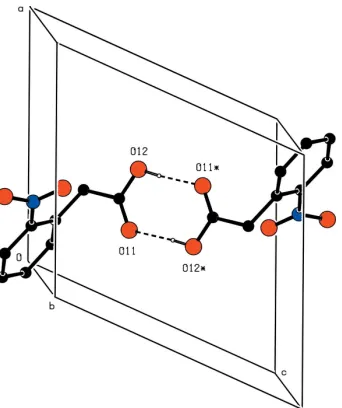
2(8) (Bernsteinet al., 1995) dimers (Fig. 2).
Two C—H O hydrogen bonds link the dimers, so forming a (100) sheet built fromR2
2(8) andR 4
4(18) rings. The resulting net
is of type (4,4) (Batten & Robson, 1998). There are no direction-specific interactions between adjacent sheets. In particular, C—H (arene) hydrogen bonds and aromatic– stacking interactions are both absent.
Experimental
A commercial sample of (I) (Acros) was crystallized from ethanol (m.p. 412–413 K).
Crystal data
C8H7NO4 Mr= 181.15
Monoclinic, P21=c a= 9.3182 (3) A˚
b= 9.4466 (2) A˚
c= 9.9733 (3) A˚
= 114.7990 (17) V= 796.95 (4) A˚3
Z= 4
Dx= 1.510 Mg m
3
MoKradiation
= 0.12 mm1
T= 120 (2) K Lath, colourless 0.520.260.10 mm
Data collection
Bruker Nonius KappaCCD area-detector diffractometer
’and!scans
Absorption correction: multi-scan (SADABS; Sheldrick, 2003)
Tmin= 0.949,Tmax= 0.988
8852 measured reflections 1829 independent reflections 1682 reflections withI> 2(I)
Rint= 0.031
max= 27.7
Refinement
Refinement onF2 R[F2> 2(F2)] = 0.037
wR(F2) = 0.108 S= 1.16 1829 reflections 120 parameters
H-atom parameters constrained
w= 1/[2(F
o2) + (0.0444P)2
+ 0.341P]
whereP= (Fo2+ 2Fc2)/3
(/)max< 0.001
max= 0.34 e A˚ 3
min=0.27 e A˚ 3
Extinction correction:SHELXL97
(Sheldrick, 1997)
Extinction coefficient: 0.103 (10)
organic papers
o1916
Wardellet al. C [image:2.610.311.566.70.226.2]8H7NO4 Acta Cryst.(2006). E62, o1915–o1917
Figure 1
The molecular structure of (I), showing the atom-labelling scheme. Displacement ellipsoids are drawn at the 30% probability level.
Figure 2
Part of the crystal structure of (I), showing the formation of a centrosymmetricR2
2(8) dimer. For the sake of clarity, H atoms bonded
[image:2.610.92.266.73.289.2]to C atoms have been omitted. Atoms marked with an asterisk (*) are at the symmetry position (1x, 1y, 1z).
Figure 3
A stereoview of part of the crystal structure of (I), showing the formation of a (100) sheet ofR2
2(8) andR44(18) rings. For the sake of clarity, H atoms
[image:2.610.311.563.303.472.2]bonded to aromatic C atoms have been omitted.
Figure 4
[image:2.610.91.262.334.538.2]Table 1
Selected torsion angles ().
C2—C1—C11—C12 83.34 (16) C1—C11—C12—O12 159.51 (11)
C1—C2—N2—O21 29.64 (17)
Table 2
Hydrogen-bond geometry (A˚ ,).
D—H A D—H H A D A D—H A
O12—H12 O11i
0.84 1.83 2.6622 (14) 173 C11—H11A O21ii
0.99 2.35 3.1758 (16) 140 C11—H11B O22iii
0.99 2.54 3.4398 (19) 151
Symmetry codes: (i) xþ1;yþ1;zþ1; (ii) xþ1;y1 2;zþ
1 2; (iii) xþ1;yþ1;z.
All H atoms were located in a difference map and then treated as riding, with C—H distances of 0.95 A˚ (aromatic) or 0.99 A˚ (CH2),
and O—H distances of 0.84 A˚ , and with Uiso(H) = 1.2Ueq(C) or
1.5Ueq(O).
Data collection: COLLECT (Nonius, 1999); cell refinement: DENZO(Otwinowski & Minor, 1997) andCOLLECT; data reduc-tion:DENZOandCOLLECT; program(s) used to solve structure: OSCAIL (McArdle, 2003) and SHELXS97 (Sheldrick, 1997); program(s) used to refine structure: OSCAIL and SHELXL97 (Sheldrick, 1997); molecular graphics:PLATON(Spek, 2003); soft-ware used to prepare material for publication: SHELXL97 and PRPKAPPA(Ferguson, 1999).
The X-ray data were collected at the EPSRC X-Ray Crys-tallographic Service, University of Southampton, UK; the authors thank the staff of the Service for all their help and advice. JLW thanks CNPq and FAPERJ for financial support.
References
Allen, F. H. (2002).Acta Cryst.B58, 380–388.
Batten, S. R. & Robson, R. (1998).Angew. Chem. Int. Ed.37, 1460–1494. Bernstein, J., Davis, R. E., Shimoni, L. & Chang, N.-L. (1995).Angew. Chem.
Int. Ed. Engl.34, 1555–1573.
Ferguson, G. (1999).PRPKAPPA. University of Guelph, Canada.
Glidewell, C., Low, J. N., Skakle, J. M. S. & Wardell, J. L. (2003a).Acta Cryst.
C59, o124–o126.
Glidewell, C., Low, J. N., Skakle, J. M. S. & Wardell, J. L. (2003b).Acta Cryst.
C59, o144–o146.
Glidewell, C., Low, J. N., Skakle, J. M. S. & Wardell, J. L. (2004).Acta Cryst.
C60, o120–o124.
Glidewell, C., Low, J. N., Skakle, J. M. S. & Wardell, J. L. (2006).Acta Cryst.
C62, o5–o7.
Grabowski, S. J., Krygowski, T. M., Ha¨felinger, G. & Ritter, G. (1990).Acta Cryst.C46, 428–430.
McArdle, P. (2003). OSCAIL for Windows. Version 10. Crystallography Centre, Chemistry Department, NUI Galway, Ireland.
Nonius (1999).COLLECT. Nonius BV, Delft, The Netherlands.
Otwinowski, Z. & Minor, W. (1997). Methods in Enzymology, Vol. 276,
Macromolecular Crystallography, Part A, edited by C. W. Carter Jr & R. M. Sweet, pp. 307–326. New York: Academic Press.
Sheldrick, G. M. (1997). SHELXS97 and SHELXL97. University of Go¨ttingen, Germany.
Sheldrick, G. M. (2003).SADABS. Version 2.10. University of Go¨ttingen, Germany.
Spek, A. L. (2003).J. Appl. Cryst.36, 7–13.
Wardell, J. L., Skakle, J. M. S., Low, J. N. & Glidewell, C. (2005).Acta Cryst.
E61, o3849–o3851.
organic papers
Acta Cryst.(2006). E62, o1915–o1917 Wardellet al. C
supporting information
sup-1
Acta Cryst. (2006). E62, o1915–o1917
supporting information
Acta Cryst. (2006). E62, o1915–o1917 [https://doi.org/10.1107/S1600536806013274]
2-Nitrophenylacetic acid: hydrogen-bonded sheets of
R
22(8) and
R
44(18) rings
James L. Wardell, John N. Low and Christopher Glidewell
2-Nitrophenylacetic acid
Crystal data
C8H7NO4
Mr = 181.15
Monoclinic, P21/c Hall symbol: -P 2ybc
a = 9.3182 (3) Å
b = 9.4466 (2) Å
c = 9.9733 (3) Å
β = 114.7990 (17)°
V = 796.95 (4) Å3
Z = 4
F(000) = 376
Dx = 1.510 Mg m−3
Mo Kα radiation, λ = 0.71073 Å Cell parameters from 1848 reflections
θ = 2.9–27.5°
µ = 0.12 mm−1
T = 120 K Lath, colourless 0.52 × 0.26 × 0.10 mm
Data collection
Bruker Nonius KappaCCD area-detector diffractometer
Radiation source: Bruker Nonius FR591 rotating anode
Graphite monochromator
Detector resolution: 9.091 pixels mm-1
φ and ω scans
Absorption correction: multi-scan (SADABS; Sheldrick, 2003)
Tmin = 0.949, Tmax = 0.988 8852 measured reflections 1829 independent reflections 1682 reflections with I > 2σ(I)
Rint = 0.031
θmax = 27.7°, θmin = 3.2°
h = −12→11
k = −11→12
l = −11→12
Refinement
Refinement on F2 Least-squares matrix: full
R[F2 > 2σ(F2)] = 0.037
wR(F2) = 0.108
S = 1.16 1829 reflections 120 parameters 0 restraints
Primary atom site location: structure-invariant direct methods
Secondary atom site location: difference Fourier map
Hydrogen site location: inferred from neighbouring sites
H-atom parameters constrained
w = 1/[σ2(F
o2) + (0.0444P)2 + 0.341P] where P = (Fo2 + 2Fc2)/3
(Δ/σ)max < 0.001 Δρmax = 0.34 e Å−3 Δρmin = −0.27 e Å−3
Extinction correction: SHELXL97 (Sheldrick, 1997), Fc*=kFc[1+0.001xFc2λ3/sin(2θ)]-1/4 Extinction coefficient: 0.103 (10)
Fractional atomic coordinates and isotropic or equivalent isotropic displacement parameters (Å2)
x y z Uiso*/Ueq
supporting information
sup-2
Acta Cryst. (2006). E62, o1915–o1917
C11 0.40523 (15) 0.29551 (14) 0.18979 (14) 0.0196 (3) C12 0.44276 (14) 0.38895 (14) 0.32300 (14) 0.0191 (3) O11 0.34184 (11) 0.43438 (11) 0.35993 (11) 0.0260 (3) O12 0.59458 (11) 0.41254 (11) 0.39762 (11) 0.0254 (3) C2 0.18252 (14) 0.41302 (13) −0.03303 (14) 0.0161 (3) N2 0.28971 (12) 0.52395 (11) −0.03878 (12) 0.0181 (3) O21 0.39974 (11) 0.56059 (10) 0.07718 (11) 0.0238 (3) O22 0.26447 (12) 0.57548 (11) −0.15940 (11) 0.0287 (3) C3 0.02718 (15) 0.42023 (14) −0.13979 (14) 0.0191 (3) C4 −0.07970 (15) 0.31978 (15) −0.13815 (15) 0.0233 (3) C5 −0.03015 (16) 0.21349 (15) −0.03267 (16) 0.0252 (3) C6 0.12610 (16) 0.20739 (14) 0.07154 (15) 0.0221 (3)
H11A 0.4257 0.1958 0.2228 0.024*
H11B 0.4775 0.3199 0.1435 0.024*
H12 0.6100 0.4558 0.4761 0.038*
H3 −0.0046 0.4929 −0.2123 0.023*
H4 −0.1866 0.3235 −0.2090 0.028*
H5 −0.1035 0.1442 −0.0315 0.030*
H6 0.1581 0.1327 0.1418 0.027*
Atomic displacement parameters (Å2)
U11 U22 U33 U12 U13 U23
C1 0.0180 (6) 0.0173 (6) 0.0159 (6) 0.0001 (4) 0.0071 (5) −0.0028 (5) C11 0.0185 (6) 0.0190 (6) 0.0184 (6) 0.0023 (4) 0.0048 (5) 0.0015 (5) C12 0.0175 (6) 0.0211 (6) 0.0163 (6) 0.0006 (5) 0.0047 (5) 0.0043 (5) O11 0.0179 (5) 0.0390 (6) 0.0195 (5) −0.0005 (4) 0.0064 (4) −0.0050 (4) O12 0.0159 (5) 0.0363 (6) 0.0202 (5) −0.0015 (4) 0.0038 (4) −0.0065 (4) C2 0.0156 (6) 0.0167 (6) 0.0168 (6) −0.0007 (4) 0.0076 (5) −0.0030 (5) N2 0.0160 (5) 0.0173 (5) 0.0202 (5) 0.0021 (4) 0.0069 (4) 0.0010 (4) O21 0.0209 (5) 0.0234 (5) 0.0228 (5) −0.0064 (4) 0.0049 (4) −0.0045 (4) O22 0.0255 (5) 0.0333 (6) 0.0250 (5) 0.0000 (4) 0.0082 (4) 0.0130 (4) C3 0.0177 (6) 0.0211 (6) 0.0166 (6) 0.0024 (4) 0.0054 (5) −0.0031 (5) C4 0.0153 (6) 0.0282 (7) 0.0234 (7) −0.0015 (5) 0.0052 (5) −0.0098 (5) C5 0.0225 (7) 0.0258 (7) 0.0295 (7) −0.0083 (5) 0.0132 (6) −0.0075 (6) C6 0.0252 (7) 0.0204 (6) 0.0219 (7) −0.0031 (5) 0.0109 (5) −0.0002 (5)
Geometric parameters (Å, º)
C1—C2 1.3931 (18) C2—N2 1.4653 (16)
C1—C6 1.3947 (18) N2—O22 1.2256 (15)
C1—C11 1.5092 (17) N2—O21 1.2303 (14)
C11—C12 1.5089 (18) C3—C4 1.3807 (19)
C11—H11A 0.99 C3—H3 0.95
C11—H11B 0.99 C4—C5 1.386 (2)
C12—O11 1.2226 (17) C4—H4 0.95
C12—O12 1.3121 (15) C5—C6 1.3907 (19)
supporting information
sup-3
Acta Cryst. (2006). E62, o1915–o1917
C2—C3 1.3930 (17) C6—H6 0.95
C2—C1—C6 116.14 (11) O22—N2—O21 123.51 (11)
C2—C1—C11 124.64 (11) O22—N2—C2 117.97 (10)
C6—C1—C11 119.19 (11) O21—N2—C2 118.51 (10)
C12—C11—C1 113.77 (10) C4—C3—C2 118.74 (12)
C12—C11—H11A 108.8 C4—C3—H3 120.6
C1—C11—H11A 108.8 C2—C3—H3 120.6
C12—C11—H11B 108.8 C3—C4—C5 119.64 (12)
C1—C11—H11B 108.8 C3—C4—H4 120.2
H11A—C11—H11B 107.7 C5—C4—H4 120.2
O11—C12—O12 123.51 (12) C4—C5—C6 120.56 (12)
O11—C12—C11 123.17 (11) C4—C5—H5 119.7
O12—C12—C11 113.29 (11) C6—C5—H5 119.7
C12—O12—H12 109.5 C5—C6—C1 121.51 (13)
C3—C2—C1 123.40 (12) C5—C6—H6 119.2
C3—C2—N2 116.23 (11) C1—C6—H6 119.2
C1—C2—N2 120.37 (10)
C2—C1—C11—C12 83.34 (16) C3—C2—N2—O21 150.02 (12)
C6—C1—C11—C12 −98.81 (14) C1—C2—N2—O21 −29.64 (17)
C1—C11—C12—O11 22.60 (18) C1—C2—C3—C4 0.71 (19)
C1—C11—C12—O12 −159.51 (11) N2—C2—C3—C4 −178.94 (11)
C6—C1—C2—C3 0.43 (19) C2—C3—C4—C5 −0.97 (19)
C11—C1—C2—C3 178.34 (12) C3—C4—C5—C6 0.1 (2)
C6—C1—C2—N2 −179.94 (11) C4—C5—C6—C1 1.1 (2)
C11—C1—C2—N2 −2.03 (19) C2—C1—C6—C5 −1.32 (19)
C3—C2—N2—O22 −29.86 (16) C11—C1—C6—C5 −179.35 (12)
C1—C2—N2—O22 150.48 (12)
Hydrogen-bond geometry (Å, º)
D—H···A D—H H···A D···A D—H···A
O12—H12···O11i 0.84 1.83 2.6622 (14) 173
C11—H11A···O21ii 0.99 2.35 3.1758 (16) 140
C11—H11B···O22iii 0.99 2.54 3.4398 (19) 151