inorganic papers
i44
David R. Allan 2Na+O8S22 doi:10.1107/S1600536806004302 Acta Cryst.(2006). E62, i44–i46
Acta Crystallographica Section E Structure Reports Online
ISSN 1600-5368
Sodium peroxodisulfate
David R. Allan
School of Chemistry, The University of Edinburgh, King’s Buildings, West Mains Road, Edinburgh EH9 3JJ, Scotland
Correspondence e-mail: [email protected]
Key indicators
Single-crystal X-ray study T= 150 K
Mean(O–O) = 0.003 A˚ Rfactor = 0.038 wRfactor = 0.034
Data-to-parameter ratio = 10.5
For details of how these key indicators were automatically derived from the article, see http://journals.iucr.org/e.
Received 18 January 2006 Accepted 3 February 2006
#2006 International Union of Crystallography
All rights reserved
The asymmetric unit of disodium peroxodisulfate 2Na+S2O8
2
consists of a single Na+ cation and half of a peroxodisulfate dianion, the latter lying across a crystal-lographic inversion centre. The crystal structure is isostruc-tural with that of potassium peroxodisulfate and it is composed of layers of molecules, partitioned by the Na+ cations, parallel to the (011) plane of the triclinic cell. Neighbouring molecules within each layer are bridged end-to-end by pairs of short S O intermolecular contacts [S O = 3.074 (2) A˚ ].
Comment
Sodium peroxodisulfate, (I), readily forms SO4radicals in hot aqueous solution. It is a powerful oxidizing and bleaching agent and it can also be used as a polymerization promoter, as well as providing a cleaner alternative to ferric chloride for copper etchant solutions (Serguchevet al., 1980).
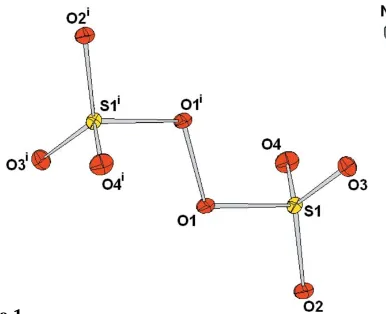
[image:1.610.227.420.569.726.2]Compund (I) crystallizes from aqueous solution in the triclinic space groupP1, with one Na+cation and half of the peroxodisulfate dianion, the latter located across an inversion centre, in the asymmetric unit (Fig. 1). Its crystal structure,
Figure 1
which is isostructural with that of potassium peroxodisulfate, K2S2O8, [Naumov et al., 1997; ICSD (Belsky et al., 2002) refcode 54024] is composed of layers of peroxodisulfate anions, which are aligned parallel to the (011) plane and partitioned by corrugated layers of Na+cations (Fig. 2).
The intramolecular S—O distances and O—S—O bond angles for the peroxodisulfate dianion are very simlar to those reported for the potassium analogue (see Table 1). However, the cation environments for the two analogues are quite different. In potassium peroxodisulfate, the K+ cations are coordinated by nine O atoms with interatomic distances ranging from 2.751 (3) to 3.347 (3) A˚ . For sodium peroxo-disulfate, the Na+cations are coordinated by six O atoms, with Na—O interatomic distances between 2.340 (2) and 2.596 (2) A˚ . It is interesting to note that in the sodium
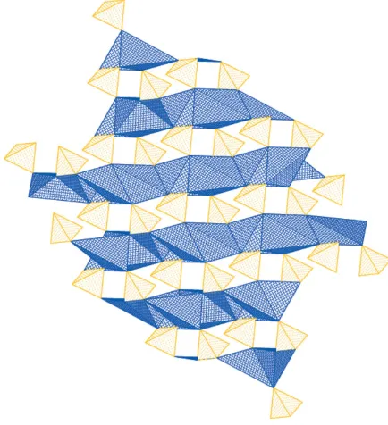
analogue, the O atom involved in the intramolecular peroxo bond, O1, is not involved in the cation coordinate environ-ment, although it does exhibit the shortest Na—O distance outside this range [Na1—O1 = 3.167 (2) A˚ ]. The K1—O1 distance in the potassium analogue [3.089 (3) A˚ ] is of an intermediate length compared with the other O atoms defining the coordination environment. The overall effect of the Na— O coordination environment in sodium peroxodisulfate is the formation of a three-dimensional network. This is indicated by the polyhedral plot shown in Fig. 3. The Na+ cations form layers of distorted edge-sharing octahedra (shown as the blue polyhedra in Fig. 3), while the tetrahedra formed by each end of the dianions (shown as the yellow polyhedra in Fig. 3) form corner-sharing bridges between the layers.
Perhaps the most striking difference between the two structures concerns the S O intramolecular contact distances. Within the layers, neighbouring anions are aligned end-to-end so that pairs of relatively short S O contacts are formed. In the sodium analogue these contacts are extremely short [S1 O3 = 3.074 (2) A˚ ] (Fig. 4), while in the crystal structure of the potassium analogue these contacts are significantly longer [S1 O3 = 3.417 (3) A˚ ].
Experimental
The sample of sodium peroxodisulfate was prepared from anhydrous starting material (of 99% purity, as received from Aldrich) and recrystallized from an aqueous solution by slow evaporation. A suitable crystal was selected from the resulting batch. The sample was cooled to 150 K using an Oxford Cryosystems low-temperature device (Cosier & Glazer, 1986) during data collection.
Crystal data
2Na+O 8S22
Mr= 238.11
Triclinic,P1 a= 4.780 (2) A˚ b= 5.575 (2) A˚ c= 6.091 (3) A˚ = 101.871 (7)
= 103.337 (7)
= 97.418 (7)
V= 151.89 (11) A˚3
Z= 1
Dx= 2.603 Mg m
3 MoKradiation Cell parameters from 680
reflections = 7–57
= 1.02 mm1 T= 150 K Needle, colourless 0.200.050.05 mm
Data collection
Bruker SMART diffractometer ’and!scans
Absorption correction: multi-scan SADABS(Sheldrick, 2004) Tmin= 0.67,Tmax= 0.95 1326 measured reflections 696 independent reflections
590 reflections withI> 2(I) Rint= 0.021
max= 28.9
h=6!6 k=7!7 l=8!7
inorganic papers
Acta Cryst.(2006). E62, i44–i46 David R. Allan 2Na+O
[image:2.610.44.296.72.215.2]8S22
i45
Figure 3A polyhedral representation of the crystal structure of sodium peroxodisulfate. The structure is viewed along thecaxis, with theaaxis directed to the right and thebaxis directed upwards. The blue polyhedra indicate the layers of distorted edge-sharing NaO6octahedra while the
[image:2.610.315.565.72.117.2]yellow tetrahedra indicate the SO4groups of the peroxodisulfate dianion.
Figure 4
The dashed lines indicate short S O contacts in sodium peroxodisulfate. This view is approximately perpendicular to (011).
Figure 2
[image:2.610.59.276.260.498.2]Refinement
Refinement onF R[F2> 2(F2)] = 0.038 wR(F2) = 0.034 S= 1.06 590 reflections 56 parameters
Modified Chebychev polynomial (Watkin, 1994; Prince, 1982) with the coefficients 1.89,1.11, 1.17 (/)max< 0.001
max= 0.48 e A˚
3
min=0.42 e A˚
3
Table 1
Selected geometric parameters (A˚ ,).
S1—O1 1.6392 (19)
S1—O2 1.4389 (18)
S1—O3 1.4408 (19)
S1—O4 1.4396 (19)
O1—O1i
1.479 (3)
Na1—O3 2.376 (2)
Na1—O3ii
2.380 (2) Na1—O2iii
2.389 (2) Na1—O2iv
2.476 (2) Na1—O4v
2.340 (2) Na1—O4vi
2.596 (2)
O1—S1—O2 97.30 (11)
O1—S1—O3 105.92 (11)
O1—S1—O4 106.87 (11)
O2—S1—O3 115.78 (12)
O2—S1—O4 115.65 (12)
O3—S1—O4 113.08 (11)
O1i—O1—S1 106.26 (17)
Symmetry codes: (i)xþ1;yþ2;zþ1; (ii)x;yþ1;z; (iii)x1;y1;z; (iv)xþ1;yþ1;z; (v)xþ1;yþ1;zþ1; (vi)x1;y;z.
Indexing withGEMINI(Sparks, 1999) revealed that the sample was twinned non-merohedrally with two domains. The data set was integrated using the orientation matrix of the stronger subset of reflections, corresponding to the larger domain. During refinement, the ROTAXprocedure, as implemented in theCRYSTALS refine-ment package (Cooper et al., 2002), was used to identify the rela-tionship between the two domains. This could be expressed by the matrix (100, 010, 0.667 0.523 1), which corresponds to a twofold rotation about thec* axis. Subsequent refinement indicated that the twin fraction of the second domain was 0.379 (8).
Data collection:SMART(Bruker Nonius, 2001); cell refinement:
SAINT; data reduction:SAINT(Bruker Nonius, 2003); program(s)
used to solve structure:SIR92 (Altomare et al., 1994); program(s) used to refine structure: CRYSTALS (Betteridge et al., 2003); molecular graphics:CAMERON(Watkinet al., 1996); software used to prepare material for publication: CRYSTALS and PLATON
(Spek, 2003).
We thank Dr F. P. A. Fabbiani for her help during the data collection and Professor A. J. Blake for his help in preparing this manuscript. We also thank the EPSRC for funding both this project and DRA’s Advanced Research Fellowship.
References
Altomare, A., Burla, M. C., Camalli, G., Cascarano, G., Giacovazzo, C., Guagliardi, A. & Polidori, G. (1994).J. Appl. Cryst.27, 435.
Belsky, A., Hellenbrandt, M., Karen, L.V. & Luksch, P. (2002).Acta Cryst.B58, 364–369.
Betteridge, P. W., Carruthers, J. R., Cooper, R. I., Prout, K. & Watkin, D. J. (2003).J. Appl. Cryst.36, 1487.
Bruker–Nonius (2001). SMART. Bruker–Nonius AXS Inc., Madison, Wisconsin, USA.
Bruker–Nonius (2003). SAINT. Bruker–Nonius AXS Inc., Madison, Wisconsin, USA.
Cooper, R. I., Gould, R. O., Parsons, S. & Watkin, D. J. (2002).J. Appl. Cryst.
35, 168–174.
Cosier, J. & Glazer, A. M. (1986).J. Appl. Cryst.19, 105–107. Naumov, D. Yu. & Virovets, A. V. (1997).J. Struct. Chem.38, 772–778. Prince, E. (1982).Mathematical Techniques in Crystallography and Materials
Science. New York: Springer–Verlag.
Serguchev, Yu. A. & Beletskaya, I. P. (1980).Usp. Khim.49, 2257–2285. Sheldrick, G. M. (2004).SADABS. University of Gottingen, Germany. Sparks, R. A. (1999).GEMINI. Bruker AXS Inc, Madison, Wisconsin, USA. Spek, A. L. (2003).J. Appl. Cryst.36, 7–13.
Watkin, D. J., (1994).Acta Cryst.A50, 411–437.
Watkin, D. J., Prout, C. K. & Pearce, L. (1996). CAMERON. Chemical Crystallography Laboratory, University of Oxford, England.
inorganic papers
i46
David R. Allan 2Na+Osupporting information
sup-1 Acta Cryst. (2006). E62, i44–i46
supporting information
Acta Cryst. (2006). E62, i44–i46 [https://doi.org/10.1107/S1600536806004302]
Sodium peroxodisulfate
David R. Allan
disodium peroxodisulfate
Crystal data
2Na+·O 8S22−
Mr = 238.11
Triclinic, P1 Hall symbol: -P 1
a = 4.780 (2) Å
b = 5.575 (2) Å
c = 6.091 (3) Å
α = 101.871 (7)°
β = 103.337 (7)°
γ = 97.418 (7)°
V = 151.89 (11) Å3
Z = 1
F(000) = 118
Dx = 2.603 Mg m−3
Mo Kα radiation, λ = 0.71073 Å Cell parameters from 680 reflections
θ = 7–57°
µ = 1.02 mm−1
T = 150 K Needle, colourless 0.20 × 0.05 × 0.05 mm
Data collection
Bruker SMART diffractometer
Graphite monochromator
φ and ω scans
Absorption correction: multi-scan SADABS (Sheldrick, 2004)
Tmin = 0.67, Tmax = 0.95 1326 measured reflections
696 independent reflections 590 reflections with I > 2σ(I)
Rint = 0.021
θmax = 28.9°, θmin = 3.6°
h = −6→6
k = −7→7
l = −8→7
Refinement
Refinement on F
Least-squares matrix: full
R[F2 > 2σ(F2)] = 0.038
wR(F2) = 0.034
S = 1.06 590 reflections 56 parameters 0 restraints
Primary atom site location: structure-invariant direct methods
Modified Chebychev polynomial (Watkin, 1994; Prince, 1982) with the coefficients 1.89, -1.11, 1.17
supporting information
sup-2 Acta Cryst. (2006). E62, i44–i46
Special details
Refinement. ABSTM02_ALERT_3_C The ratio of expected to reported Tmax/Tmin(RR′) is < 0.90 T min and Tmax reported: 0.670 0.950 T min(prime) and Tmax expected: 0.814 0.950 RR(prime) = 0.824 Please check that your absorption correction is appropriate. PLAT061_ALERT_3_C Tmax/Tmin Range Test RR′ too Large ···. 0.82 The crystal was a fine needle and the absorption correction correspondingly anisotropic.
PLAT029_ALERT_3_C _diffrn_measured_fraction_theta_full Low ···. 0.97 This value is only marginally smaller than the ideal (0.99).
PLAT432_ALERT_2_C Short Inter X···Y Contact S1.. O3.. 3.07 A ng.
This short intermolecular contact bridges persulfate molecules end-on to form infinite chains. This interaction is similar to that observed by Naumov et al. (1997) for the isostructural potassium persulfate where the molecules are also bridged by short S···O intermolecular bonds. In this instance, however, the distance is 3.417 A ng.
Fractional atomic coordinates and isotropic or equivalent isotropic displacement parameters (Å2)
x y z Uiso*/Ueq
Na1 0.0359 (2) 0.31397 (19) 0.20685 (18) 0.0131 S1 0.61154 (13) 0.78124 (11) 0.24744 (11) 0.0080 O1 0.5785 (4) 1.0388 (3) 0.4180 (3) 0.0143 O2 0.7722 (4) 0.8959 (3) 0.1113 (3) 0.0113 O3 0.3176 (4) 0.6526 (3) 0.1263 (3) 0.0111 O4 0.7701 (4) 0.6451 (3) 0.3949 (3) 0.0130
Atomic displacement parameters (Å2)
U11 U22 U33 U12 U13 U23
Na1 0.0141 (5) 0.0136 (5) 0.0102 (5) 0.0005 (4) 0.0012 (4) 0.0032 (4) S1 0.0088 (3) 0.0082 (3) 0.0068 (3) 0.00165 (19) 0.0025 (2) 0.00122 (19) O1 0.0223 (10) 0.0094 (8) 0.0132 (9) −0.0009 (7) 0.0125 (8) 0.0006 (7) O2 0.0135 (9) 0.0106 (8) 0.0112 (8) 0.0013 (7) 0.0063 (7) 0.0031 (6) O3 0.0099 (9) 0.0106 (8) 0.0114 (8) 0.0004 (7) 0.0016 (7) 0.0017 (7) O4 0.0119 (9) 0.0165 (9) 0.0116 (9) 0.0046 (7) 0.0013 (7) 0.0065 (7)
Geometric parameters (Å, º)
S1—O1 1.6392 (19) Na1—O3ii 2.380 (2) S1—O2 1.4389 (18) Na1—O2iii 2.389 (2) S1—O3 1.4408 (19) Na1—O2iv 2.476 (2) S1—O4 1.4396 (19) Na1—O4v 2.340 (2) O1—O1i 1.479 (3) Na1—O4vi 2.596 (2) Na1—O3 2.376 (2) Na1—Na1ii 3.565 (2)
supporting information
sup-3 Acta Cryst. (2006). E62, i44–i46
O4v—Na1—O2iii 97.98 (8) Na1ii—Na1—O3 41.50 (5) O3ii—Na1—O2iii 83.62 (7) Na1vii—O2—Na1iv 102.44 (7) O4v—Na1—O2iv 126.29 (8) Na1vii—O2—S1 127.66 (11) O3ii—Na1—O2iv 78.77 (7) Na1iv—O2—S1 127.67 (11) O2iii—Na1—O2iv 77.56 (7) Na1—O3—Na1ii 97.08 (8) O4v—Na1—O4vi 77.36 (7) Na1—O3—S1 128.52 (11) O3ii—Na1—O4vi 79.00 (7) Na1ii—O3—S1 134.38 (11) O2iii—Na1—O4vi 113.94 (8) Na1v—O4—Na1viii 102.64 (7) O2iv—Na1—O4vi 153.52 (7) Na1v—O4—S1 137.82 (12) O4v—Na1—Na1ii 139.91 (7) Na1viii—O4—S1 117.76 (11) O3ii—Na1—Na1ii 41.42 (5)
Symmetry codes: (i) −x+1, −y+2, −z+1; (ii) −x, −y+1, −z; (iii) x−1, y−1, z; (iv) −x+1, −y+1, −z; (v) −x+1, −y+1, −z+1; (vi) x−1, y, z; (vii) x+1, y+1, z; (viii)