CLINICOPATHOLOGICAL STUDY OF SOFT TISSUE TUMORS
1,*
Dr. Murali
1
Tutor, Department of Pathology,
2
Professor, Department of Pathology, Vijayanagar
ARTICLE INFO ABSTRACT
Background:
nonepithelial, extra skeletal structures. Soft tissue includes the fibrous tissue, adipose tissue, skeletal muscle and smooth muscle tissue, peripheral nervous tissue and vascul
tissue.Objectives
clinicopathological correlation of soft tissue tumors with respect to incidence of age, sex, site distribution & histopathological types of soft t
study conducted in the Department of Pathology, Vijayanagar Institute of Medical Sciences, Ballari, Karnataka. The study evaluated all the soft tissue tumors received between January 2017 to june 2018. Me
and processed through paraffin wax embedding method. Sections were cut at 5 micrometer thickness and stained by Hematoxylin and Eosin (H&E) stain. All tumors were ex
microscope and classified as per WHO classification.
90.8% were benign, 3.06% were intermediate and 6.12% were malignant. Male to female ratio was 1.04:1. Most common sites were extremities
benign lesions, the commonest was lipoma (37.7%) followed by hemangioma (22.4%). Other benign tumors include fibroblastic (3.06%), fibro
Intermediate grade s
case (1.02%) inflammatory myofibroblastic tumor. Malignant tumors were pleomorphic liposarcoma (1.02%), fibrosarcoma (2.04%), monophasic synovial sarcoma (1.02%) and undiffe pleomorphic sarcoma (2.04%).
tumors. A thorough clinical history, gross, and light microscopic evaluation of hematoxylin and eosin stained sections are fundamental aspects in the diagno
clinicopathological evaluation is still the gold standard for the diagnosis of soft tissue neoplasms.
Copyright © 2019, Murali Krishna and Shanth. This unrestricted use, distribution, and reproduction in any medium,
INTRODUCTION
Soft tissue is defined as the no nepithelial, extra skeletal tissue exclusive of the reticuloendothelial system, glial
supporting tissue of various parenchymatous organs. By convention it also includes the peripheral nervous system because tumors arising from the nerves present as soft tissue masses (Goldblum, 2013). There are more than 100 histological subtypes of Soft tissue sarcoma, which are often associated with unique clinical, prognostic and therapeutic features (Fletcher, 2013). Hence these are divided into four categories: benign, intermediate (locally aggressive), intermediate (rarely metastasising) and malignant
*Corresponding author: Dr. Murali Krishna, E.
Tutor, Department of Pathology, Vijayanagar Institute of Medical Sciences, Ballari, Karnataka, India.
ISSN: 0975-833X
Article History:
Received 04th August, 2019
Received in revised form
28th September, 2019
Accepted 25th October, 2019
Published online 26th November, 2019
Citation:Dr. Murali Krishna, E. and Dr. Shanthi, M.
(11), 8422-8426.
Key Words:
Clinicopathological, Benign soft Tissue Tumor,
Malignant soft Tissue Tumors, Sarcoma.
RESEARCH ARTICLE
CLINICOPATHOLOGICAL STUDY OF SOFT TISSUE TUMORS
Dr. Murali Krishna, E. and
2Dr. Shanthi, M.
Tutor, Department of Pathology, Vijayanagar Institute of Medical Sciences, Ballari, Karnataka, India
Professor, Department of Pathology, Vijayanagar Institute of Medical Sciences, Ballari, Karnataka, India
ABSTRACT
Background: Soft tissue includes supportive connective tissue of various organs and other nonepithelial, extra skeletal structures. Soft tissue includes the fibrous tissue, adipose tissue, skeletal muscle and smooth muscle tissue, peripheral nervous tissue and vascul
Objectives: To study the incidence of soft tissue tumors in our institute. To study the clinicopathological correlation of soft tissue tumors with respect to incidence of age, sex, site distribution & histopathological types of soft tissue neoplasms. Materials and Methods
study conducted in the Department of Pathology, Vijayanagar Institute of Medical Sciences, Ballari, Karnataka. The study evaluated all the soft tissue tumors received between January 2017 to june 2018. Method of statistics used was descriptive analysis. All the specimens were fixed in formalin and processed through paraffin wax embedding method. Sections were cut at 5 micrometer thickness and stained by Hematoxylin and Eosin (H&E) stain. All tumors were ex
microscope and classified as per WHO classification. Result: Out of 98 cases of soft tissue tumors, 90.8% were benign, 3.06% were intermediate and 6.12% were malignant. Male to female ratio was 1.04:1. Most common sites were extremities (64.2%) followed by head and neck (21.4%).Of all benign lesions, the commonest was lipoma (37.7%) followed by hemangioma (22.4%). Other benign tumors include fibroblastic (3.06%), fibro-histiocytic (2.04%) and pericytic tumors (1.02%). Intermediate grade soft tissue tumors include 2 cases (2.04%) of well differentiated liposarcoma and 1 case (1.02%) inflammatory myofibroblastic tumor. Malignant tumors were pleomorphic liposarcoma (1.02%), fibrosarcoma (2.04%), monophasic synovial sarcoma (1.02%) and undiffe pleomorphic sarcoma (2.04%). Conclusion: Benign soft tissue tumors outnumbered malignant tumors. A thorough clinical history, gross, and light microscopic evaluation of hematoxylin and eosin stained sections are fundamental aspects in the diagnosis of soft tissue tumors. The clinicopathological evaluation is still the gold standard for the diagnosis of soft tissue neoplasms.
This is an open access article distributed under the Creative Commons medium, provided the original work is properly cited.
Soft tissue is defined as the no nepithelial, extra skeletal tissue exclusive of the reticuloendothelial system, glial and the supporting tissue of various parenchymatous organs. By convention it also includes the peripheral nervous system because tumors arising from the nerves present as soft tissue . There are more than 100 of Soft tissue sarcoma, which are often associated with unique clinical, prognostic and therapeutic . Hence these are divided into four categories: benign, intermediate (locally aggressive), malignant (Jain, 2014).
Dr. Murali Krishna, E.,
Tutor, Department of Pathology, Vijayanagar Institute of Medical
An attempt is being made to study the incidence, and clinical presentation of these soft tissue tumors
attempts to study the histomorphology of this complex group of tumors, taking into account the relevant age, sex and site related information.
MATERIALS AND METHODS
The present study is a prospective study for one and half year. All the cases diagnosed clinically as soft tissue tumors and that were surgically excised and sent to the Department of Pathology, VIMS, Ballari, are taken for present study, during the period from January 2017 to June 2018. Data for the prospective study were obtained from clinical records of surgery department and records from histopathology section. All the surgically resected specimens with clinical diagnosis of soft tissue tumors along with detailed clinical parameters are subjected for the present study.
International Journal of Current Research Vol. 11, Issue, 11, pp.8422-8426, November, 2019
DOI: https://doi.org/10.24941/ijcr.37193.11.2019
Krishna, E. and Dr. Shanthi, M. 2019. “Clinicopathological study of soft tissue tumors”, International Journal of Current Research
Available online at http://www.journalcra.com
z
CLINICOPATHOLOGICAL STUDY OF SOFT TISSUE TUMORS
, Ballari, Karnataka, India
, Ballari, Karnataka, India
Soft tissue includes supportive connective tissue of various organs and other nonepithelial, extra skeletal structures. Soft tissue includes the fibrous tissue, adipose tissue, skeletal muscle and smooth muscle tissue, peripheral nervous tissue and vascular endothelial : To study the incidence of soft tissue tumors in our institute. To study the clinicopathological correlation of soft tissue tumors with respect to incidence of age, sex, site
Materials and Methods: Prospective study conducted in the Department of Pathology, Vijayanagar Institute of Medical Sciences, Ballari, Karnataka. The study evaluated all the soft tissue tumors received between January 2017 to june thod of statistics used was descriptive analysis. All the specimens were fixed in formalin and processed through paraffin wax embedding method. Sections were cut at 5 micrometer thickness and stained by Hematoxylin and Eosin (H&E) stain. All tumors were examined under light : Out of 98 cases of soft tissue tumors, 90.8% were benign, 3.06% were intermediate and 6.12% were malignant. Male to female ratio was (64.2%) followed by head and neck (21.4%).Of all benign lesions, the commonest was lipoma (37.7%) followed by hemangioma (22.4%). Other benign histiocytic (2.04%) and pericytic tumors (1.02%). oft tissue tumors include 2 cases (2.04%) of well differentiated liposarcoma and 1 case (1.02%) inflammatory myofibroblastic tumor. Malignant tumors were pleomorphic liposarcoma (1.02%), fibrosarcoma (2.04%), monophasic synovial sarcoma (1.02%) and undifferentiated : Benign soft tissue tumors outnumbered malignant tumors. A thorough clinical history, gross, and light microscopic evaluation of hematoxylin and eosin sis of soft tissue tumors. The clinicopathological evaluation is still the gold standard for the diagnosis of soft tissue neoplasms.
Commons Attribution License, which permits
An attempt is being made to study the incidence, and clinical f these soft tissue tumors. The present study attempts to study the histomorphology of this complex group of tumors, taking into account the relevant age, sex and site
MATERIALS AND METHODS
The present study is a prospective study for one and half year. All the cases diagnosed clinically as soft tissue tumors and that were surgically excised and sent to the Department of Pathology, VIMS, Ballari, are taken for present study, during from January 2017 to June 2018. Data for the prospective study were obtained from clinical records of surgery department and records from histopathology section. All the surgically resected specimens with clinical diagnosis of
ith detailed clinical parameters are subjected for the present study.
INTERNATIONAL JOURNAL OF CURRENT RESEARCH
The resected specimens were fixed in 10% buffered formalin and detailed gross examination of the tumor is carried out. Sections of 3-5 micron thickness were made with rotary microtome and stained with routine Hematoxylin and Eosin. Special histochemical stains like Vangeison’s, PAS, Masson’s trichome and phosphotungstic acid hematoxylin (PTAH) were carried out wherever necessary.
RESULTS
During the period from January 2017 to June 2018 a total of 4851 specimens were received at the Department of Pathology, Vijayanagar Institute of Medical Sciences, Ballari. Soft tissue tumors, which included benign, intermediate and malignant tumors accounted for a total number of 98 cases which comprised 2% of the received specimens and 12% of all neoplasms.
Distribution of tumors: The 98 cases of soft tissue tumors comprised of 89 benign, 3 intermediate (locally agressive) and 6 malignant tumors.
Age incidence: The age incidence ranged from a 1 year to 73
years. The average age was 32.8 years. The different soft tissue tumors in different age groups showed that the commonest age group was the 3rd decade followed by 4th decade of life, comprising of 27.5% and 21.14% respectively.
Benign tumors extended over the entire age range, with the peak occurring in the third decade of life. The sarcomas were distributed between the fourth and sixth decades with an average of 45 years.
Sex incidence: Of the 98 soft tissue tumors, 50 occurred in males (51.02%) and 48 in females (48.97 %). With the male female ratio of 1.04:1. In females benign tumors constituted 43.8% of and malignant tumors constituted of 5.1%. The sex ratio among benign tumors was 1.09:1 and among the malignant was 1:5.
Site distribution: The sites of various soft tissue tumors were based in anatomical sites and were categorized into, head and neck, upper limb and lower limbs (extremities), abdomen and back. Majority of the tumors were seen in upper limb and lower limbs (extremities) comprising of 63 cases (64.2%) of all soft tissue tumors, followed by head and neck, 21 cases (21.4%). Least commonly involved was the abdomen, with only 6 cases (6.1%) (Table 2). The benign tumors were predominatly seen invloving extremities in 53 cases (54%), in keeping with the overall picture. The extremities were also the site affected most often among the intermediate and malignant tumors (Chart 3)
Clinical presentation: In the present study majority (74.4%) of the tumors measured ≤5cm. Most tumors presented solitary swelling and few presented as multiple swellings such as lipomas and neurofibromas. Majority of the tumors are painless, slow growing asymptomatic masses 79 cases (80.6%). Pain was a symptom in some sarcomas and some cases of neurofibroma and few cases of lipomas. Most of the tumors are in subcutaneous plain about 86 cases (87.7%) (Table.1)
[image:2.595.315.557.48.195.2]Chart 1. Site distribution of 98 tumors within benign, intermediate and malignant
Table 1. Clinical presentation of soft tissue tumors
Clinical features No. of cases Percent
Size of the swelling ≤5cm
>5 cm
73 25
74.4% 25.5% Number of swellings
Solitary Multiple
91 7
92.8% 7.14% Pain
Present Absent
19 79
19.3% 80.6% Plain of the swelling
Subcutaneous Intramuscular
86 12
[image:2.595.317.548.251.560.2]87.7% 12.2%
Fig 1. Well differentiatdliposarcoma with mature adipocytes, atypical cells showing large irregular hyperchromatic nuclei, vacuolated cytoplasm (H&E.400x)
Fig.2. Monophasic synovial sarcoma showing monotonous cells with ovoid to spindled vesicular nuclei intervening stroma &
[image:2.595.322.542.617.759.2]Fig. 3. Gross image showing extensive necrosis and infiltration into deep muscular Plain
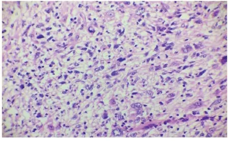
Fig. 4. Undifferentiated pleomorphic sarcoma showing irregular fascicles, pleomorphic and bizarre tumor cells with foamy cytoplasm, atypical mitotic figures andgaint cells (H&E 400x)
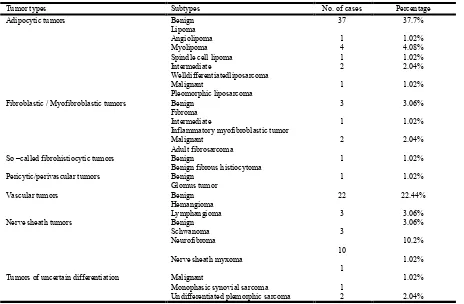
Distribution of individual tumors: Out of 98 soft tissue tumors most common tumor is lipoma with 37 tumors (37.7%) followed by hemangioma 22 cases (22.44%). Neurofibromas were the third most common tumor with 10 cases (10.2%) (Table 2).
DISCUSSION
Soft tisue tumors are extraskeletal tumors derived from cells of mesenchymal origin. Soft tissue tumors comprise a wide array of entities, ranging from the commonly occuringlipomas to rarlyoccruing sarcomas
Incidence of soft tissue tumors: Since many benign tumors, especially lipomas go unresected, an accurate estimate of the prevalence of soft tissue tumors in the general population is difficult to determine. Thus, data obtained from hospital-based studies may not reflect the true prevalence of soft tissue tumors in the general population. While the incidence of benign tumors is generally difficult to estimate, sarcomas have been reported to account for less than 1% of all neoplasms. Of late, a rising trend has also been noted. The present study encountered 98 cases of soft tissue tumors during the period January 2017 to June 2018. This represented 2% of all the specimens received during the same period. This closely approximates with 2.4 % and 1.3% incidence reported by Pramila et al and Janaki et al respectively. Benign soft tissue tumors, in general, vastly outnumber the malignant ones. Enzinger and Weiss in their textbook on soft tissue tumors quotes ratio as high as 100: 1. However Pramila et al. (2014), Janaki et al. (2015) and Megha Sharma et al. (2015) found the ratio to be 9.5:1, 21.4:1 and 8.8:1 respectively.
In the present study the ratio obtained was 14.8:1 which is similar to study by Agravat et al.
Age distribution: No age is exempt from soft tissue tumors. The age incidence in soft tissue tumors vary according to the histologic types. Tumors like hemangiomas and lipomas were common in the young whereas intermediate grade and sarcomas were common in older age group. In the present study the ages ranged from 1 year to 73 years, the mean being 32.5 years. Benign tumors had a peak incidence in the third decade of life while the malignant lesions were distributed in the fourth and sixth decades. Janaki et al and Venkatraman et al observed a peak incidence of benign in fourth and fifth decade of life respectively, among benign lesions. The age of sarcomas ranged from 38 to 53 years with incidence of cases distributed in the fourth and sixth decades. Umarani et al also had a similar range. However Pramila et al and Janaki et al detected peak incidence only in sixth decade.
Sex incidence: Overall, there was almost equal incidence among males and females, the male female ratio being 1.04:1. The difference was not much within the category of benign tumors, but among the intermediate males constituted 2 out of 3 cases and in malignant lesions, females constituted as much as 83 % of the tumors. Janaki et al. (2015) observed male: female ratio of 1: 1 similar to the present study. The male: female ratio among sarcomas noted in the present study was 1:5 unlike other studies which had male predominance. The variation could be accounted by the fact that only few cases of malignant tumors were documented in the present study, or by the low incidence of malignant tumors in our geographical area thereby perhaps masking the true nature of the sex distribution. Other studies, however did detect a slightly higher incidence in males.
Site distribution: Soft tissue tumors may occur anywhere in the body. In the present study extremities were the most commonly affected region (64.2%) followed by head and neck, abdomen and back. Similar pattern of occurrence were witnessed by Agravat et al and other authors.
Clinical features of soft tissue sarcoma: The present study had six cases of sarcomas, of these 4 cases (66.6%) presented with pain and 2 cases (33.3%) were painless. Of the 6 cases of sarcomas, majority (83.3%) were presented in deeper plain. Johnson CJ et al9 studied 223 cases of sarcomas, in their study 59.1% cases were painless and 93.7% cases had swelling in deeper plain.
Distribution of histologic subtypes
Benign tumors: The present study found lipomas to be the commonest type comprising 37 of all tumors. In JanakiM et al5 series lipomas turned out to be the single most common benign tumor. Other studies conducted by Agravat et al and Pramila et al had 29% and 47% cases of lipomas respectively.
Intermediate tumors: In present study intermediate grade soft tissue consists of 3.06% of cases of all the 98 tumors. In Janaki et al reported similar values, 3.8% intermediate grade tumors and most common being fibroblastic tumors.
[image:3.595.52.276.236.375.2]certain studies reported adipocytic tumors, fibroblastic tumors while other studies show skeletal muscle tumors, tumors of uncertain differentiation were the usual finding. However, Umarani et al. (2015) also had tumors of uncertain differentiation as the most common soft tissue sarcoma in their series similar to present study. The present study had not come across tumors from smooth muscle and skeletal muscle origin within the period of the study. The commonest single group of soft tissue tumors found were adipocytic tumors (46.9%), which included lipomas, angiolipomas, myolipoma, spindle cell lipoma and liposarcomas (Fig 1). The most common benign tumor was lipomas (37,7%), followed by hemangiomas with 22 cases (22.4%). Hemangiomas were commonest in the younger age group and occurred in the region of the head and neck. Lipomas were seen most commonly over extremities.
The other benign tumors included fibroma (3 cases), benign fibrous histiocytoma (2 cases), glomus tumor (1 case), lymphangioma (3 cases), schwannoma (3 cases), neurofibroma (10 cases) and nerve sheath myxoma (1 case). The intermediate soft tissue tumors in this study were, well differentiated liposarcoma (2 cases) and inflammatory myofibroblastic tumor (1 case). Among sarcomas, most common were fibrosarcoma and undifferentiated pleomorphic sarcoma each were 2 in number. All the sarcomas were located in the extremities. The other soft tissue sarcomas encountered in this study were, pleomorphic liposarcoma (1 case) (Fig 4) and monophasic synovial sarcoma (1 case) (Fig 3).
Conclusion
The present study comprised 98 cases of soft tissue tumors. Soft tissue tumors are highly heterogenous group of tumors. Benign soft tissue tumors outnumbered the malignant tumors by a difference of 14.8:1. In the present study, 89 cases were diagnosed as benign tumors, 3 cases as borderline malignant tumors and 6 cases as malignant tumors.
Male to female ratio was 1.04:1. Overall the results are almost in accordance with the figures recorded in the various studied literatures. A careful gross examination of the specimen and adequate sampling of the tumor is essential. Immunohistochemistry and Special stains are helpful in addition to the routine Hematoxylin and eosin for the proper diagnosis of Soft tissue tumors. Availability of a modern, more logical histopathologic classification and standard nomenclature now offers a better clinico pathological co-relation. In conclusion, clinicopathological evaluation is still the gold standard for the proper diagnosis of soft tissue tumors. It can be said that this study recapitulated the diverse nature of lesions that is encompassed under the term 'soft tissue tumors’!
Conflict of Interest: Nil
Funding: Nil
REFERENCES
Agravat AH., Dhruva GA., Parmar SA. 2010. Histopathology Study of Human's Soft Tissue Tumors And Tumor Like Lesions. Journal of cell and Tissue Research., 10(2):2287. Fletcher CD. 2013. Soft tissue tumors: Epidemiology, clinical
features, histopathological typing and grading. Lyon:
International Agency for Reasearch on Cancer Press., p2-9 Gogi, AM., Ramanujam R. 2013. Clinicopathological study and management of peripheral soft tissue tumors. Journal of clinical and diagnostic research: 7(11):2524..
Goldblum JR., Weiss SW., Folpe AL. 2013. Enzinger and Weiss's Soft Tissue Tumors; General considerations. 6thed. Philadelphia: Mosby Elsevier., p1-11
Jain P., Shrivastava A., Malik R. 2014. Clinicomorphological assessment of soft tissue tumors. Sah J App Med Sci.,
[image:4.595.65.529.77.381.2]2(2D):886-90. Table 2. Distribution of individual tumors
Tumor types Subtypes No. of cases Percentage
Adipocytic tumors Benign
Lipoma
37 37.7%
Angiolipoma 1 1.02%
Myolipoma 4 4.08%
Spindle cell lipoma 1 1.02%
Intermediate
Welldifferentiatedliposarcoma
2 2.04%
Malignant
Pleomorphic liposarcoma
1 1.02%
Fibroblastic / Myofibroblastic tumors Benign
Fibroma
3 3.06%
Intermediate
Inflammatory myofibroblastic tumor
1 1.02%
Malignant Adult fibrosarcoma
2 2.04%
So –called fibrohistiocytic tumors Benign
Benign fibrous histiocytoma
1 1.02%
Pericytic/perivascular tumors Benign
Glomus tumor
1 1.02%
Vascular tumors Benign
Hemangioma
22 22.44%
Lymphangioma 3 3.06%
Nerve sheath tumors Benign
Schwanoma 3
3.06%
Neurofibroma
10
10.2%
Nerve sheath myxoma
1
1.02%
Tumors of uncertain differentiation Malignant
Monophasic synovial sarcoma 1
1.02%
Janaki M., VijayaSatish A. 2015. Morphological study of soft tissue tumors, International Journal of Research in Health Sciences. Vol-3, Issue-2
Johnson CJ., Pynsent PB., Grimer RJ. 2001. Clinical features of soft tissue sarcomas. Annals of the Royal College of
Surgeons of England., 83(3):203.
Sharma M., Khajuria A., 2015. Pattern of Soft Tissue Tumors- A Histopathological study JK Science.2015:1:17(2):63
Umarani MK., Lakra PS., Barathi M. 2015. Histopathological spectrum of soft tissue tumors in a teaching hospital.
International Organization of Science Research Journal of
Dental and Medical Sciiences.,1(14):74-80.
Venkatraman J., Rathna S., Dhananjay SK., Govindaraj T. 2014. Clinicopathological study of benign soft tissue tumors. International Journal of Current Research and Review., 6(8):57-62.