RESCUE AND CHARACTERISATION OF OROPOUCHE
VIRUS IN MAMMALIAN CELL LINES
Natasha Louise Tilston-Lunel
A Thesis Submitted for the Degree of PhD
at the
University of St Andrews
2016
Full metadata for this thesis is available in
St Andrews Research Repository
at:
http://research-repository.st-andrews.ac.uk/
Please use this identifier to cite or link to this thesis:
http://hdl.handle.net/10023/12769
This item is protected by original copyright
Oropouche virus in mammalian
cell lines
Natasha Louise Tilston-Lunel
BSc, MSc
This thesis is submitted in partial fulfilment for the degree of
Doctor of Philosophy
at the
University of St Andrews
Rescue and characterisation of
Oropouche virus in mammalian
cell lines
Natasha Louise Tilston-Lunel
Biomolecular Sciences Research Complex
University of St Andrews
St Andrews, UK
A thesis submitted for the degree of Doctor of Philosophy
in
Molecular Virology
November 2015
Supervisors:
Professor Richard M. Elliott
University of Glasgow, Centre for Virus Research
Professor Richard E. Randall
Abstract
Oropouche virus (OROV) is a medically important orthobunyavirus, which causes frequent outbreaks of a febrile illness in the northern parts of Brazil. However, despite being the cause of an estimated half a million human infections since its first isolation in Trinidad in 1955, details of the molecular biology of this tripartite, negative-sense RNA virus remain limited. The work presented in this thesis has re-determined the nucleotide sequences of OROV strain BeAn19991 (GenBank accession numbers: L, KP052850; M, KP052851 and S, KP052852), and demonstrates that the S segment is significantly longer than the published sequence with an additional 204 nucleotides at the 3' end. Data analysis revealed that there is a critical nucleotide mismatch at position 9 within the base-paired terminal panhandle structure of each genomic segment. Using a combination of deep sequencing and Sanger sequencing the complete genome sequences of 10 field isolates of OROV were also determined for the first time, and led to the identification of a novel OROV reassortant virus. Phylogenetic analysis of these sequences and of published sequences showed that there are two genotypes of OROV, rather than the four genotypes previously proposed. Further work led to the development of a T7-RNA polymerase-driven minigenome and virus-like particle (VLP) production systems for OROV; the information from these was subsequently used to develop a reverse genetics system for OROV. Using reverse genetics, OROV mutants that lack either the non-structural proteins NSm or NSs were generated. In vitro
growth properties of the OROV mutant lacking NSm were indistinguishable from the wild-type virus, but the NSs mutant was attenuated in growth, particularly in interferon (IFN) competent cells. Further work demonstrated NSs as a viral IFN antagonist and that it’s C-terminus is required for this activity. Interestingly, OROV is more resistant to IFN-α treatment than Bunyamwera virus, but this is not related to its NSs protein.
Publications based on this thesis
Published
Acrani GO* and Tilston-Lunel NL*., Spiegal M., Weidmann M., Silva D., Nunes MRT., Elliott RM. (2014). Establishment of a minigenome system for Oropouche virus reveals the S genome segment to be significantly longer than reported previously.
Journal of General Virology, 95, 513-523.
Tilston-Lunel NL., Hughes J., Acrani GO., da Silva DE., Azevedo RS., Rodrigues SG., Vasconcelos PF., Nunes MR., Elliott RM. (2015). Genetic analysis of members of the species Oropouche virus and identification of a novel M segment sequence. Journal of
General Virology, 96(7):1636-1650.
Tilston-Lunel NL*., Acrani GO*., Randall RE., Elliott RM. (2015). Generation of recombinant Oropouche viruses lacking the nonstructural protein NSm or NSs, Journal
of Virology. In Press, Dec 23. pii: JVI.02849-15.
In preparation
Natasha L. Tilston-Lunel, Xiaohong Shi, Richard M. Elliott and Gustavo Olszanski
Acrani. Minigenome analysis suggests that Oropouche and Schmallenberg orthobunyaviruses would be capable of reassortment.
Contributed to, but not part of this thesis
Mariana Varela, Ilaria M. Piras, Catrina Mullan, Xiaohong Shi, Natasha L.
Tilston-Lunel, Rute Maria Dos Santo Pinto, Stephen R. Welch, Felix Kreher, Aislynn Taggart,
Stuart J.D. Neil, Richard M. Elliott and Massimo Palmarini. BST-2 is One of the Determinants of Orthobunyavirus Host Range (in preparation).
Acknowledgments
Richard I did it! At 7 pages it’s not quite as short as you said it would be! Wish you were here to tell me what you think, but Steve did a good job of reminding me of you and things you probably would have said whilst walking into the office shaking your head at me! I hope you can read it now and be proud. I miss you and I want to thank you for constantly pushing me to do better and encouraging me whenever I doubted myself. I want to thank you for all the times when I came knocking at your office door whether it was for science or for my confusions in life, your support has brought me to where I am today and for that I am truly grateful. I am proud to have been your PhD student Professor Elliott!
I would like to thank Professor Rick Randall, your help and your encouraging words have been crucial for me in these last few months. I thank my collaborator Dr. Gustavo Olszanski Acrani, you my Oropouche buddy! have made this PhD fun. I have enjoyed all the conversations we have had and all the crazy ideas we have shared! You have become a true friend and I will never forget your constant support and kind words. I wish you the very best of luck with starting your own group.
I would like to acknowledge the people who have read this thesis, my supervisor Professor Rick Randall and Dr. Gustavo Olszanski Acrani. Thank you Dr. Isabelle Dietrich for reading my introduction and Dr. Steve Welch for the critical review of my work. Mr. Mark Tilston, the best dad in the world! thank you for putting up with me and proof reading this entire thesis!
questions I had and Agnieszka for always saying ‘there is no such thing as a dumb question’. Angela thank you for always being there to talk to, hopefully that bottle is still OK to drink! I am going to miss all of you and I wish you all the very best in everything.
I also want to acknowledge our collaborators Professor Manfred Weidmann for initial data before I started on this project. Dr. Marcio Roberto Teixeira Nunes and Professor Pedro Vasconcelos for Oropouche virus samples and for resources I used during my stay in your lab. Dr. Daisy da Silva, thank you for helping me in the lab and teaching me how to use the sequencers. I thank everyone at the Evandro Chagas Institute, for trying so very hard to teach me Portuguese! for showing me your culture and most of all making me feel welcome. A special shout-out to the Nunes group - Dr. Daisy da Silva, Dr. Clayton Lima, Dr. Janaina Vasconcelos, Dr. João Vianez, Jedson Cardoso, Keley Nunes, Layanna Oliveira, Rodrigo de Oliveira and Dr. Sandro Patroca Silva. Sandro, I’m sorry I still can’t dance the Samba!
I would like to thank members of my PhD committee Dr. David Jackson and Dr. Rona Ramsay for critical appraisal during my four and nine month review meetings. I thank the Medical Research Council for funding me, and Professor Massimo Palmarini for the support you have given me.
I would like to thank my adopted Brazilian family Daisy da Silva, Regina Andrade, Fcisco da Silva, Tulio da Silva and Katia Andrade for the amazing time I had in Brazil. I’m not exactly sure how I would have survived there without you!
I am humbled by the love and encouragement I’ve constantly received from all my family and friends, whilst there are so many of you to list a special thanks must be given to Aruna Tilston (ma), Mark Tilston (dad), Chad Tilston, Andrew Tilston-Lunel, Lokajini Kasinathar, Steve Welch, Lisa Monteith and Sumita Sharma. I can’t remember a day where I’ve felt low during these last few months and not received a message of some kind from any one of you. I love you all.
Chad your positivity has always been my motivation. Jini, my soul mate, you have helped me keep my sanity. Lisa, thank you for being such an awesome big sister. Alasdair Monteith, unfortunately I don’t think I’ll be swimming in money like Beyoncé any time soon! Paris puppy a woof-woof to you too! Mum and dad, I don’t even know where to begin, the sacrifices you have made, and your unconditional love and support have made me the person I am today. Thank you.
Andrew Tilston-Lunel, husband I finished first! Now I’m going to miss our “writing-up” coffee breaks to ASDA and our “writing-“writing-up” chapatti and fried-egg lunches. It’s been fun writing this together and thank you for putting up with me and answering all the science confusions I’ve had! We’ve come a long way since we first met in 2004 and I would like to thank you for everything. I wish you nothing but happiness. I love you.
Narendra Kumar Sithiah (11.11.1977 – 07.01.2016)
To,
My parents, Mark and Aruna Tilston And
My mentor and father figure Richard M. Elliott
Declaration
s
1. Candidate’s declarations:
I, Natasha Louise Tilston-Lunel, hereby certify that this thesis, which is approximately
46,288 words in length, has been written by me, and that it is the record of work carried out by me, or principally by myself in collaboration with others as acknowledged, and that it has not been submitted in any previous application for a higher degree.
I was admitted as a research student in September 2011 and as a candidate for the degree of PhD in Molecular Virology in September 2012; the higher study for which this is a record was carried out in the University of St Andrews between 2012 and 2015.
Date ………. Signature of candidate ………
2. Supervisor’s declaration:
I hereby certify that the candidate has fulfilled the conditions of the Resolution and Regulations appropriate for the degree of ………. in the University of St Andrews and that the candidate is qualified to submit this thesis in application for that degree.
Date………. Signature of supervisor ………
3. Permission for publication:
or research use unless exempt by award of an embargo as requested below, and that the library has the right to migrate my thesis into new electronic forms as required to ensure continued access to the thesis. To the best of my knowledge, no third-party copyright permissions are required in order to allow such access and migration, and I have requested the appropriate embargo below.
The following is an agreed request by candidate and supervisor regarding the publication of this thesis:
PRINTED COPY
Embargo on all print copy for a period of 1 year on the following ground:
• Publication would preclude future publication.
Date………
Signature of candidate ……… Signature of supervisor………
ELECTRONIC COPY
Embargo on all electronic copy for a period of 1 year on the following ground:
• Publication would preclude future publication.
Date………
Table of Contents
Abstract ... II
Publications based on this thesis ... III
Acknowledgments ... IV
Declarations ... X
List of abbreviations ... XIX
Chapter I. General Introduction ... 2
1.1 The family Bunyaviridae ... 3
1.1.1 The bunyavirus virion and genome ... 6
The protein coding regions ... 10
The untranslated regions ... 16
1.1.2 The life-cycle of a bunyavirus particle in mammalian cells ... 18
Attachment and entry ... 18
Membrane fusion ... 19
Transcription and Replication ... 19
Release ... 21
1.1.3 Host responses to bunyavirus infection ... 24
The type 1 interferons and bunyaviruses ... 24
Bunyaviruses and the antiviral response ... 31
Programmed death of a bunyavirus-infected cell ... 32
1.2 Oropouche virus ... 33
1.2.1 Epidemiology ... 33
1.2.2 Clinical profile ... 38
1.2.3 Molecular Epidemiology ... 38
1.2.4 Pathogenesis ... 39
1.2.5 Virus-host interaction ... 41
1.2.6 Antivirals and OROV ... 41
1.3 Reverse genetics ... 42
1.3.1 A brief history ... 43
1.3.2 Bunyavirus reverse genetics ... 43
1.4 Aims ... 49
Chapter II. Materials and Methods ... 52
2.1 Materials ... 52
2.1.1 Bacterial strains ... 52
2.1.2 Eukaryotic cell lines ... 52
2.1.3 Viruses ... 54
2.1.6 Reagents ... 66
2.1.6.1 Bacterial Culture ... 66
2.1.6.2 Tissue Culture ... 66
2.1.6.3 Fixing and staining solutions ... 67
2.1.6.4 Virus plaque assay overlay ... 67
2.1.6.5 Transfection reagents ... 67
2.1.6.6 DNA analysis ... 67
2.1.6.7 RNA analysis ... 68
2.1.6.8 Protein analysis ... 68
2.1.7 Antibodies ... 69
2.1.8 Enzymes ... 69
2.2 Methods ... 71
2.2.1 Cell culture ... 71
2.2.1.1 Cell maintenance ... 71
2.2.1.2 Transfection of mammalian cells ... 71
2.2.1.3 Oropouche virus rescue ... 73
2.2.1.4 Preparation of working viral stocks ... 73
2.2.1.5 Virus titration by plaque assay ... 73
2.2.1.6 Plaque purification ... 74
2.2.1.7 Virus yield assay ... 74
2.2.1.8 Virus growth curve ... 75
2.2.1.9 Interferon-based assays ... 75
2.2.2 Protein analysis ... 76
2.2.2.1 Sodium Dodecyl Sulfate Poly-Acrylamide Gel Electrophoresis (SDS-PAGE) ... 76
2.2.2.2 Western blotting ... 76
2.2.2.3 Immunofluorescence ... 77
2.2.2.4 Metabolic labelling of mammalian cells ... 77
2.2.2.5 Luciferase assay ... 78
2.2.3 Viral RNA Extraction ... 78
2.2.4 Nucleic acid manipulation ... 79
2.2.4.1 Polymerase Chain Reaction (PCR) ... 79
2.2.4.2 Cloning ... 80
2.2.5 Viral genome sequencing ... 83
2.2.5.1 Amplification of viral sequences for Sanger sequencing ... 83
2.2.5.2 RACE analysis ... 83
2.2.5.3 RNA ligation ... 83
2.2.5.4 Deep sequencing ... 84
2.2.6 Genetic analysis ... 84
2.2.6.1 Phylogenetic analysis ... 84
2.2.6.2 Reassortant and genetic divergence analysis ... 85
2.2.7 Data analysis ... 85
Chapter III. Results ... 88
Section 1: Genetic analysis of members of the species Oropouche virus and identification of a novel M segment sequence ... 88
3.1.1 Introduction and Aims ... 88
3.1.2 A brief look at deep sequencing technology ... 89
3.1.5 Complete sequence of a novel Simbu virus M segment ... 96
3.1.6 Phylogenetic analysis ... 100
3.1.7 Genetic relationships among members of the species Oropouche virus .... 107
3.1.8 Discussion ... 114
3.1.9 Summary ... 118
Section 2: Establishment of a minigenome system for Oropouche virus reveals the S genome segment to be significantly longer than reported previously ... 119
3.2.1 Introduction and Aims ... 119
3.2.2 Cloning and sequence determination of the genome of Oropouche virus strain BeAn19991 ... 120
3.2.3 Establishment of an OROV minigenome system ... 129
Optimisation of OROV minigenome activity ... 132
Functionality analysis of published OROV UTRs and UTRs from this study ... 134
3.2.4 A comparison of two different OROV S segment UTRs ... 137
3.2.5 Establishment of a virus-like particle assay for OROV ... 139
3.2.6 Analysis of OROV promoter strength ... 141
3.2.7 Analysis of OROV NSs ... 143
Effect of OROV NSs on minigenome activity ... 143
Effect of OROV NSs on CMV promoter ... 143
Cellular localization of OROV NSs ... 145
3.2.8 Discussion ... 148
3.2.9 Summary ... 150
Section 3: Minigenome analysis suggests that Oropouche and Schmallenberg orthobunyaviruses would be capable of reassortment ... 151
3.3.1 Introduction and Aims ... 151
3.3.2 SBV L and N are capable of replicating and transcribing OROV minigenomes ... 152
3.3.3 Viral Like Particles (VLPs) confirm minigenome results ... 160
3.3.5 The importance of UTR positions 8 and 9 ... 162
3.3.6 Analysis of SBV L UTR ... 165
3.3.7 Analysis of minigenome activity within the Simbu serogroup ... 168
3.3.8 Discussion ... 172
3.3.9 Summary ... 176
Section 4: Generation of recombinant Oropouche viruses lacking the non-structural proteins NSm or NSs ... 177
3.4.1 Introduction and Aims ... 177
3.4.2 Recovery of wild-type OROV strain BeAn 19991 ... 177
3.4.3 Growth of recombinant OROV in mammalian cell lines ... 181
3.4.4 Generation of OROV mutants ... 183
3.4.5 Growth properties of recombinant viruses in mammalian cell-lines and their effect on host-protein synthesis ... 188
3.4.6 OROV NSs protein inhibits type I IFN production in A549 cells ... 193
3.4.7 Creation of an additional OROV delNSs mutant ... 195
3.4.8 OROV is less sensitive to IFN-α treatment than BUNV ... 198
3.4.9 Replication of rOROV in ISG-expressing cell-lines ... 204
3.4.12 Summary ... 212
Chapter IV. General Discussion ... 216
4.1. Fulfilment of project aims ... 216
4.2. Worth of the project in a wider context and potential for future research .... 218
4.3. Conclusions ... 222
Chapter V. Appendices ... 224
Chapter VI. References ... 230
List of Tables
Table 1. 1. Bunyavirus protein size ... 14
Table 1. 2. The terminal UTR consensus sequence of bunyaviruses ... 16
Table 1. 3. Examples showing the 3’ and 5’ UTR lengths for different orthobunyaviruses (antigenome sense) ... 17
Table 1. 4. Recorded Oropouche fever outbreaks in South America ... 36
Table 2. 1. Common sequencing Primers ... 55
Table 2. 2. List of primers used in chapter 3, section 1 ... 56
Table 2. 3. List of primers used for OROV BeAn19991 genome sequencing and cloning in Chapter 3, section 2. ... 57
Table 2. 4. List of primers used for cloning in Chapter 3, section 2. ... 58
Table 2. 5. Oligonucleotides used to generate plasmids for M-UTR analysis in Chapter 3, section 3. ... 60
Table 2. 6. Oligonucleotides used to create minigenome plasmids in Chapter 3, section 3. ... 61
Table 2. 7. Oligonucleotides used in Chapter 3, section 4. ... 63
Table 2. 8. Plasmids based on pTM1 backbone ... 64
Table 2. 9. Plasmids based on pTVT7 backbone ... 64
Table 2. 10. Minigenome plasmids ... 65
Table 3.1. 1. Information about samples sequenced in this study ... 93
List of Figures
Figure 1. 1. Phylogenetic relationship of RNA viruses. ... 5
Figure 1. 2. Bunyavirus virion structure. ... 8
Figure 1. 3. The different coding strategies used by bunyaviruses. ... 9
Figure 1. 4. Crystal structure of SBV N. ... 15
Figure 1. 5. Life cycle of the bunyaviruses. ... 22
Figure 1. 6. Electron micrographs of bunyavirus entry and exit from the host cell. ... 23
Figure 1. 7. Schematic of the type I IFN pathway. ... 29
Figure 1. 8. Bunyavirus and the host cell transcriptional machinery. ... 30
Figure 1. 9. Epidemiology of OROV. ... 35
Figure 1. 10. Clinical symptoms of OROV. ... 40
Figure 1. 11. Bunyavirus rescue system. ... 47
Figure 1. 12. Schematic of a minigenome and VLP assay. ... 48
Figure 3.1. 1. Stages involved in 454 sequencing. ... 91
Figure 3.1. 2. Location of samples sequenced in this study. ... 94
Figure 3.1. 3. Comparison of UTR sequences. ... 98
Figure 3.1. 4. Phylogenetic trees of the Simbu serogroup viruses. ... 103
Figure 3.1. 5. Amino acid comparisons among viruses comprising the species Oropouche virus. ... 106
Figure 3.1. 6. Phylogenetic trees of viruses comprising members of the species Oropouche virus. ... 109
Figure 3.1. 7. Reassortment among viruses comprising the species Oropouche virus. 113 Figure 3.2. 1. Cloning OROV BeAn19991. ... 124
Figure 3.2. 2. Alignment of newly sequenced OROV L segment with published sequences. ... 125
Figure 3.2. 3. Sequencing results of OROV L and M segment UTRs. ... 126
Figure 3.2. 4. Sequencing results of OROV S segment UTRs. ... 127
Figure 3.2. 5. Analysis of the OROV S segment. ... 128
Figure 3.2. 6. pTM1OROV-N. ... 130
Figure 3.2. 7. Schematic representation for cloning OROV minigenome plasmids. .... 131
Figure 3.2. 8. Optimisation of OROV minigenome activity. ... 133
Figure 3.2. 9. Minigenome assay. ... 135
Figure 3.2. 10. Comparison of the published and the revised OROV BeAn19991 UTR sequences shown as a panhandle structure (antigenomic sense). ... 136
Figure 3.2. 11. Comparison of OROV S segment UTRs. ... 138
Figure 3.2. 12. VLP production assay. ... 140
Figure 3.2. 13. OROV promoter strength. ... 142
Figure 3.2. 14. Effect of NSs on minigenome activity. ... 144
Figure 3.2. 15. Effect of NSs on CMV-driven reporter gene expression. ... 144
Figure 3.2. 16. eGFP-tagged OROV NSs protein. ... 146
Figure 3.2. 17. V5-tagged OROV NSs protein. ... 147
Figure 3.3 2. Minigenome activity. ... 156
Figure 3.3 3. Titration of OROV L-N on SBV M-minigenome. ... 157
Figure 3.3 4. OROV UTRs. ... 159
Figure 3.3 5. VLP assay. ... 161
Figure 3.3 6. Analysis of the Simbu M UTR. ... 163
Figure 3.3 7. Analysis of the terminal UTR nucleotides. ... 164
Figure 3.3 8. Analysis of Simbu L segment UTRs. ... 166
Figure 3.3 9. Analysis of Simbu S segment UTRs. ... 167
Figure 3.3 10. Schematic representation of cloning strategy used for LENV, OYAV and Perdoes virus M-minigenomes. ... 170
Figure 3.3 11. Simbu M-Minigenome comparison ... 171
Figure 3.4. 1. Rescue of recombinant OROV strain BeAn19991. ... 179
Figure 3.4. 2. Growth comparison of recombinant OROV with the wild-type virus. ... 180
Figure 3.4. 3. Characterization of recombinant OROV. ... 182
Figure 3.4. 4. Creation of OROV mutant lacking NSm. ... 185
Figure 3.4. 5. Creation of OROV NSs mutants. ... 186
Figure 3.4. 6. Creation of OROV BeAn19991 S-segment mutant. ... 187
Figure 3.4. 7. Growth properties of recombinant viruses. ... 190
Figure 3.4. 8. Host-cell protein shut-off. ... 191
Figure 3.4. 9. Growth properties of recombinant viruses in A549 cells. ... 192
Figure 3.4. 10. Biological interferon production assay. ... 194
Figure 3.4. 11. Generation and characterization of rOROVdelNSs2 virus. ... 197
Figure 3.4. 12. Sensitivity of OROV to IFN-α treatment. ... 200
Figure 3.4. 13. Sensitivity of rOROV to IFN-α treatment pre-treatment. ... 201
Figure 3.4. 14. OROV recombinants and IFN-α. ... 202
Figure 3.4. 15. Plaque phenotype in IFN-α treated cells. ... 203
Figure 3.4. 16. OROV growth in ISG-expressing cell-lines. ... 205
Figure 3.4. 17. Plaque morphology of OROV on ISG-expressing cell-lines. ... 206
Figure 3.4. 18. Growth kinetics in mosquito cells. ... 207
List of abbreviations
°C degrees Celsius (temperature)
3’ UTR 3’ untranslated region
5’ UTR 5’ untranslated region
aa amino acid
BHK baby hamster kidney
bp base pair
BSA bovine serum albumin
cDNA complementary DNA
CPE cytopathic effect
CTD carboxy-terminal domain
DC-SIGN Dendritic Cell-Specific Intercellular adhesion molecule-3-Grabbing
Non-integrin
DAPI 4’, 6-diamidino-2-phenylindole
DNA deoxyribonucleic acid
dNTP deoxyribonucleotide triphosphate
DMEM Dulbecco’s modified Eagle medium
DMSO dimethyl sulphoxide
DTT dithiothreitol
dsRNA double-stranded RNA
ECL enhanced chemoluminescence
EDTA ethylene diamine tetra-acetic acid
eGFP enhanced GFP
EM electron microscopy
ER endoplasmic reticulum
FBS foetal bovine serum
g gram
Gn/Gc glycoproteins Gn and Gc
GMEM Glasgow modified Eagles media
HRP horseradish peroxidase
ICTV International Committee on Taxonomy of Viruses
IFN interferon
IRES internal ribosomes entry site
IRF interferon regulatory factor
ISG interferon stimulated gene
kDa kilo Dalton
L L protein (viral polymerase) or large segment
M medium segment
M molar concentration (moles per litre)
MDA5 melanoma differentiation-associated protein 5
MEM modified Eagles media
mg milli-gram
min minutes
ml milli-litre
mM milli-molar
M-MLV RT Moloney murine leukemia virus reverse transcriptase
MOI multiplicity of infection
mRNA messenger RNA
N nucleoprotein
NCS newborn calf serum
ng nano-gram
NSm non-structural medium (non-structural protein encoded on the M segment)
NSs non-structural small (non-structural protein encoded on the S segment)
nt nucleotide
ORF open reading frame
PAMPs pathogen-associated molecular patterns
PBS phosphate-buffered saline
PCR polymerase chain reaction
pfu particle forming unit
pH -log10[H+]
p.i. post-infection
PKR protein kinase R
PRR pattern recognition receptors
p.t. post-transfection
PVDF polyvinylidene difluoride
RACE rapid amplification of cDNA end
RdRp RNA-dependant RNA-polymerase
RIG-1 retinoic acid inducible gene-1
rpm revolutions per minute
RNA ribonucleic acid
RNAP RNA polymerase
RNP ribonucleocapsid
RT room temperature
RT-PCR reverse-transcription PCR
SDS-PAGE sodium dodecyl sulphate polyacrylamide gel electrophoresis
STAT1 signal transducers and activators of transcription 1
TFIIH transcription initiation factor II H
Tm melting temperature
TPB typtose phosphate broth
Tris tris-hygroxymethyly-aminomethane
T7RNAP T7 RNA polymerase
UTR un-translated regions
UV ultra-violet
VLP virus like particle
vRNA viral RNA
wt wild-type
Virus abbreviations:
AKAV Akabane virus
BUNV Bunyamwera virus
CCHFV Crimean-Congo haemorrhagic fever virus
CEV California encephalitis virus
DENV Dengue virus
DOBV Dobrava virus
DUGV Dugbe virus
EMCV Encephalomyocarditis virus
HTNV Hantaan virus
IQTV Iquitos virus
JATV Jatobal virus
LACV La Crosse virus
LENV Leanyer virus
MDDV Madre de Dios virus
OROV Oropouche virus
OYAV Oya virus
PHV Prospect Hill virus
PTV Punta Toro virus
PUUV Puumala virus
SAV San Angelo virus
SBV Schmallenberg virus
SEOV Seoul virus
SFTSV Severe fever with thrombocytopenia syndrome virus
SNV Sin Nombre virus
TAHV Tahyna virus
TSWV Tomato spotted wilt virus
Nothing in Biology makes sense except in the light of Evolution
Chapter I. General Introduction
athogenic viruses have a huge impact on human, animal and plant health causing significant morbidity and mortality, as well as placing a costly burden on economies. Global development in the past century has caused tremendous change bringing the human population in much closer proximity to each other and the surrounding environment than ever before. Human behaviour has altered ecosystems, accelerated climate change and increased our chances of coming into contact with pathogens and their vectors. Some examples of infectious virus spill-overs into the human population include Hendra virus in Australia in 1994 (Murray et al., 1995), Nipah virus in Malaysia in 1997 (Uppal, 2000), H1N1 influenza in Mexico in 2009 (Dawood et al., 2009), severe acute respiratory syndrome coronavirus (SARS-CoV) in China in 2002 (Holmes & Rambaut, 2004), Severe fever with thrombocytopenia syndrome virus (SFTSV) in China in 2010 (Li, 2013) and the Middle East respiratory syndrome coronavirus (MERS-CoV) in Saudi Arabia in 2012 (Raj et al., 2014). This is a trend that is likely to continue. With SARS-CoV and the recent (2013 – 2015) Ebola virus outbreak in West Africa (Gatherer, 2014) we have learnt the potential devastation that these viruses can have once introduced into a naive population. It is therefore of paramount importance that we continue to elucidate the molecular biology of these emerging and re-emerging pathogens, and understand their evolution in our complex society.
The focus of this study has been on the human pathogen Oropouche virus (OROV). OROV belongs to the Bunyaviridae family, which also contains the recent emergent viruses Schmallenberg virus (SBV) (Hoffmann et al., 2012) and SFTSV (Zhang et al., 2013). OROV has a geographic distribution in South America where it causes recurring outbreaks of flu-like illness in the Amazonian regions of Brazil. Over half a million OROV infections have occurred in over 30 outbreaks since its isolation in 1955. In an urban environment, OROV is transmitted to humans by the midge Culicoides paraensis. The natural host of the virus is the pale-throated three-toed sloth and possibly other non-human primates (Anderson et al., 1961; Pinheiro, 1962). Recently however, OROV has
while OROV reassortant viruses capable of infecting humans have been isolated in Peru and Venezuela (Aguilar et al., 2011; Ladner et al., 2014), indicating that OROV may be circulating more widely in South America than previously appreciated. This thesis describes the establishment of a reverse genetics system for OROV and its initial characterisation that will enable us to study this important yet poorly understood emerging viral zoonosis.
The literature review that now follows describes our current understanding of the
Bunyaviridae family and how these viruses manipulate the human host cell. This is then
followed by research so far published on OROV. Developments in bunyavirus reverse genetics are also discussed followed by the specific aims of this PhD project.
1.1
The family
Bunyaviridae
The Bunyaviridae family is one of the largest groups of segmented RNA viruses (Figure
1.1) consisting of over 350 known pathogenic and non-pathogenic members. Based on serological cross-reactivity of each virus, biochemical characteristics and conserved terminal untranslated regions (UTR) in their genome these viruses are further sub-divided into the genera Orthobunyavirus, Hantavirus, Nairovirus, Phlebovirus and Tospovirus (Hunt & Calisher, 1979; Elliott & Blakqori, 2011; Elliott & Schmaljohn, 2013). All viruses in general are referred to as bunyaviruses. With the exception of hantaviruses all other members are transmitted to their host primarily by arthropods, mainly sandflies [eg. sandfly fever Sicilian virus (SFSV) in the Mediterranean basin], midges (eg. OROV in South America), mosquitoes [eg. Rift Valley fever virus (RVFV) in Africa or La Crosse encephalitis virus (LACV) in the United States] or ticks [eg. Crimean-Congo haemorrhagic fever virus (CCHFV) in Africa, Asia, Eastern Europe and the Middle East]. Hantaviruses for an unknown evolutionary advantage tend to cause persistent asymptomatic infection in rodents and are transmitted onwards to humans via aerosolised rodent urine/faeces; examples include Hantaan virus (HTNV), Seoul virus (SEOV), Puumula virus (PUUV) and Sin Nombre virus (SNV). The
Tomato spotted wilt virus (TSWV) (Elliott & Blakqori, 2011; Elliott & Schmaljohn, 2013).
Bunyaviruses became recognised as a separate family of viruses by the International Committee on Taxonomy of Viruses (ITCV) in 1975 (Elliott & Blakqori, 2011) and it was using a bunyavirus, Bunyamwera virus (BUNV), that in 1996 Bridgen and Elliott made the first breakthrough for segmented negative-sense RNA virus recovery via plasmid DNA alone (Bridgen & Elliott, 1996). BUNV has since become the prototype virus of the Bunyaviridae family and initial discoveries made on BUNV by Elliott and colleagues have been crucial to our understanding of how these viruses replicate and manipulate their host cell.
Figure 1. 1. Phylogenetic relationship of RNA viruses.
(A) Unrooted maximum likelihood tree based on the viral polymerase protein. Bunyaviruses are highlighted in yellow and cluster with other segmented RNA virus families (in red). The number of genome segments for each genus is presented in brackets. Chuviridae (in blue) consists of viruses that have either a circular genome, or both circular and segmented genome or only linear genomes. (B) Transmission cycles. The image illustrates the various transmission cycles through which arboviruses circulate in nature. These figures were taken from a recent study by Li et al. on the diversity of RNA viruses in arthropods (Li et al., 2015).
1.1.1 The bunyavirus virion and genome
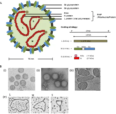
All bunyaviruses share a similar genomic organisation and replication strategy. The genome is composed of three single-stranded negative/ambi-sense RNA segments named according to their sizes large (L), medium (M) and small (S). Each segment consists of a coding region flanked by UTRs, which have terminal ends that are partially complementary and give the genome its pseudo-circular appearance (Figure 1.2). It was recently demonstrated that these complementary regions of both 5’ and 3’ UTRs interact on separate sites in the viral polymerase, a feature now thought to be conserved in all negative strand RNA virus polymerases (Gerlach, 2015). The secondary structure of the UTR functions as a promoter for viral genome replication and transcription (von Bonsdorff & Pettersson, 1975; Raju & Kolakofsky, 1989; Dunn et al., 1995; Elliott & Weber, 2009; Elliott & Blakqori, 2011).
Figure 1. 2. Bunyavirus virion structure.
(A) Schematic diagram showing a bunyavirus virion. Electron microscopy of OROV data estimates that OROV virions are ≈70 nm in diameter (Personal communication, Dr. Gustavo Olszanski Acrani, University of Sao Paulo). On the right is the coding strategy used by OROV along with its genomic segments and expressed proteins. The size of the segments and proteins are given in brackets. (B) Cryo-electron micrographs of virus particles: (i) LACV (Talmon et al., 1987); (ii) RVFV (Huiskonen et al., 2009); (iii) Tula virus (Huiskonen et al., 2010), the image shows an elongated tubular particle; (iv) electron micrographs showing UUKV RNA molecules with panhandle (Hewlett et al., 1977).
A
B
(i) (ii) (iii)
L M S
(iv)
Gc glycoprotein Gn glycoprotein
N protein
L protein (viral polymerase)
L
S
M
RNA
RNP
(RiboNucleoProtein)
70 nm
L (6.8 kb) = L (270 kDa)
M (4.4 kb) =
N (27 kDa) NSs (11 kDa) S (0.95 kb) =
Coding strategy:
3’ 5’
Figure 1. 3. The different coding strategies used by bunyaviruses.
A schematic representation of the various coding strategies of the L, M and S bunyavirus segments. RNA molecules are in the negative-sense orientation 3’ – 5’. Representative viruses include Bunyamwera virus (BUNV), Rift Valley fever virus (RVFV), Dugbe virus (DUGV), Hantaan virus (HTNV) and tomato spotted wilt virus (TSWV). mRNA orientation is in the 5’ – 3’ direction (black arrows). Viral proteins (coloured boxes) include L, polymerase; Gn and Gc, glycoproteins; N, nucleocapsid protein; and the non-structural proteins, NSm and NSs (Elliott & Schmaljohn, 2013).
3’# 5’#
RNA segment:
mRNA
Representative virus
L# all bunyaviruses
Gn# NSm# Gc# BUNV
Gn#
NSm# Gc# RVFV
78#KDa# L:
M:
Gn# Gc# DUGV/HTNV
Gn# Gc# TSWV
NSm#
N# BUNV
N# RVFV/TSWV
NSs#
N# DUGV/HTNV
The protein coding regions
L
The L gene on the L segment encodes an RNA dependent RNA-polymerase (RdRp) that the virus requires in order to transcribe and replicate its genome (Jin & Elliott, 1991). Work in the early 1990’s by Jin and Elliott established that the bunyavirus polymerase domain resides in the central region of the protein (Jin & Elliott, 1992, 1993b). This work succeeded in the initial identification of a probable “polymerase module” that appeared to remain evolutionarily conserved in every available RdRp sequence (Poch et al., 1989). Segmented negative strand virus RdRps additionally exhibit endonuclease activity and for bunyaviruses this was identified by isolating positive/coding-sense RNA that contained 5 – 15 nucleotide long host-cell derived capped oligonucleotides at the 5’ termini (Patterson et al., 1984; Jin & Elliott, 1993b, a; Garcin et al., 1995). This endonuclease activity termed “cap-snatching” was first identified in Influenza virus (Krug et al.; Plotch et al., 1979) and is a transcription initiation method where by short 5’ caps from the host pre-mRNAs are cleaved for the purpose of priming viral mRNA synthesis (Reich et al., 2014). The cap-snatching endonuclease domain of the bunyavirus L protein is present at its N-terminus (Muller et al., 1994), and is supported by structural data on LACV (Reguera et al., 2010). Nairoviruses have an Ovarian Tumor (OTU)-domain additionally present before the endonuclease domain around amino acids 29 – 158 (Honig et al., 2004; Kinsella et al., 2004; Capodagli et al., 2011; Devignot et al., 2015). Interestingly this domain is also present in RdRps of several positive-sense RNA viruses, and is implicated in antagonising the host innate immune pathway as discussed in 1.1.3.
M
similar to that of the class II fusion proteins of alphaviruses and flaviviruses, further strengthening the evidence for Gc in fusion (Dessau & Modis, 2013).
In addition to Gn/Gc some bunyaviruses also encode a non-structural protein (NSm) on the M segment (Figure 1.3 and Table 1.1) (Fazakerley et al., 1988; Matsuoka et al., 1988), and from early work using BUNV it was shown that this protein also localises to the Golgi (Nakitare & Elliott, 1993; Shi et al., 2006). A MAGV mutant with two-thirds of its NSm C-terminus missing suggested that the C-terminus of this protein is not crucial for the virus in cell culture (Pollitt et al., 2006). This is also true for SBV, as a large internal deletion in the NSm C-terminus does not affect the virus infectivity even
in vivo (Kraatz et al., 2015). What role the protein may play in the virus life-cycle in the
case of these orthobunyaviruses is still unclear. However, for the phlebovirus RVFV the NSm protein is important for infection in mosquitoes by allowing the virus to cross the midgut barrier (Crabtree et al., 2012; Kading et al., 2014). Additionally, in RVFV the NSm can stay fused to Gn, producing a protein called P78 (Figure 1.3), which also has a role in virus circulation in mosquitoes (Kreher et al., 2014). In tospoviruses the NSm protein has been shown to be important for virus cell-to-cell spread (Kormelink et al., 1994; Storms et al., 1995; Soellick et al., 2000).
S
The S segment mainly encodes the N protein. However, some ortho- and hanta- viruses have evolved to take advantage of the leaky scanning of ribosomes. In these viruses N is encoded from the first AUG site, whilst an additional protein called NSs is encoded from a downstream AUG site on the same mRNA transcript (Fuller & Bishop, 1982; Fuller et al., 1983; Vera-Otarola et al., 2012). In contrast to this, phlebo- and tospo- viruses employ an ambisense coding strategy so that N and NSs can be translated on separate mRNA transcripts from the same genomic segment. Nairoviruses are not known to encode NSs (Elliott & Schmaljohn, 2013). The size of various bunyavirus S segment proteins are listed in Table 1.1.
(Weber et al., 2001; Elliott & Schmaljohn, 2013; Brennan et al., 2015). The N protein on the other hand is important in the viral life-cycle, interacting with L protein during genome replication and with Gn-Gc during virus particle formation. Due to this, high quantities of N are present in both virions and in the virus infected host cells (Elliott & Schmaljohn, 2013). N is responsible for encapsidating and protecting each vRNA segment in the form of RNPs, and though virions contain genome and antigenome segments there appears to be a preference for genomic encapsidation (Richmond et al., 1998; Osborne & Elliott, 2000). N does this via a signal in the 5’ UTR panhandle structure (Osborne & Elliott, 2000; Severson et al., 2001; Mir & Panganiban, 2004) and encapsidation begins via N-N interaction. Using reverse genetics these interacting regions in BUNV N were mapped to residues 1 to 10 of the N-terminus and residues 216 to 233 of the C-terminus, along with a central domain encompassing amino acids 94 to 158 (Leonard et al., 2005; Eifan & Elliott, 2009). Similar N-N oligomerisation and RNA binding have also been suggested for viruses in the tospo- and hanta- virus genera (Uhrig et al., 1999; Kaukinen et al., 2001; Severson et al., 2001; Kaukinen et al., 2003). Protein structural data for several orthobunyavirus N proteins have now confirmed the original BUNV mutagenesis studies. N protein structures for BUNV (Li
et al., 2013), leanyer virus (LEAV) (Niu et al., 2013), SBV (Dong et al., 2013b; Dong
et al., 2013a) and LACV (Reguera et al., 2013) demonstrate that N forms a tetrameric
its N-terminus, which can result in the cleavage of the N protein. This site was shown to be conserved in several CCHFV strains, although what significance it holds in terms of the virus life-cycle is yet to be determined (Karlberg et al., 2011; Carter et al., 2012).
An interesting deviation from the typical NSs-encoding orthobunyaviruses is Brazoran virus. Its putative NSs ORF is encoded from the first initiation codon prior to N, although using a weak Kozak sequence. This putative protein is 19.6 kDa in size which is larger than the “typical” ≈10 kDa orthobunyavirus NSs protein size. In addition, the C-terminus of N contains a glutamine-rich domain not present in other orthobunyavirus N proteins (Lanciotti et al., 2013). At present it is unclear what the significance of any of these observations are; future functional and structural work on these proteins will however prove useful to understanding the evolution of these diverse bunyaviruses.
Table 1. 1. Bunyavirus protein size
The various proteins (sizes in kDa) encoded by representative members of each genus.
Genus Virus L Gn Gc NSm N NSs
Hantavirus Hantaan 247 70 35 0 48 0 Nairovirus Dugbe 459 35 73 0 50 0 Orthobunyavirus Bunyamwera 259 32 110 11 26 11
Figure 1. 4. Crystal structure of SBV N.
(A) Protomer structure of SBV N showing the N terminal arm (NTA; blue), a C-terminal domain (CTD) and the C C-terminal arm (CTA; red). Taken from (Dong et al., 2013a). (B) Tetrameric structure of SBV N–RNA complex. RNA (42 nucleotide, stick form, orange) bound inside the tetrameric SBV N ring, formed by four protomers (blue, green, yellow and cyan). The dotted line shows a gap in the bound RNA. Taken from (Dong et al., 2013b).
A
The untranslated regions
The bunyavirus UTR contains signals for genome replication, packaging and encapsidation. RNA secondary structures are crucial for such functions. In bunyaviruses, the terminal 3’ and 5’ ends of each segment are complementary and based on available sequences the first 8 to 11 nucleotides of all three segments are highly conserved within a given genus (Table 1.2). Beyond the conserved nucleotides the sequences and UTR lengths begin to vary and become unique within a segment and a specific virus (Elliott et al., 1991; Kohl et al., 2004a; Barr et al., 2005; Elliott & Schmaljohn, 2013).
Table 1. 2. The terminal UTR consensus sequence of bunyaviruses
Sequences are represented in genomic sense. Some orthobunyaviruses differ from the prototype sequence at positions 8 and 9, which are highlighted in bold. The mismatch at position 9 is highlighted in red.
Genus Virus Terminal nucleotides
Hantavirus Hantaan 3'- AUCAUCAUCUG... 5'- UAGUAGUAUGC...
Nairovirus Dugbe 3'- AGAGUUUCU... 5'- UCUCAAAGA...
Orthobunyavirus Bunyamwera 3'- UCAUCAC5'- AGUAGUGUAGUGA... CU...
Phlebovirus Rift Valley fever 3'- UGUGUUUC... 5'- ACACAAAG...
Tospovirus Tomato spotted wilt 3'- UCUCGUUA...
5'- AGAGCAAU...
viruses lacking large portions of these sequences are viable (Lowen & Elliott, 2005; Mazel-Sanchez & Elliott, 2012). The minimum requirement for a viable BUNV S segment mutant is a 22-nucleotide 5’-UTR and at least 112 nucleotides at the 3’-terminus (Lowen & Elliott, 2005). Further work on BUNV M revealed that nucleotides 20 to 33 at both termini are important for genome packaging (Kohl et al., 2006). Also the segment-specific sequences appear to have a role in regulation of packaging and co-packaging of each segment into a single virion (Kohl et al., 2006; Terasaki et al., 2011). Recent work on BUNV has now demonstrated that the L protein evolves to some degree in order to accommodate mutations in these UTRs (Mazel-Sanchez & Elliott, 2015)
Table 1. 3. Examples showing the 3’ and 5’ UTR lengths for different orthobunyaviruses (antigenome sense)
Virus Serogroup L M S Accession No.s GenBank
5' 3' 5' 3' 5' 3'
Oropouche Simbu 43 50 31 91 44 218 KP052850-52 Schmallenberg Simbu 27 90 23 138 31 106 KC355457-59 Leanyer Simbu 68 180 40 141 67 179 HM627179-81 Brazoran unclassified 44 126 58 230 71 272 NC_022038-39,
KC854416 Bunyamwera Bunyamwera 50 108 56 100 85 174 NC_001925-27 La Crosse California encephalitis 61 127 61 140 81 195 NC_004108-10
1.1.2 The life-cycle of a bunyavirus particle in mammalian cells
Attachment and entry
Similar to other enveloped viruses a bunyavirus virion utilises its glycoproteins Gn and Gc to attach and enter into a host cell. The type of cellular receptor the virion binds to will determine the cell tropism of that virus. Current understanding of host cell receptors for bunyaviruses are limited, however knowledge of the varied cell tropism of some bunyaviruses indicate that these viruses may have evolved to interact with a number of mammalian cell receptors (Elliott & Schmaljohn, 2013). Phleboviruses RVFV, SFTSV, TosV, Punta Toro (PTV) and UUKV, and orthobunyavirus Germiston virus were shown to interact with host cell receptor DC-SIGN (DC [dendritic cell] - specific ICAM [intercellular adhesion molecule] - 3 grabbing non-integrin). DC-SIGN is a receptor present on immature DCs, which reside in peripheral tissues and are likely the first cells to encounter incoming viruses. DC-SIGN is a type II membrane protein with a calcium-dependent lectin extracellular domain likely capable of interacting with the glycosylated sites on the viral glycoproteins (Lozach et al., 2011; Hofmann et al., 2013). This interaction would then trigger a response in the DC causing it to mature into an antigen-presenting cell (Tan & O'Neill, 2005). As number of viruses appear to have evolved to use DC-SIGN as entry into mammalian cells they have also evolved mechanisms of blocking the maturation of these cells (Rogers & Heise, 2009). Phleboviruses RVFV and TosV have also been shown to interact with the proteoglycan heparin sulfate receptor (Jin et al., 2002; de Boer et al., 2012b). Whilst, pathogenic hantaviruses can interact with integrins β1, β2 (CR3 and CR4) and β3 (Gavrilovskaya et
al., 1998; Gavrilovskaya et al., 1999; Raftery et al., 2014), as well as Decay-accelerating factor (DAF)/CD55 and gC1qR/p32 (Choi et al., 2008; Krautkramer & Zeier, 2008) for attachment to endothelial and epithelial cells.
1.5 A, step 2). Interestingly, UUKV appears to predominantly use a clathrin-independent pathway to enter A549 (human) and BSC-40 (money) cell-lines (Lozach et al., 2010). Akabane virus (AKV) on the other hand uses the clathrin-independent pathway in bovine-derived cell-lines, whereas in non-bovine cells it seems to use the CME pathway (Bangphoomi et al., 2014).
Membrane fusion
The endocytic pathway follows a pH gradient that can be detected by bunyavirus glycoproteins triggering a conformational change in their structure (Överby et al., 2008). This pH sensing is possible due to protonation of the histidine residues typically found on the viral fusion protein (Kampmann et al., 2006; Mueller et al., 2008). As discussed previously, Gc likely functions as a fusion protein. This is further strengthened by work on RVFV, where certain conserved histidine residues on Gc were found to be important for virus infectivity (de Boer et al., 2012a). Conformational changes in Gc would then mediate fusion between viral and endosomal membranes, hence allowing release of viral RNP into the cell cytoplasm (Figure 1.5 A, step 3) (Mercer et al., 2010; Cosset & Lavillette, 2011). Several enveloped viruses are known to use this mode of penetration (White et al., 1981; Kielian et al., 2010). Detailed work by Lozach et al. demonstrated that UUKV is transported from the early endosomes (pH <6.3) to late endosomes (pH <5.3) before infection occurs (Figure 1.6 B). The authors demonstrated that infection was pH dependent since neutralisation of vesicular pH inhibited infection, whilst acidification of the external cell environment was sufficient to trigger fusion of the viral and cell plasma membranes (Lozach et al., 2010). Cell-to-cell fusion has been shown using different bunyaviruses, where over-expression as well as infection induces syncytium formation (Jacoby et al., 1993; Hacker & Hardy, 1997; Ogino et al., 2004; Plassmeyer et al., 2005; Shi et al., 2007).
Transcription and Replication
this using BUNV and RVFV demonstrated that both L and N are together required for active transcription (Dunn et al., 1995; Lopez et al., 1995). As N also encapsidates the genome, structural data suggests that N could potentially expose the UTRs using its flexible arms allowing L to bind (Elliott, 2014). Recent structural data on LACV L bound to vRNA revealed that the terminal 3’ and 5’ UTR sequences are crucial and that they each bind specific regions in the L molecule, confirming all prior in vitro
mutagenesis work. The overall crystal structure of L indicates a main globular core, which harbours the RdRp and RNA-binding domains connected to a flexible endonuclease domain by a linker region. The template entry and exit tunnels and the nascent RNA exit tunnels are located in the main globular core (Gerlach et al., 2015).
Transcription occurs prior to replication and unlike transcription, replication occurs in a cap-independent manner. The exact mechanism for switching from cap-dependent to cap-independent initiation is still uncertain, but the possibilities of host cell translation shut-off forcing this switch; or that viral and/or host proteins may be involved in the process have been proposed (Guu et al., 2012; Elliott & Schmaljohn, 2013). In transcription the nascent mRNA terminates upstream of the 5’ end, however in replication the nascent strand is processed right to the very end of the 5’ termini. Transcription termination signals ubiquitous to all three segments have not been found, but a pentanucleotide sequence 5’-UGUCG-3’ in BUNV S segment appears to be able to signal termination (Barr et al., 2006; Ikegami et al., 2007; Blakqori et al., 2012).
Release
Figure 1. 5. Life cycle of the bunyaviruses.
(A) Schematic diagram of the various stages in a bunyavirus life-cycle. EE, early endosomes; LE, late endosomes; ER, endoplasmic reticulum; SV, secretary vesicles. (B) Transcription and replication of the bunyavirus genome. The genome is in a negative-sense orientation and is transcribed into a replicative intermediate known as the antigenome. Red circles depict the N protein encapsidating the genome/antigenome. The red dashed-box highlights that the mRNA contains 10 – 15 nt long host-derived primers/caps. Numbers
5’ 3’
3’ 5’
6. Genome replication
5’ 3’
host-derived cap 5. Translation 4. Transcription Protein RNA species Antigenome (+) Genome (-) mRNA (+) bunyavirus Nucleus ER Golgi L S M L S M L = S = M = L S M L S M L S M L S M L S M
1. Attachment to cell receptor
L S M SV L S M L S M L S M 2. Endocytosis L S M 3. Fusion EE LE Lysosome 4. Transcription 5. Translation 6. Genome replication
7. Assembly 8. Migration 9. Fusion 10. Egress 5’# 3’# 3’# 5’#
5’ 3’
5’# 3’# 3’# 5’#
Figure 1. 6. Electron micrographs of bunyavirus entry and exit from the host cell.
(A) OROV (arrowhead) entering HeLa cells via clathrin-coated pits (arrow). Image
taken from (Santos et al., 2008). (B) UUKV inside early (EE#2) and late (LE)
endosomal vesicles in A549 cells. Image taken from (Lozach et al., 2010). (C) Viral
factories of BUNV in the BHK-21 cells. G, Golgi; V, virus particle. The arrows show various tubular and globular structures that form part of the viral factories. Image taken
from (Fontana et al., 2008). (D) BUNV inside the Golgi in BHK-21 cells (arrows),
post-Golgi area (arrowhead). (E) BUNV exiting BHK-21 cells by secretary vesicles (SV) (F). Exited BUNV attached to the cell surface. Images (D), (E) and (F) were taken from
(Salanueva et al., 2003), where the cryosections were labelled with anti-Bunyamwera
A B
C D
E F
150 nm
100 nm 100 nm
100 nm
1.1.3 Host responses to bunyavirus infection
Vertebrate and invertebrate cells have evolved to recognise molecular signatures, thereby enabling them to distinguish between “self” and “non-self”. In terms of a virus infection, certain distinct features on the viral genome or on their replication intermediate are recognised by “pattern” recognition receptors (PRRs) on or within the host cell. These foreign molecular features are known distinctively as pathogen-associated molecular patterns (PAMPs) and they can set in motion a series of events all geared towards preventing onwards spread of the infection (Ausubel, 2005; Randall & Goodbourn, 2008; Iwasaki, 2012).
The type 1 interferons and bunyaviruses
Mammalian cells can sense the presence of foreign RNA through cellular receptors (PRRs) such as transmembrane toll-like receptors (TLR), and cytosolic receptors RIG-I (retinoic acid-inducible gene 1) and MDA5 (melanoma differentiation-associated protein 5). TLRs are mainly expressed in lymphocytes, dendritic cells, macrophages and epithelial cells. For instance, TLR3 is highly expressed in the endosomes of myeloid
dendritic cells and can recognise dsRNA following endocytosis of the virus (Schaefer et
al., 2004). RIG-1 and MDA5 on the other hand are more widely expressed. RIG-I
receptors recognise short dsRNA with triphosphorylated 5’ (5’ppp) ends, whereas
MDA5 can recognise long dsRNA structures (Goodbourn et al., 2000; Randall &
Goodbourn, 2008; Reikine et al., 2014; Schneider et al., 2014; Hoffmann et al., 2015;
Weber, 2015). Recognition by RIG-1 and MDA5 triggers phosphorylation of transcription factors such as the interferon regulatory factors (IRF) IRF-3 and IRF-7,
and NF-κB (nuclear factor kappa-light-chain enhancer of activated B cells) via
activation of mitochondrial adaptor molecule called Cardif/VISA/MAVS/IPS-1 (CARD
adaptor inducing IFN-β/Virus-induced signalling adaptor/Mitochondrial antiviral
signalling protein/IFN-β promoter stimulator protein-1). The phosphorylation of the
transcriptional factors leads to their translocation into the nucleus in order to induce the
production of type 1 interferons (IFNs), IFN-α and IFN-β (Figure 1.7). Translocation
NF-κB is exposed after phosphorylation of its inhibitor molecule (Inhibitor of NF-κB,
IκB) that is subsequently degraded by the proteasome. Tank Binding Kinase 1 (TBK1)
and IKKε are essential kinases which are also involved in activating IRF-3 and IRF-7.
IRF-3 activation can also occur via TLR3 signalling through adaptor molecule
Toll-interleukin (IL)-1-resistance (TIR) domain-containing adaptor inducing IFN-β (TRIF)
and kinases TBK1 and IKKε (Goodbourn et al., 2000; García-Sastre & Biron, 2006;
Randall & Goodbourn, 2008; Schneider et al., 2014; Hoffmann et al., 2015).
IFN-α and IFN-β are cytokines that are expressed widely by a number of cell-types, and
are encoded on chromosome 9 along with other type 1 IFNs (IFN-ε, IFN-Κ and IFN-ω)
(Reikine et al., 2014). The produced IFN-α/β use the JAK-STAT signalling pathway to
signal the expression of a number of proteins that have antiviral effects and/or are positive or negative regulators of the pathway (IFN-stimulated genes, ISGs)
(Goodbourn et al., 2000; Randall & Goodbourn, 2008; Schneider et al., 2014;
Hoffmann et al., 2015). The JAK-STAT pathway is activated when IFN-α/β bind to the
type 1 IFN heterodimeric receptor complex (IFNAR1 and IFNAR2), both on the cell they were produced in and on adjacent cells. The IFNAR receptors are expressed on almost all cells and when activated lead to a conformational change in the bound IFN receptor chain with Janus activated Kinase 1 (JAK1) and non-receptor tyrosine kinase 2 (TYK2). JAKs are ubiquitously present in cells and activated JAKs lead to phosphorylation of transcriptional activator proteins STAT1 and STAT2 (signal transducer and activator of transcription). STAT1 is phosphorylated by JAK1 on tyrosine 701 and Tyk2 phosphorylates STAT2 on tyrosine 690. This causes the dimerization of STAT1 and STAT2 hence forming a NLS, further STAT1/STAT2 interact with IRF9 to form a heterotrimeric transcription factor complex ISG factor 3 (ISGF3), which translocates to the nucleus and binds to an IFN-stimulated response element (ISRE) in the genome. Activation of ISRE then leads to induction of over 100
ISGs (Figure 1.7) (Goodbourn et al., 2000; García-Sastre & Biron, 2006; Randall &
Goodbourn, 2008; Reikine et al., 2014; Schneider et al., 2014; Hoffmann et al., 2015).
have evolved to encode proteins that function specifically as antagonists against this
system (Goodbourn et al., 2000; Randall & Goodbourn, 2008; Schneider et al., 2014;
Hoffmann et al., 2015). Bunyaviruses tend to target the cells’ early response stage. New
York-1 hantavirus (NY-1) for instance, uses its Gn-CT tail to inhibit RIG-1 as well as
TBK1 (Helgason et al.; Fitzgerald et al., 2003; Alff et al., 2006). Whilst, NSs-encoding
bunyaviruses mainly use the NSs protein to target the transcriptional machinery of the
host cell hence inhibiting IFN induction (Weber et al., 2002; Elliott & Weber, 2009;
Elliott & Blakqori, 2011). The first evidence of NSs being an IFN antagonist was
demonstrated in 2001 when Bridgen et al. created a BUNV mutant lacking a
functioning NSs ORF (rBUNdelNSs) (Bridgen et al., 2001). This mutant displayed a
reduced ability to shut-off host cell protein synthesis, was attenuated in mice and induced high levels of type I IFN in cultured cells. Similar results were soon shown
with the phlebovirus RVFV (Bouloy et al., 2001). RVFV mutants Clone-13, with a
large in-frame NSs deletion of 549 nucleotides, and MP12 with a single-amino acid
substitution at position 513 in NSs are attenuated in vivo and in vitro. When both these
mutant NSs ORFs were replaced with that of wild-type (wt) RVFV the viruses behaved similar to the wt in terms of IFN induction and replication efficiency in IFN-competent
mice. The absence of IFN-β mRNA in both the wt RVFV and wt BUNV infected cells
demonstrated that NSs was blocking IFN-β production at the transcription level (Bouloy
et al., 2001; Weber et al., 2002). Similar results were obtained with other bunyaviruses
such as LACV, AKV, SBV and UUKV (Blakqori & Weber, 2005; Ogawa et al., 2007;
Elliott et al., 2013; Rezelj et al., 2015) demonstrating that at least in these viruses NSs
is non-essential for virus replication, but rather enhances replication by antagonising the host cells response.
We are still in the process of understanding all NSs interactions within a mammalian
cell. From work on BUNV and RVFV we know that its ability to inhibit IFN-β
activation occurs downstream of transcriptional activation through disruption of the
DNA-dependent RNA polymerase II (RNAPII) activity (Weber et al., 2002; Kohl et al.,
2003b; Billecocq et al., 2004). RNAPII is an enzyme that transcribes all protein- coding
genes of the eukaryotic genome and is regulated by multiprotein complex Mediator
cell stimuli signalling the complex formation, which consists of modules head, middle and tail. The head module is involved in stabilizing the RNAPII initiation complex
(Plaschka et al., 2015) and BUNV NSs was found to interact with sub-unit Med8 of this
module (Figure 1.8) (Leonard et al., 2006). Med8 is one of the sub-units that interact
with the carboxy-terminal domain (CTD) of RNAPII. The CTD is made up of a heptad
repeat sequence (Y1S2P3T4S5P6S7), of which there are 52 in the mammalian RNAPII,
and its phosphorylation is essential for RNAPII function. Phosphorylation at serine (Ser) 5 is required for initiation and recruitment of capping enzymes, while Ser2 phosphorylation is required for elongation and 3’-end processing of the nascent mRNA
transcript, Figure 1.8 (Robinson et al., 2012; Corden, 2013; Eick & Geyer, 2013). Cells
infected with BUNV show a significant reduction in RNAPII Ser2 phosphorylation. This was initially thought to be due to an interaction of the BUNV NSs C-terminus (amino acids 83 – 91) with Med8, however a BUNV NSs mutant lacking a N-terminus of 21 amino acids was also unable to degrade RNAPII, indicating that both the C- and
the N- terminus are important for NSs function (Thomas et al., 2004; Leonard et al.,
2006; van Knippenberg et al., 2010). LACV and SBV NSs target RNAPII for
degradation by the proteasome and C-terminal mutations of SBV NSs have been shown
to affect the protein’s ability to degrade RNAPII (Blakqori et al., 2007; Verbruggen et
al., 2011; Barry et al., 2014). The NSs protein of RVFV on the other hand interacts with
subunits of the general transcription factor TFIIH (Figure 1.8), which also has a role in
RNAPI transcription (Assfalg et al., 2012). With RVFV NSs preventing subunits p44
and XPD from interacting, it presumably affects the activity of Cdk7 (cyclin-dependent kinase) of CAK (Cdk activating kinase), which is responsible for Ser5 phosphorylation
on RNAPII, Figure 1.8 (Le May et al., 2004).
RVFV NSs is intriguing as so far it is the only known bunyavirus NSs to form filamentous structures in the nucleus. It is also so far the only bunyavirus NSs shown to
directly target the IFN-β promoter by interacting with SAP30 (Sin3A associated protein
30) and transcriptional repressor protein YY1. This interaction maintains the
YY1/SAP30/NCor/HDAC/Sin3A co-repressor complex in a silent state on the IFN-β
promoter thereby directly inhibiting IFN-β transcription (Le May et al., 2008). With
virus uses to capture kinases TBK1 and IKKε, and the proteins STAT1 and STAT2
(Ning et al., 2014; Ning et al., 2015). Viruses from the Anopheles A, B and Tete
serogroups encompass some of the non-NSs encoding orthobunyaviruses, and as
expected show no inhibition of IFN-β production (Mohamed et al., 2009). For rodent
viruses TULV and PUUV, only putative NSs ORFs have been predicted, and
over-expression of these in reporter assays have shown an inhibition of IFN-β, NF-kB and
IRF-3 activity (Jaaskelainen et al., 2007). HTNV, Dobrava (DOBV) and SEOV rodent
viruses on the other hand have been shown to use their N protein to inhibit NF-kB
activation, by targeting the signalling protein tumor necrosis factor-α (TNF-α) (Taylor
et al., 2009). Nairovirus CCHFV however, seems to use its L protein to antagonise the
host immune system. For the TNF-α mediated NF-kB pathway to be activated its
inhibitor IκB must be ubiquitinated, and with an OTU-domain present on the CCHFV L
protein the virus is able to inhibit protein ubiquitination in the cell. OTU-domains have protease activity and these domains are also present in other nairoviruses such as


